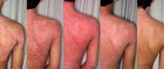
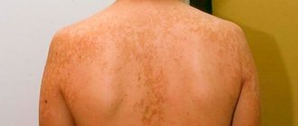
Pityriasis rosea, or Gibert's disease, is a common skin pathology that manifests itself as spotty rashes of varying sizes and locations. Although the disease often goes away on its own, ignoring its symptoms can lead to skin inflammation and other problems.
Cost of services in our clinic
| Appointment with a dermatologist, candidate of medical sciences | 1500 rub. |
| Consultation with a dermatologist (KMS) when removing 2 tumors | 0 rub. |
| Removal of a neoplasm (wart, mole) using the radio wave method | 500 rub. |
| Make an appointment by phone: 8-800-707-15-60 (toll-free) |
| *The clinic is licensed to remove tumors |
The popular belief that the disease can be “caught” from animals is incorrect. Infection occurs only from humans.
Diagnosis of the disease
Determining the correct diagnosis is sometimes difficult due to the absence of a pathogen. The disease can be confused with other lichen and toxicoderma.
The examination is a visual inspection. A qualified dermatovenerologist will take into account the characteristic placement of spots on the body, their appearance, shape, and based on this will make a diagnosis.
Tests that may also be prescribed:
- general urine and blood tests;
- microprecipitation reaction (diagnosis of syphilis);
- scraping from affected areas.
After diagnosis, the specialist will determine how to treat pityriasis rosea and will advise the patient in detail regarding lifestyle.
Make an appointment
Lichen planus
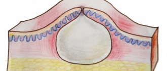
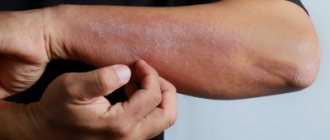
Cutaneous manifestations of lichen planus are a rash that looks exactly the same, located on various parts of the body. The lesions have the form of peculiar compacted elevations on the skin, which have an irregular shape; their surface appears shiny when examined. They have a pinkish-purple color and a slight depression in the central part. Sometimes the color of the formations can be crimson-red. Their sizes are most often small - from 2 to 3 mm. The shine of the surface of the lesions is the most characteristic feature. It is very visible if you look at the patient's skin at a slightly angle, directing the light from the lamp in the same way.
Although the size of the lesions is relatively small, they may increase in the future. Individual lesions are capable of merging with each other, resulting in large plaques forming on the patient’s skin. Their surface begins to peel off. The scales are small in size, which is a fairly characteristic feature. For a more accurate diagnosis, there is also a simple test. Its essence is that a small layer of vegetable oil is applied to the skin in the area of pathological lesions. As a result, visible signs appear on the surface of the affected skin. Some of them are small white dots. Others look like white stripes, which seem to be located in the thickness of the skin and shine through it. After the name of the scientist who discovered it, this phenomenon is called Wickham’s sign. It is due to the fact that in the area of pathological formations there is a thickening of the stratum corneum of the skin, but this occurs unevenly. After the patient recovers and the lesions disappear, more intensely colored areas of skin remain in their place. They are quite persistent and can last for a long time. Skin rashes with lichen planus are accompanied by subjective sensations on the part of the patient. This manifests itself in the form of a feeling of itching and pain. The pain syndrome is constant and can be intense, as a result of which sleep, appetite and daily life are disrupted, and non-vrosis-like symptoms can develop.
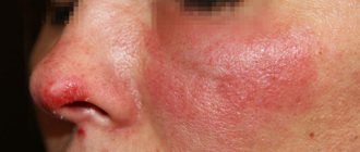
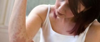
Lichen planus has, so to speak, a “favorite” location on the skin, which is located in the area of the flexor surfaces of the forearms. Other common sites of localization may be the areas of the wrist joints, the inner surfaces of the thighs, and the extensor surfaces of the legs. Often plaques are found in the groin, armpits, and oral cavity. The skin of the face and scalp, palms, and soles, on the contrary, is almost never affected. In some cases, the elements of the rash are located in an interesting way: they appear as stripes that alternate with areas of normal skin. This is most often observed on the extremities. The appearance of the patient's skin in these cases is so unique that it is almost impossible to confuse the pathology with any other. Sometimes there is not a single pathological focus on the patient’s skin, while the mucous membranes are affected to a very significant extent. This occurs in a quarter of all patients with lichen planus.
The mucous membranes of various locations are affected: in the mouth, glans penis in men and the vestibule of the vagina in women. Lesions on the mucous membrane of the cheeks look characteristic. Here they have a grayish color and are represented by very small nodules, almost the size of a pinhead. They are always arranged in groups that have the most bizarre shapes: in the form of rings, nets, openwork lace. The lesions located on the surface of the tongue are completely different in appearance. They are white and similar to the rashes associated with leukoplakia. These are plaques protruding above the surface of the mucous membrane, which have clearly defined jagged contours. Rashes with lichen planus can also be located on the red border of the lips. In these cases, the color of the lesions is purple; they also appear as plaques with a scaly surface. Another characteristic feature is that the plaques are covered with a grayish-white mesh. The lower lip is affected much more often than the upper lip. Some patients with lichen planus have changes in the nail plates. A clearly visible longitudinal striation appears on them, which is sometimes so pronounced that it looks like scallops. The nail bed becomes red due to an inflammatory reaction of allergic origin. Spots of cloudiness form on the nail plates of the fingers and toes.
Another argument in favor of the allergic origin of the disease is that lesions of lichen planus appear mainly after exposure to irritating factors on the skin. Moreover, the focus is formed strictly in the place where the impact was applied. For example, often lesions resulting from scratching have the appearance of characteristic stripes.
The course of the disease is long, sometimes even for years and months. Sometimes the lesions occupy not only the above areas, but also spread to all skin integuments. This form of the disease is called generalized. This is a severe and long-lasting pathology, which is often complicated by the addition of another disease - secondary erythroderma.
The so-called typical course of lichen planus was described above. However, making a diagnosis is a simple task due to the extremely characteristic manifestations of the pathology. But this does not happen in all cases. There are a number of atypical forms of the disease that develop as new types of allergens, in the presence of concomitant diseases of the skin and other organs, and in cases of disorders in the immune system and metabolism in the body.
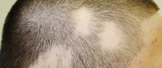
Hypertrophic warty form. As the name implies, this form of the disease is based on the appearance of warty growths of the skin. Plaques appear that are purple or brownish-brown in color. Their surface is covered with a large number of small warts, in the area of which there is a thickened stratum corneum of the skin. Around these plaques and warts there may be small nodules quite typical for ordinary lichen planus. The process most often affects the front surfaces of the legs. Lesions can also occur in other places, but this is much less common. Atrophic and sclerotic forms of the disease. At the very beginning, the disease proceeds quite typically, with plaques and nodules characteristic of lichen planus appearing on the skin. However, later, when they disappear, areas of atrophy and thickening (sclerosis) of the skin remain on the patient’s skin in these places. A very unique and interesting picture develops when the process is localized simultaneously on the scalp. In this case, spot baldness develops, which may resemble some other pathologies in appearance. In this case, compaction and thickening of the stratum corneum of the skin on the extensor surfaces of the extremities is almost always observed. This peculiar picture was named after the authors who discovered and described it, the Little-Lassouer symptom.
Pemphigoid, or vesicular, variety. This is a very rare form of the disease. It manifests itself in the form of the formation of lesions on the skin in the form of small bubbles filled with clear liquid, sometimes mixed with blood. The bubbles are small in size, often no larger than a pea or cherry. Blisters can appear on the skin either without prior visible irritation or against the background of nodules, plaques, etc. The process is located in most cases in the area of the legs or feet. In some cases, lesions characteristic of both the simple form of lichen and the blistering form may appear simultaneously on the skin of the same patient.
Moliniform variety of lichen planus. This form of pathology is also rare. Quite large lesions appear on the skin, which are arranged in chains and seem to be strung in the form of a necklace. They are most often the size of a cherry pit. The lesions are nodules. They rise dome-shaped above the skin and have a rounded appearance. Sometimes they are quite small, dense, located close to each other in the form of strips, which can create the appearance of linear scars. In some cases, they clearly resemble beads in appearance. Such rashes usually affect large areas of the skin. There are especially many of them in the forehead, on the back of the ears, on the neck, in the area of the elbows, on the back of the hands, on the stomach, and in the buttocks. In most patients, the lower areas of the face - cheeks, nose - remain completely unaffected; areas of skin above the collarbones, between the shoulder blades. The palms and soles, as well as the genitals, are never affected. A number of authoritative researchers are inclined to consider this pathology not as a type of lichen planus, but as a completely independent disease that has its own causes.
Pointed, or perifollicular, form. With this type of lichen planus, lesions characteristic of its simple form, and others that are slightly different, appear on the patient’s skin. The nodules have a conical shape and contain a small spike at their top. This picture develops when the hair follicles are predominantly affected by the pathological process. It is clear that the most common localization of rashes is the scalp. After the lesions disappear, small scars remain in these places, which seem to sink in comparison with the surrounding skin.
Ring-shaped variety of the disease. It develops when the lesions of lichen planus very quickly increase in size, at the same time they begin to heal from the central part. The result is like a ring. In the center after healing there is always a brighter colored area of skin. In addition to rings, pathological formations on the skin in this form can be in the form of garlands, arcs, or half rings. They can occur both during the growth of one lesion, and during the merging of several. This type of disease develops much more often in males, and the formations are located mainly in the genital area. Sometimes the lesions can very closely resemble in their appearance papules of syphilitic origin, which leads to an incorrect diagnosis and incorrect therapy.
Linear lichen planus and zosterimorphic lichen planus . They are very similar forms of the disease to each other, so they are combined into one group. In this case, the lesions are located on the skin along the course of large nerves and resemble herpetic rashes.
Erosive-ulcerative form. The disease affects the mucous membranes. This is the most severe and difficult to treat type of lichen planus. Defects appear on the mucous membranes of the oral cavity and on the lips, which in some cases can even turn into ulcers. Around these formations there are areas of redness of the skin, in the area of which lesions typical of lichen planus occur. Defects in the mucous membranes have irregular contours and are often covered with films and plaques. If these films are removed with a spatula, bleeding easily occurs. In most patients, these lesions are few in number and small in size, while the patient does not experience any subjective sensations or make any complaints. The difficulty of treating this form is that during the therapy itself, most ulcers disappear on their own without a trace. However, after stopping treatment, the rash almost always develops in the same or different place. The process can continue for a long time.
Methods of treating the disease
With uncomplicated pityriasis rosea, the patient needs to change his lifestyle:
- avoid skin irritation in the rash area;
- use a soft washcloth when bathing, and it is better to reduce the water procedures themselves to a minimum;
- follow a hypoallergenic diet;
- exclude the use of cosmetics for the skin;
- Do not wear synthetics and woolen items until recovery.
In case of a protracted process and complications, the doctor may prescribe:
- antiviral agents;
- local antibacterial agents - ointments (in case of infection);
- treating stains with salicylic alcohol.
After healing, the skin on the affected area may darken or become lighter, but will soon return to normal.
In case of incorrect treatment:
- using products that irritate the skin;
- improper bathing;
- with hyperhidrosis;
- the presence of allergies - rashes can persist for a long time and cover the entire body.
Therefore, it is important not to self-medicate. The diagnosis of pityriasis rosea must be confirmed by a dermatovenerologist.
Contraindications
Patients with pityriasis rosea, depending on the degree of the disease, may be contraindicated for prolonged water procedures - hot baths, showers. This is due to the fact that the spots are predisposed to swelling, and water can provoke swelling. If water is regularly exposed, damaged tissue may respond poorly to treatment.
However, it is impossible to completely abandon the treatment of inflammation - bacterial microflora will cause a deterioration in the condition of damaged tissues. In this case, a quick rinse in cool or slightly warm water is recommended.
The patient must avoid skin irritation so as not to provoke an exacerbation of the disease. Irritants include:
- rough linen, wool or synthetic clothing;
- ultraviolet irradiation;
- cosmetic and medicinal preparations with tar and sulfur;
- hypothermia.
The disease can be complicated if the diet contains:
- citrus;
- smoked meats;
- dairy products;
- marinade, hot seasonings and sauces;
- fish, seafood;
- carbonated drinks;
- dried fruits.
Only a doctor can give precise nutritional recommendations.
If you notice symptoms of pityriasis rosea, immediately consult a dermatovenerologist.
Other articles by the author
- Pyoderma
- Herpes zoster
- Pityriasis rosea
- Shingles
Methods of infection and risk groups
At the moment, there is no reliable evidence of the transmission of this disease from a sick person to a healthy one. There are only infrequent cases when someone in the environment fell ill with lichen upon contact, but in all cases there were provoking factors. These include:
- presence of herpes virus in the body;
- weakened immune system;
- recently suffered viral and colds;
- frequent hypothermia of the body.
The risk group mainly includes women under 40 years of age and children. Men suffer from this disease much less frequently. Almost no cases of infection are detected in infants and people over 40 years of age. In order to reduce the risk of contracting Gibert's disease, it is worth strengthening your immune system. It is reliably known that in most cases the disease appeared against the background of a weakening of the body’s protective functions.