- What is nail hyperkeratosis?
- Causes
- Treatment methods for hyperkeratosis of the nail plate
Hyperkeratosis of the nail bed is an excessive thickening of the stratum corneum against the background of slow desquamation of dead cells. As a result, the nail takes on a spherical shape, its color becomes yellow and uneven, and the nail plate becomes brittle and inelastic. The disease occurs in a chronic form. The pathological focus can spread to neighboring structures - affecting other nails and skin of the foot. Against the background of mycoses, itching and an unpleasant odor are possible.
Effective procedures for the treatment of hyperkeratosis
Phototherapy
Plasma therapy
NeoGen procedure
Mesotherapy
Peeling
Medical pedicure
One of the fairly common types of skin diseases is hyperkeratosis. This is an excessive thickening of the stratum corneum of the epidermis. With the development of such a pathology, horn cells begin to rapidly divide and desquamation is impaired, which ultimately leads to thickening of the skin. Moreover, the thickening can reach several centimeters. According to statistics, hyperkeratosis is observed in approximately 20% of men and 40% of women. As a rule, the problem occurs in individual areas and leads to a significant deterioration in the appearance of the skin.
In children, the pathology can be observed at the age of 1–3 years, and it mainly affects the face and hands. In adults, changes in the stratum corneum are more common on the feet and toenails, as well as on the face and scalp. There are several types of the disease, which have their own symptoms and causes of development. Let's figure out what hyperkeratosis is and whether such a pathology is dangerous for health.
What causes?
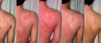
The main risk factor for the development of psoriasis is hereditary disposition. In 40% of patients, immediate relatives suffer from the same disease.
Other causes of psoriasis of the toenails and fingernails include:
- nervous stress, emotional instability;
- living in a cool, dry climate;
- past infectious diseases;
- bad habits;
- malnutrition;
- frequent mechanical damage to the nail plate.
Nail psoriasis is a chronic disease that cannot yet be cured.
It consists of periods of remissions and exacerbations. Exacerbation attacks are caused by certain medications (lithium drugs, antidepressants, antimalarials, anticonvulsants, antibiotics), dietary errors, alcohol intake, nervous stress, and infections.
The risk of developing the disease does not depend on gender or age. It is not uncommon for a child to develop nail psoriasis, and even a newborn can suffer. In 75% of cases, parents or doctors discover the first signs of pathology in childhood before the age of 5.
Features of the pathology
The concept of hyperkeratosis comes from two Greek words: hyper – “many” and keratosis – “formation of keratin”. In fact, hyperkeratosis is not an independent disease - most often it is just a symptom of other diseases. For example, such a problem occurs with lichen, ichthyosis or erythroderma. However, thickening of the stratum corneum can sometimes appear in a healthy person: for example, the feet, elbows and knees are often susceptible to this problem.
Hyperkeratosis is considered a potentially life-threatening disease, but treatment is still recommended. Pathology can cause serious cosmetic defects, which often lead to social maladaptation and decreased self-esteem.
The basis of the stratum corneum of the skin is made up of horny plates, which contain the protein keratin. Typically, the layer includes 15–20 layers of horny scales; when the epidermis thickens, their number can reach 100.
Essentially, the stratum corneum is the end result of the division of keratinocytes of the epidermis, which is why it has a specific structure. It is based on horn cells held together by intercellular lipids. Normally, this layer of the epidermis is constantly renewed: the horny scales are peeled off and replaced with new cells.
The desquamation process starts in the basal layer of the skin, where keratinocytes are produced. After this, the cells gradually move to the upper layers, losing their nucleus and organelles and turning into flat scales, which develop into the stratum corneum.
Normally, the epidermal renewal cycle lasts about 21–28 days; with age it slows down and can reach 45–72 days. In addition, the state of cellular metabolism is influenced by various factors such as lifestyle, nutrition, and hormonal levels of the body.
With hyperkeratosis, the patient's production of proteolytic enzymes, which regulate the processes of destruction of protein bonds between the cells of the stratum corneum, is disrupted. As a result, there is no desquamation of old cells, and due to the accumulation of keratinocytes, a significant thickening of the epidermis occurs.
Diagnostics
The primary diagnostic complex includes:
- dermatoscopy;
- scraping microscopy;
- mycological culture (to exclude the fungal nature of the disease).
In parallel, differential diagnosis is carried out with the exception of such a disease as “ribbed nail”, which occurs due to a violation of the general metabolism or the functioning of individual systems and organs.
In this situation, our podologists recommend consultation with related specialists who are competent in correcting the underlying pathology.
Types of pathology
The following forms of hyperkeratosis are distinguished:
- Follicular. This type of pathology is characterized by damage to the hair follicles. Most often, follicular hyperkeratosis appears in areas with dry, dehydrated skin. The reasons are heredity, vitamin deficiency, inflammatory skin diseases or neglect of personal hygiene. There are two forms of pathology: with follicular hyperkeratosis of type I, spiky nodules and plaques appear around the hair follicle, with type II - a hemorrhagic rash caused by a lack of vitamin C and K. Very often, the ducts of the hair follicles are clogged with blood or red pigment.
- Lenticular. This form is characterized by the appearance of small “plugs”, most often on the hair follicles, which look like keratinized spots. The reasons for the development are not exactly known, but most often the problem occurs in elderly patients. As a rule, the pathology is chronic, but without a tendency to regression.
- Disseminated. With this form, polymorphic elements appear on the skin, which outwardly resemble short and thick hair. They are isolated from each other and have no tendency to grow together.
- Seborrheic. This type of pathology is typical for oily skin and appears as a result of metabolic disorders. At an early stage, characteristic yellow-brown spots of a round shape appear, which gradually fade and turn into plaques. The lesions can be quite large in diameter.
- Warty. Externally, the warty form resembles warts, but their appearance is not caused by papillomavirus.
- Plantar. Hyperkeratosis of the feet can occur on the entire sole or only on the heel area. As a rule, this form is characterized by severe dryness and roughness of the skin, as well as noticeable thickening in the affected areas. There is a dry callus (a local lesion with clear boundaries of thickening of the stratum corneum), a core callus (a sharply limited area of thickening of a round shape of small size) and a soft callus (appears between the fingers as a result of increased humidity).
- Subungual. Quite often, subungual hyperkeratosis occurs, resulting from traumatic onychia or onychomycosis. It is characterized by rapid growth from the distal edge, which causes an enlargement of the nail plate.
Types of onychodystrophy
Nail dystrophy can be congenital, arising against the background of burdened heredity or gene abnormalities, and acquired. The first group includes:
- anonychia (complete or partial absence of nails);
- platonychia (flattening of the nail plate);
- koilonychia (spoon-shaped depression in the central part of the nail);
- onychomadesis (separation of the plate from the bed on the side of the cuticle);
- micronychia (abnormally short nail plate);
- Hippocratic fingers (flask-shaped thickening of the terminal phalanges of the fingers with convex nail plates in the shape of watch glasses)
Acquired trophic disorders:
- onycholysis (detachment of the entire nail from the bed);
- onychoschisis (transverse splitting of the anterior (free) edge of the nail plate);
- onychogryphosis (thickening and curvature, like a bird's claw);
- onychorrhexis (a condition in which the nail cracks along the plate);
- onychauxis (loss of transparency, darkening, even blackening);
- Keller's canaliform dystrophy (formation of a longitudinal groove with branches “herringbone symptom”);
- Beau-Reilly lines (arched transverse grooves, the so-called “ribbed nail”);
- thimble syndrome (presence of small pinpoint indentations on the plate, more common in psoriasis);
- scleronychia (hypertrophy, thickening, compaction);
- hapalonychia (softening);
- trachyonychia (roughness, dullness);
- dyschromia (pointed, stripe-like or total color changes).
Reasons for development
Pathogenesis varies depending on the specific type of disease. Very often, the development of pathology is facilitated by exogenous (external) causes. It could be:
- pressure on the skin;
- wearing uncomfortable clothes/shoes;
- exposure to aggressive agents.
This provokes the body’s protective functions, which, in turn, cause increased cell division. As a result, the natural process of cell desquamation is disrupted, which leads to a thickening of the skin layer.
The endogenous (internal) causes of the disease include the following factors:
- hereditary skin diseases;
- gastrointestinal diseases;
- disruption of the regeneration process after injury;
- skin neoplasms;
- fungal infections;
- hormonal imbalances;
- hypovitaminosis;
- inflammatory diseases;
- metabolic disorders in the body.
Excessive roughening of the skin is often explained by other ongoing diseases:
- psoriasis;
- ichthyosis;
- keratoderma;
- lichen;
- diabetes.
If we talk about foot hyperkeratosis, this form of pathology is often caused by improper gait or flat feet, excess body weight, and wearing tight and uncomfortable shoes.
Symptoms of hyperkeratosis
The main symptom of the pathology (and its main characteristic) is thickening of the skin in the affected areas . Other symptoms may vary depending on the specific form of the disease.
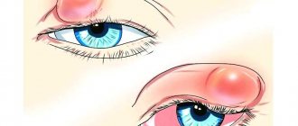
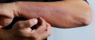
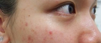
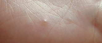
With follicular hyperkeratosis, “goose bumps” appear, that is, dense reddish pimples with a spiky shape. They are located at the base of the hair follicles. Most often, all this is observed on the side or back of the thigh, buttocks, arms, and face. In rare cases, the patient may experience itching.
Plantar hyperkeratosis is characterized by roughening of the skin on the entire sole of the foot or only in the heel area. Calluses and bleeding cracks often occur, which causes discomfort and pain.
With the seborrheic form, characteristic pigmented formations appear, which over time lose color and become colorless plaques.
The symptoms of hyperkeratosis are more pronounced in adults; in children, the pathology is often similar to dermatitis. At the initial stages of development there are no symptoms.
Stages of development
The following stages of disease development are distinguished:
- First . In some areas, the skin turns pale and becomes dry. No thickening of the stratum corneum has yet been observed.
- Second. When touching the skin, particles falling off are noticeable. Slight thickening is possible.
- Third . The skin peels off greatly and loses its smoothness, becoming rough and rough to the touch.
- Fourth. At this stage, multiple complications arise that lead to serious changes in the appearance of the epidermis. The thickening of the stratum corneum is visible to the naked eye.