Hallus valgus is a condition in which damage occurs to the joint of the big toe. This condition is commonly called bunion. Bunions are caused by the formation of a bone spur in response to injury. In reality, Hallus valgus is not just a reaction to injury. Interestingly, this disease is almost never found in cultures that do not wear shoes. Tight shoes, such as high heels and cowboy boots, can contribute to the development of hallux valgus. Wide shoes with room for the toes reduce the chances of developing deformities and help reduce irritation to the area of the bunion, if present.
The term hallus valgus actually describes what happens to the big toe. Hallus is the medical term for the big toe, and valgus is an anatomical term that means that the deformity occurs away from the midline of the body. So valgus deformation of the big toe beyond the foot. As this process progresses, other changes appear. One of those changes is that the bone that sits above the big toe, the first metatarsal bone, begins to deviate more in the other direction. This bone is called primus varus. Primus means the first metatarsal bone, and varus is a medical term that means that the deformity is a deviation from the midline of the body. This creates a situation where the first metatarsal bone and big toe now form an angle with respect to a line running along the inner edge of the foot. Bunion, which develops first, is actually a reaction to pressure from shoes. Initially, the reaction to trauma is an area of irritated, swollen tissue that constantly rubs between the shoe and the bone located under the skin. Over time, constant pressure can cause the bone tissue to thicken, which increases swelling and further friction from the shoe.
Causes
Many problems that arise in the legs are the result of abnormal pressure or friction. The easiest way to determine the presence of the consequences of pathological pressure is to examine the leg. The leg is a hard bone covered with skin. In most cases, symptoms develop gradually as the skin and soft tissues absorb the excess impact on the leg. Any protruding bone or injury aggravates the already existing consequences of the injury. The skin reacts to friction and pressure by forming a callus. The soft tissues located under the skin react to excess stress. Both the callus and the thickened soft tissue underneath the callus become painful and inflamed. Reducing pain helps to reduce pressure. Pressure can be reduced externally through looser shoes or internally through surgery and removal of excess tissue.
Risk factors
- Shoes influence the incidence of hallux valgus (it is lower in adults who do not wear shoes). However, this does not mean that only shoes cause this disease. Tightening shoes can cause pain and pinching of the foot nerves along with the formation of hallux valgus. Fashionable shoes may be too tight and too narrow for the 'foot to look aesthetically pleasing.' High heels increase stress on the foot, further exacerbating the problem. Moreover, fashion trends are followed not only by young people, but also by people in the older age group. Risk factors can be divided into the main ones:
- There is a high probability of hallux valgus deformity in females. The cause may be shoes.
- Ballerinas who spend a lot of time on blocks dancing on their toes and thus may be expected to have a higher likelihood of developing hallux valgus
- Age. The incidence of deformity increases with age, with rates of 3% in people aged 15–30 years, 9% in people aged 31–60 years, and 16% in those over 60 years of age 1
- Genetic factors play a role
- Associated diseases
There are specific causes of biomechanical instability, including neuromuscular disorders. This may be associated with various types of arthritis. These associated diseases include:
- Gout.
- Rheumatoid arthritis.
- Psoriatic arthropathy.
- Joint hypermobility associated with diseases such as Marfan syndrome, Down syndrome.
- Multiple sclerosis.
- Sharot's disease
- Cerebral paralysis.
Women over 30 years of age most often come to our Clinic with this problem. Main reasons:
- heredity
- wearing uncomfortable shoes
- excessive physical activity
- overweight
- hormonal changes (including after pregnancy)
- inflammatory processes in the joints
- consequences of injuries or unsuccessful surgical interventions.
At first, protruding bones seem to be a cosmetic defect; over time, pain appears, the motor ability of the foot is impaired, and as a result, arthrosis of the knee and hip joint, osteochondrosis and other pathologies associated with the musculoskeletal system develop. Depending on the severity of the deformity, various surgical techniques are used for treatment. At the initial stages of deformation, treatment must begin with conservative methods.
Establishing diagnosis
Before prescribing treatment, our specialists conduct a thorough diagnosis of the patient; during this diagnosis, the etiology (origin of the disease) and severity are identified. There are international standards by which all orthopedic traumatologists classify pathology according to the same principles, namely:
- Static.
The deformation occurred due to poor posture - Congenital.
The disorder is associated with a genetic feature of the position of one of the bones of the foot - Compensatory.
In this case, the deformation is a response to the anatomical feature of the tendon, as a result of which the tibia changes position and deforms the ankle joint; - Paralytic deformity.
Occurs after polio or other inflammatory processes in the central nervous system; - Spastic
. It is a complication of muscle spasms; - Hypercorrectional.
In case of improper treatment of foot pathologies; - Traumatic.
It is a complication of fractures and ligament injuries.
The significance of this classification is that treatment of pathology is impossible without addressing the cause. The operation will relieve the patient of deformity and lead to a normal lifestyle. And influencing the etiology of the disease will help avoid the reappearance of deformity.
Depending on the severity, hallux valgus deformity can be:
- Easy
- Average
- Heavy
The method of treating the patient depends on the severity of the disease.
Symptoms
Symptoms of hallux valgus are mainly caused by bunions. Bunion is quite painful. With severe hallux valgus, a cosmetic problem also appears. Moreover, choosing shoes becomes difficult, especially for women who want to be fashionable and for them, wearing fashionable shoes becomes a real challenge. Finally, increasing deformity begins to move the second toe and may create conditions for the second toe to rub against the shoe.
Causes of bunions in the foot
The following factors can provoke the development of hallux valgus:
- osteoporosis;
- genetic predisposition;
- flat feet;
- hormonal disorders;
- wearing tight shoes with narrow toes;
- diseases of cartilage tissue;
- excess body weight.
According to statistics, a protruding bunion bothers approximately 40% of women over 30 years of age. In men, the deformity is extremely rare. This is due to the greater elasticity of the ligaments in the fairer sex.
Diagnostics
Disease history
- The patient may experience pain in the big toe when walking or making any movements. This may indicate degeneration of intra-articular cartilage.
- The pain may be aching in the metatarsal area due to wearing shoes. Possible increase in deformation. .
- It is necessary to find out what physical activities increase the pain and what relieves the pain (maybe just taking off your shoes).
- History of injury or arthritis.
- It is quite rare to experience sharp pain or tingling in the dorsal region of the bursa of the thumb, which may indicate traumatic neuritis of the middle dorsal cutaneous nerve.
- The patient may also describe symptoms caused by the deformity, such as a painful second toe, interdigital keratosis, or ulcer formation.
Visual inspection
- It is necessary to observe the patient's gait. This will help determine the degree of pain and possible gait disturbances associated with problems in the legs.
- The position of the big toe in relation to the other toes. Distortion of the joint can be in different projections.
- Prominent joint position. Erythema or swelling indicates shoe pressure and irritation.
- Range of motion of the big toe at the metatarsal joint. Normal posterior flexion is 65-75° with plantar flexion less than 15°. Moreover, it is necessary to pay attention to whether pain and crepitus are present. Pain without crepitus suggests the presence of synovitis.
- The presence of any keratosis, which suggests pathological chafing from abnormal gait..
- Associated deformities may include hammertoes and flexible or rigid flat feet. These deformities can cause hallux valgus to progress more rapidly as the lateral support of the foot is reduced.
Changes in movement of the thumb joint:
- Increased abduction of the big toe in the transverse and frontal planes.
- Increased average toe prominence.
- Change in backward flexion of the joint.
In addition, it is necessary to pay attention to the condition of the skin and peripheral pulse. Good blood circulation is especially important if surgical treatment is planned and normal healing of the postoperative wound is necessary.
How to recognize the disease?
The following changes indicate the development of a bone in the leg:
- the thumb deviates outward, while forming a “ball” at its base;
- there is constant aching pain in the area of deformity;
- fast fatiguability;
- difficulties with choosing shoes.
In the absence of treatment and progression of the pathology, a hammer-shaped shape of the remaining toes is observed. The appearance of the slightest signs of disease should be a reason to contact an orthopedist.
Treatment
Conservative treatment
Treatment of hallux valgus almost always begins with the selection of comfortable shoes that do not cause friction or stress. In the early stages of Hallus valgus, wearing shoes with a wide front can stop the progression of the deformity. Since the pain that results from bunions occurs due to pressure from shoes, treatment focuses on relieving the pressure that shoes place on the deformity. Wider shoes reduce pressure on bunions. Bunion pads can reduce pressure and friction from shoes. There are also numerous devices, such as spacer orthotics, that can splint the toe and change the load distribution on the foot.
Drug treatment and physiotherapy
Nonsteroidal anti-inflammatory drugs and physical therapy may be prescribed to reduce inflammation and relieve pain. In addition, corticosteroid injections are possible. Long-term physical therapy has not proven to be therapeutically effective.
Orthopedic products
It is possible to use various orthopedic products (instep supports, toe correctors, interdigital rollers). The use of orthopedic devices helps to stop further deformation in the early stages. With severe deformation, the use of orthopedic products can only slightly reduce pain. Custom insoles help correct damaged arches.
If the deformity is caused by a metabolic disorder or a systemic disease, then it is necessary to carry out treatment aimed at correcting the underlying disease with the involvement of a rheumatologist or endocinologist.
Kinds
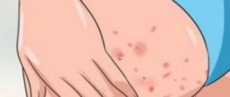
Manifestations of hyperkeratosis form on the legs, arms, torso, scalp, palms, and feet. Their location and appearance depend on the reasons for their appearance.
The most common types of thickenings are:
- Calluses, corns on the soles of the feet and palms. Formed in areas subject to prolonged friction or pressure;
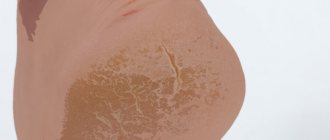
- Actinic keratosis. Develops due to excessive exposure of the skin to ultraviolet rays. Appears as flat, rough patches of yellow, brown or red color;
- Follicular hyperkeratosis (“goose bumps”). This is a rash of small multiple tubercles on the face, shoulders, legs, and buttocks. It is caused by a violation of the exfoliation of dead skin particles. This form of hyperkeratosis occurs in children; it goes away on its own in adolescence;
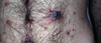
- Warts. Occurs when infected with the human papillomavirus. They appear as convex growths of various shapes and sizes. On the feet they are called plantar warts;
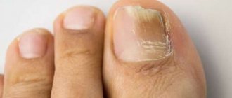
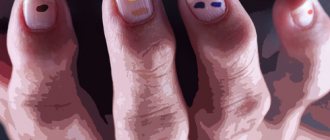
- Under nail hyperkeratosis. This is a thickening of the skin in the area of the nail bed, nail folds, which is caused by injuries, psoriasis, and onychomycosis. It leads to lifting, bending, and deformation of the nail plate.
You can see what different types of hyperkeratosis look like in the photo on the Internet. Its manifestations also include skin changes in certain skin diseases - eczema, psoriasis, lichen planus, seborrhea.
Surgery
If all conservative measures are not effective, then a decision is made on surgical treatment. Currently, there are more than 100 surgical techniques for the treatment of Hallus valgus. The main tasks in surgical treatment are as follows:
- remove bunion
- reconstruct the bones that make up the big toe
- balance the muscles around the joint so that there is no recurrence of the deformity
Removing the “growth”
In some mild cases of bunion formation, only the growth on the joint capsule may be removed during surgery. This operation is performed through a small incision on the side of the foot in the area of the bunion. Once the skin is cut, the growth is removed using a special surgical chisel. The bone is aligned and the skin incision is closed with small sutures.
It is more likely that reconstruction of the big toe will also be necessary. The main decision that must be made is whether the metatarsal bone needs to be cut and also reconstructed. To resolve this issue, the angle between the first metatarsal and the second bone is important. The normal angle is approximately nine or ten degrees. If the angle is 13 degrees or greater, the metatarsal bone will most likely need to be cut and reconstructed. When the surgeon cuts and repositions the bone, it is called an osteotomy. There are two main techniques used to perform osteotomy and reconstruction of the first metatarsal bone.
Distal Osteotomy
In some cases, the distal end of the bone is cut and moved laterally (this is called a distal osteotomy). This effectively reduces the angle between the first and second metatarsals. This type of surgery usually requires one or two small incisions in the leg. Once the surgeon has achieved satisfactory bone alignment, the osteotomy is followed by fixation of the bones using metal pins. After surgery and healing, the pins are removed (usually they are removed 3-6 weeks after surgery).
Procymal osteotomy
In other situations, the first metatarsal bone is cut at the proximal end of the bone. This type of surgery usually requires two or three small incisions in the leg. Once the skin is cut, the surgeon performs an osteotomy. The bone undergoes reconstruction and is temporarily fixed with metal pins. This operation also reduces the angle between the metatarsal bones. In addition, the tendon of the adductor big toe muscle is released. Therefore, after the operation, a special bandage is put on.
Rehabilitation after surgery
It takes an average of 8 weeks for the soft tissue and bones to heal. During this period, it is better to place the foot in shoes with a wooden sole or a special bandage in order to prevent trauma to the operated tissues and allow normal regeneration. Immediately after surgery you may need crutches.
In patients with severe bursitis, physiotherapy (up to 6-7 procedures) may be prescribed a certain time after surgery. In addition, you must wear shoes with wide fronts. It is also possible to use correctors. All this can allow you to quickly return to normal walking.
Complications of toe injuries
Broken toes can lead to dangerous health consequences even when the injury is relatively mild. Without qualified medical care, skin wounds, cracks and bone fractures can provoke the development of the following complications:
- massive hematomas;
- ankylosis (joint immobility);
- deformation of finger joints;
- formation of bone callus;
- sepsis;
- secondary infections.
Damage to the soft and hard tissues of the extremities is especially dangerous for patients who suffer from metabolic disorders and chronic diseases, in particular diabetes. For such patients, even a minor injury can pose a hidden threat.