Home / Articles / Why does the skin around a mole itch?
Moles and age spots should not cause discomfort or discomfort to a person. If at least one nevus begins to hurt or itch, becomes red or swollen, you need to make an appointment with a dermatologist. Sometimes it is enough for a specialist to examine the patient to determine why the skin around the mole and the mole itself are itching. In some cases, only examination of the tumor tissue helps to find the cause of the discomfort. Find out what causes itching nevi exist and why this symptom is dangerous.
Causes of red moles on the body
By its nature, a red mole is a cluster of vessels and capillaries, which normally perform the function of oxygen and nutritional supply to the epidermal structures. With the development of any internal pathological process or under the influence of exogenous factors, small vessels can connect, forming a kind of bundle, which in medicine is often called an angioma.
When asked by a patient regarding small red moles, “What is this?”, the specialist will most often answer – angioma. Typically, these pigmented formations are formed during the period of active growth of the body, that is, in early childhood, which is associated with a serious transformation of the human vascular system at this age.
A distinctive feature of red pigmented nevi is the loss of color intensity of the formation when pressing on it, which is associated with a vascular reaction. In addition, red moles normally do not hurt or itch, and do not change their shape and size over time.
The following etiotropic factors may be the causes of angiomas in adult patients:
- Excessive sun exposure (excessive ultraviolet radiation);
- Hormonal imbalance (puberty, pregnancy, menopause, etc.);
- Pathologies of the gastrointestinal tract, especially the pancreas;
- Injuries and mechanical damage to the epidermal integument;
- Diseases of the cardiovascular system;
- Violation of the formation or destruction of pigmented cells.
Today, doctors do not give a clear answer regarding the causes of red moles on the body, however, the factors listed above can become a provoking condition for their appearance.
How long does it take for wounds to heal after mole removal?
It all depends on the area of the base of the removed formation, that is, on the size of the wound itself, as well as on the characteristics of the skin where the mole grew (on the face, neck, and scalp, wounds heal the fastest, and in injured areas or on natural folds of the body - longer) .
On average, the skin after mole removal takes 10-14 days to heal. During this time, the wound should be covered with a dense scab-like crust. It is formed by an oncodermatologist after removal, and then gives the patient detailed instructions for care.
A strict restriction is not to wet the scab during the first 24 hours after mole removal. Then you just need to repeat the treatment after each water procedure.
The wound is considered healed when the crust has fallen off (on its own!) from the surface and the wound site is no longer wet. Healing after mole removal will proceed faster if you follow all the doctor’s care recommendations.
If after removal the mole takes a long time to heal (more than 3 weeks), you must contact the doctor who performed the removal. The sooner a problem is addressed, the less effort it will take to fix it.
Changes at the site of the removed nevus
If, after removing a mole, strange changes occur at the healing site, immediately contact the doctor who removed it.
Reasons for concern may be compaction, vascular growth, itching, burning, pain, or colored areas at the removal site. All these changes do not necessarily indicate recurrence of the nevus. These may be the same keloid changes, rough scarring or scar pigmentation (darkening of the young skin covering the scar - this often happens when the scar is exposed to sunlight immediately after it has healed.)
Types of red moles
The appearance of red moles on the body can be accompanied by various visual characteristics of the defect. Such epidermal formations can be either flat, forming in the deep dermal layers, or have a superficial nature, protruding above the skin. Moreover, according to the nature of the visual picture, all angiomas are usually divided into two groups:
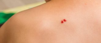
- Spot. The pigmented area has clear red borders and is a point at which a collection of vessels and capillaries is concentrated. Often, punctate angiomas are multiple in nature, manifesting themselves in the form of specific “rashes” on the patient’s skin.
- Star-shaped. Such moles are a collection of tiny thin vessels that are visible through the epidermis and converge at a central point, forming a kind of star. These structures are also associated with diseases such as rosacea.
In addition, a separate group includes especially large red moles on the body - hemangiomas, which usually significantly spoil the patient’s appearance and require cosmetic correction.
Good and bad moles
Many people worry about having a lot of moles on their body. They wonder how dangerous it could be and what it even means. But experts say that a large number of moles cannot be described as “good” or “bad.” Also, the number of moles can be inherited. And people who like to lie in the sun expose their skin to the formation of age spots or moles. Most moles on our body are acquired.
Oncologists stipulate that the appearance or damage of a flat mole is not the worst thing. It is not necessary to contact specialists right away. Treat a damaged mole, rejoice at a new one. But if a large convex mole bleeds, hurts or is damaged, run to the doctor. It is more acute inflammatory in nature. And it is not the most pleasant formation on your body. Which most people get rid of.
Also, after injury to a birthmark, you should limit yourself from exposure to ultraviolet radiation. It has a very negative effect on our skin, and even more so on the injured area.
It is worth paying attention to the color of the mole; the darker it is, the greater the danger. If you have a flat mole, it should always remain that way. As soon as a lumpiness or increase in size appears on it, a doctor is needed.
Why are red moles dangerous?
If a red mole does not bother its owner and does not change its external characteristics, it can be argued that such a formation does not pose a significant threat to the health and life of the patient. At the same time, you should get professional medical advice and resort to modern therapeutic methods if you have the following manifestations:
- The red mole grows or changes its shape;
- The patient experiences pain, itching or burning in the area where the pigmented formation is located;
- The mole is bleeding;
- Superficial structures or ulcerations appear;
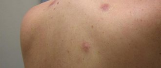
- There are more than 6 small red dots in one area of the body.
All these symptoms may indicate the development of an oncological process. In addition, the category of dangerous moles includes angiomas located in places where they can be easily injured when wearing clothes, shoes, jewelry, etc. Mechanical damage to such a formation can provoke the appearance of new red moles on the body, activate malignant transformation and leave scars or scars on the skin.
What to do if the nevus begins to grow after removal?
Why did it happen?
Maybe I should have chosen a different removal method? Maybe you need to change doctor? First of all, contact the doctor who performed the mole removal procedure. It is he who has the most reliable information about the nevus itself and his actions with it. What to do next? This is already a matter of trust. If trust in the doctor is retained, correct him; if not, you need to choose another specialist. But any intervention, especially a surgical one, should be approached soberly, balancedly and without unnecessary emotions.
With laser or radio wave removal, the risks of relapse arise not so much because of the lack of nevus cells horizontally, but because of the lack of vertical removal, that is, deep into the skin.
In different types of moles and in different areas of the skin, nevus cells can lie deep enough in the dermis and at different levels, which can cause local relapse.
If a mole recurs, you can resort to excision of the scar with recurrent growth, but in my opinion this is an overly aggressive approach. It should only be used if histological examination was not performed at the time of removal, and the recurrence looks suspicious regarding the growth of melanoma.
Is it dangerous for moles to appear at the site of removal?
No, it's not dangerous!
This is not my subjective opinion, this is data from many studies conducted. There is no evidence of an increased risk of melanoma at the site of a removed nevus. Another thing is that the removal of the mole itself was carried out as a preventive measure against the formation of melanoma, which means that if a local relapse occurs, this risk remains. Since the desired result has not been achieved, it is necessary to carefully remove the nevus.
Typically, additional removal is carried out using the same method that was used during the patient’s initial treatment. That is, as a rule, resurfacing of a recurrent nevus is carried out using a laser or radio wave method.
Is it necessary to perform a histological examination of recurrent nevus?
This can be done, but is not always necessary.
If a removed nevus has been examined histologically, then a recurrent one should be examined only if there is a suspicion of malignancy. If histology of the removed nevus was not carried out, and the primary removal was not carried out in our clinic, we will definitely send the material for histological examination. In conclusion, I want to say that, of course, the occurrence of a recurrence of a mole is an unpleasant situation for both the patient and the doctor. But it is very simple to correct this situation without any harm to health and appearance - contact a specialist to remove the recurrence of the nevus.
We professionally care about the beauty and health of your skin. If you are concerned about the appearance of moles after removal, make an appointment with an oncodermatologist
by phone, +7 (812) 318-59-90, and we will help you solve this problem.
Medical correction of red moles
The doctor, after conducting an initial examination and the necessary diagnostic measures, will make a conclusion regarding the likelihood of malignancy of the red mole. Based on this diagnosis, a strategy for further therapeutic actions is developed. If a red mole does not pose an oncological threat and is located in a closed area of the body, its removal is not necessary.
In cases where the red nevus causes aesthetic or physiological discomfort to the patient, it can be removed using modern hardware techniques, the priority among which is laser destruction. This method of removing red moles guarantees painlessness and safety for the patient, and also has a low level of trauma.
Medical offers its patients the latest expert-class medical equipment that meets the most stringent European standards. Effective therapy, high-quality medical service and affordable prices are the main principles of our clinic.
Find out the cost of the procedure “Removal of tumors”
A mole has swollen and changed color, what does a bad growth look like?
Melanoma is a skin disease that looks like a mole. It is a malignant tumor and arises from melanin pigment cells. Unfortunately, melanoma has the ability to grow quickly and affect the human body.
She looks no less nice than you can tell about her. Moreover, it cannot be immediately determined, especially if a person is not attentive to his health. All people can develop melanoma, but some are less likely to get melanoma than others.
It is determined by its species characteristics.
- A spot or raised mole is larger than 1 cm in size.
- The formation will develop from brown to black.
- The spot or mole is actively growing. The largest melanomas can reach 20 centimeters or more on any part of the body.
- Over time, the mole develops asymmetry and goes beyond its boundaries.
- It has a very soft or hard structure.
- When touching the affected area, pain is felt.
- It bleeds, burns, and hair falls out from the mole site.
The most terrible disease, the symptom of which is inflammation of a mole, is CANCER . Only an oncologist can make such a diagnosis after passing certain tests. The most important thing is to start treatment on time and believe in its success.
Remember! There is nothing more important than our health. Any deviations, be they external or internal, require research and, if necessary, treatment. It is important to study and take care of yourself throughout life . Thus, it will be possible to avoid many diseases and be confident in the future. Don’t put off your health “for later”, take care of it every day. Improve the quality of your life and the lives of those around you!
Dangerous symptoms
Only a very few black moles pose a potential threat. The risk of malignant transformation to melanoma is low, but caution should be maintained. In any case, it is advisable to show the skin lesion to a specialist. You definitely need to see a dermatologist or oncologist if you have the following symptoms:
- The black mole increases in size. Even small growth should be alarming; a rapid increase in size is an unfavorable sign.
- The surface has changed (becomes “glossy” or loose).
- The formation peels off or bleeds.
- Unpleasant sensations appear in the form of pain, burning or itching.
- The shape has changed, the edge has become striated, uneven, asymmetrical.
- The color changes: an ordinary mole becomes black, bluish or dark brown.
Any dynamics should be alarming. There is no need to panic, but you need to make an appointment with a specialist. What you definitely cannot do is try to remove the tumor yourself using traditional medicine. Injury by mechanical or chemical influence is one of the main causes of malignant degeneration.
Some information about moles
All inhabitants of the globe, without exception, have moles - birthmarks. A person is born with moles and acquires them at any age. The majority of moles appear in humans for the first time at 2-3 months of age. The active period for the appearance of new moles is also adolescence in adolescents and the period of waiting for a child in women. Birthmarks can also transform and change during a person’s life.
According to medical terminology, moles are pigmented nevi, which are classified as skin defects that can be congenital or acquired. Such a nevus consists of cells of the epidermis, or dermis, and melanocytes (cells with pigment melanin).
Not all moles are identical: there are normal birthmarks, benign ones, and there are pathological ones, which pose a significant danger to human health and life itself.
Methods for diagnosing black moles
Melanocytic tumors are one of the main problems in diagnostic dermatopathology.
Because they include benign nevi, malignant melanomas and borderline cases.
Apart from the proportion of congenital lesions, most benign nevi occur in early or adulthood.
May eventually undergo regressive changes.
Particular care should be taken if moles and nevi become black.