Symptoms
The symptoms of shingles in the initial stages are very similar to chickenpox. The incubation period is from 2 to 4 weeks. Clinical manifestations include:
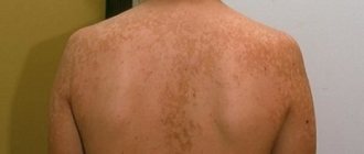
- the appearance of red spots on the skin that slowly spread throughout the body;
- redness is localized along the nerve trunks of mainly one half of the body: the area of the ribs, shoulder blades, lower back, less often on the face;
- The appearance of pain at the site of the lesion is characteristic: the pain is burning, intensifies at night.
Blisters similar to chickenpox are visualized 3-4 days after the onset of the disease. At the same time, the lymph nodes may enlarge and the temperature may rise, which lasts for several days. The contents of the vesicles become cloudy, and then they begin to dry out, forming yellow crusts. The whole process takes 6-8 days. The crusts disappear after 3 weeks. After the active stage, pain persists until complete recovery.
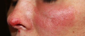
Pityriasis versicolor during pregnancy
During pregnancy, women may experience various diseases, including skin inflammation. The causes of lichen, like many ailments, in pregnant women are often a decrease in immunity or hormonal changes.
As a rule, pityriasis versicolor is not treated during pregnancy, or a gentle course with carefully selected components is prescribed so as not to harm the fetus, but to improve the quality of life of the expectant mother. Treatment is always strictly individual.
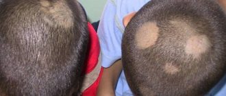
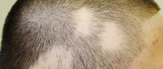
Pityriasis versicolor in children
This is a common disease in children. It usually affects the scalp, but can appear on the face and body. For young children and adolescents, as well as for pregnant women, treatment can only be prescribed externally (in particularly difficult cases, the use of medications and drugs is possible). Parents of infected children need to monitor the child’s hygiene and not allow the baby to scratch the spots in order to prevent the spread of the disease and not create a favorable background for the occurrence of other diseases.
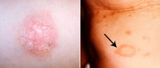
How dangerous is solar lichen? Is pityriasis versicolor contagious or not?
If the disease does not have complications and does not occur in parallel with other diseases, then it does not pose a danger to human health, except for the psychological discomfort mentioned above, especially if spots appear on the face.
The disease can be called conditionally contagious, since the fungus (causative agent) of skin inflammation is present on the skin of any person (it is part of the microflora) or can be transmitted from person to person, but the disease itself will occur only in the event of an external “irritant”.
Infection is possible:
- during personal contacts;
- through personal and hygiene items;
- in public changing rooms or fitting rooms.
How and how to treat pityriasis versicolor in humans (colored, multi-colored, etc.)?
If you notice signs of this disease, it is important to remember three simple rules:
- do not self-medicate, because the wrong medicine can only worsen the condition;
- do not wait until everything goes away on its own (the disease has a wave-like nature, that is, it can be repeated at different intervals and does not always return in a mild form);
- Do not use dubious, suggested by someone, or self-selected remedies for pityriasis versicolor.
Seeing a doctor is a sure way to not only get cured, but also strengthen your immune system, as well as get rid of annoying aesthetic skin defects that bring psychological discomfort.
Varieties
There are 5 types of herpes zoster in total.
- abortive. The mildest form, which is characterized by a rapid course of the disease. The erythematous-papular rash lasts several days, there are no vesicles.
- generalized. It is characterized by local specific manifestation and distribution of vesicles throughout the body, including mucous membranes. The duration of illness is 2-3 weeks. If symptoms persist beyond this period, then the development of immunodeficiency or a history of malignant neoplasms is possible.
- bullous Characteristic clinical manifestations are large vesicles with serous fluid. Tend to merge.
- gangrenous. Causes necrotic skin changes. This form is typical for patients with immunodeficiency and is a marker of AIDS.
- hemorrhagic. Vesicles with blood content are characteristic. After recovery, deep scars remain.
Make an appointment
Crocker-Adamson lichen spinulosus (lichen spinulosus Crocker-Adamson; spinous follicular keratosis) is a rare, predominantly genetically determined dermatosis, inherited in an autosomal dominant manner. It is characterized by the accumulation of horny plugs in the mouths of hair follicles, macroscopically manifested by follicular papules of flesh-colored or pinkish color, up to several millimeters in size with centrally located horny spines [1]. The clinical picture of the disease was first described in 1883 by the English dermatologist H. Crocker as a variant of pityriasis versicolor pilaris Devergie [2]. Lichen spinosa was identified as a separate disease in 1905 by the English dermatologist H. Adamson. He was the first to note that this dermatosis is often combined with lichen planus [3]. A. Rook, D. Wilkinson and F. Ebling (1979) considered lichen spines as a variant of keratosis pilaris. F. Gahlen and E.-I. Grussendorf (1980) used this name to designate three forms of follicular hyperkeratosis: as variants of follicular keratosis, lichen planus (Little-Lassuer syndrome) and lichenoid allergic reaction in infectious diseases (lichenoid tuberculosis, lichenoid trichophytide, salvarsan toxiderma) [1]. There are reports of diagnosing lichen spines in patients with alcoholism, diphtheria, syphilis, HIV infection, and Crohn's disease [1, 4]. American dermatologist S. Friedman in 1990 published data on 35 cases of lichen spines, which were diagnosed during an examination of 7435 patients at a dermatology clinic in the Philippines. The incidence was 5 cases per 1000 dermatological patients [5].
The etiology of lichen spines is unknown. The development of the disease is associated with a deficiency of certain vitamins (primarily A and C), the influence of exogenous factors (contact with coal tar products), and is also considered as a symptom of other genodermatoses (keratoderma, ectodermal dysplasia, congenital pachyonychia, monilethrix, etc.) . In one of the reports there is a description of lichen spines in four generations of a family [6], which once again confirms its genetic determination. Mostly children, more often boys, are affected [1]. In a study by S. Friedman [5], the disease was described in 14 men and 21 women (mean age 16.2±10.1 years).
The rashes, as a rule, are located symmetrically on the back of the neck, the extensor surface of the shoulders, in the back, abdomen, buttocks, thighs and are represented by small, often follicular, spiny horny papules, grouped in the form of foci of various shapes, the skin in the area of which is pinkish-colored. reddish color with a bluish tint, usually not pronounced. There may be abundant flour-like peeling, due to which the lesion becomes white, as if sprinkled with powder. Due to the presence of spines on the papules, when running the palm over the surface of the lesion, the impression of touching a grater is created. After removing the papule, a small depression is found containing the remains of broken hair. The rash is usually subjectively asymptomatic; some patients report slight itching. There are no lesions of internal organs [5, 7]. Diagnosis is based on the clinical picture and the results of pathomorphological examination, revealing moderately severe hyperkeratosis, horny plugs at the mouths of dilated follicles and mild lymphocytic infiltration in the dermis around the hair follicles [5, 8]. Differential diagnosis is carried out with lichenoid tuberculosis of the skin, pityriasis versicolor pilaris of Devergie, follicular mucinosis, squamous follicular keratosis of Doha [5].
Treatment includes vitamin A preparations and external therapy with keratolytics (salicylic acid, lactic acid, resorcinol, urea, barium sulfide, strontium sulfide) [9]. The combined external use of retinoids and keratolytics turned out to be highly effective [10]. Treatment of lichen spines with topical steroids is ineffective [9]. The prognosis for recovery is favorable, as there is a tendency for spontaneous recovery. Spontaneous remissions occur within 1-2 years [5].
Currently, only a few dozen cases of Crocker-Adamson lichen spines have been described in the literature, however, errors in the diagnosis of this disease can lead to long-term ineffective treatment. In this regard, each clinical case of the disease is of undoubted interest for dermatologists. Here is our observation.
Patient M., born in 1962, resident of Moscow. He considers himself sick for about a year, when, after a stressful situation, rashes characteristic of lichen planus first appeared on the skin of the torso and limbs, subjectively accompanied by severe itching. He was treated on an outpatient basis with a temporary positive effect. A month ago, a multiple follicular rash with spines appeared all over the skin against the background of congestive-hyperemic skin.
Life story.
He grew and developed normally. Heredity is not burdened. Denies bad habits.
Accompanying illnesses.
Hypertonic disease.
Allergy history is not burdened.
General status.
Upon admission, he complained of the appearance of rashes on the skin of the torso and limbs, severe itching. The patient has the correct physique and increased nutrition. General condition is satisfactory. There is vesicular breathing in the lungs, no wheezing. Blood pressure 130/80 mm Hg. Heart sounds are sonorous, rhythmic, heart rate 65 per minute. The abdomen is soft and painless on palpation. Peripheral lymph nodes are not enlarged. The osteoarticular system is externally unchanged. Physiological recovery is normal.
Local status.
The pathological skin process is chronic and widespread. Multiple rashes are represented by dark brown and dark brown pigment spots of irregular oval shape, isolated or merging with each other, as well as various bluish papules (polygonal, oval) and dark brown plaques of elongated oval shape, located in the lower abdomen and in the pubic area (Fig. 1, a).
Figure 1. Lichen planus, classic and linear form.
Crocker-Adamson lichen spines (a-d). On the inner-lateral surface of the right thigh and lower leg there is a linearly located focus of dark brown hyperpigmentation up to 1 cm wide, which passes into the ankle area into an elongated infiltrate of dense consistency with a bumpy surface protruding above the level of apparently healthy skin by 0.3-0. 5 cm (Fig. 1, b-c). Based on clinical manifestations, we diagnosed lichen planus, classical and linear form. In the area of the upper back, lateral surfaces of the body, in the lumbosacral region, on the buttocks, thighs and outer surfaces of the legs, there are symmetrically located foci of congestive red color, against which there is a multiple small-nodular follicular rash of a round shape with whitish thin spines on the surface (Fig. 1, a-d). We hypothesized that the described clinical symptoms are most consistent with Crocker-Adamson lichen spines.
Survey results
Clinical blood test, clinical urine test, biochemical blood test - within normal limits. Electrocardiogram - without features.
X-ray examination.
Radiographs of the bones of the hands and feet show a picture of osteoarthropathy. A chest x-ray did not reveal focal or infiltrative changes in the lungs.
During ultrasound examination
signs of fatty hepatosis were revealed in the abdominal organs.
Results from other laboratory methods
examinations: RIF, RIBT negative. Antibodies to HIV, HBs-Ag, HCV are negative. RMP is negative. Stool analysis - no helminth eggs were found.
During dermatoscopy of follicular rashes
small follicular papular elements measuring 0.5-1 mm were identified (Fig. 2, a-d).
Figure 2. Dermatoscopic picture of Crocker-Adamson lichen spines (a-d).
On the surface of the nodules there are sharp thread-like keratin protrusions emanating from the canals of the hair follicles (Figure 2, b). Some of the elements were located against the background of dark brown pigment spots (Fig. 2, c). Pathomorphological examination.
Some of the slices are oriented in the tangential plane. The epidermis is of normal thickness, the layers are differentiated. In the dermis, fragments of dilated orifices of follicles are found, in the lumens of which there are loose horny plugs. Perifollicularly, the formation of small-sized epithelioid cell granulomas with single giant multinucleated cells is noted (Fig. 3).
Figure 3. Histological specimen of Crocker-Adamson lichen spines (hematoxylin and eosin staining). a - acanthosis and moderate dystrophic changes in the epidermis; subepidermal moderate mononuclear infiltration; hair follicles with moderate hydropic degeneration of epithelial cells (×50); b — macrophage-epithelioid granuloma with single giant cells of the Pirogov-Langhans type (×50); c — developing granulomatous inflammatory reaction near the hair follicle (×200). Ziehl-Nielson staining, immunohistochemical examination and polymerase chain reaction did not detect Mycobacterium tuberculosis.
Conclusion
The identified changes can be observed in Crocker-Adamson lichen spines. The development of granulomatous inflammatory infiltration around the follicles can occur due to disruption of the integrity of the epithelial lining of the follicular infundibulum with the penetration of horny masses into adjacent areas of the dermis.
The treatment, including therapy for Crocker-Adamson lichen spines (vitamin A preparations, external keratolytics) and lichen planus (desensitizing, antihistamine, enzyme, sedative drugs, dexamethasone 1 mg/day, potassium preparations), contributed to the achievement of a pronounced improvement. Most of the rashes have completely or partially regressed.
In the clinical observation we presented, the diagnosis of Crocker-Adamson lichen spines was based on anamnesis data, the clinical picture of dermatosis, the results of dermatoscopy and biopsy. Difficulties in diagnosing it are associated with low occurrence, and therefore it is little known to dermatologists. It can be assumed that Crocker-Adamson lichen spines is pathogenetically close to lichen planus.
Treatment
Even the abortive form of this disease takes time. Seeing a doctor at the first symptoms increases the chance of a quick recovery. Shingles is treated on an outpatient basis. The measures are aimed at relieving pain and providing antiviral therapy. This will prevent the development of the disease. By affecting the nerve endings, lichen is insidious with complications. Antiviral drugs are taken from the first days until crusts appear. Severe forms are treated exclusively in a hospital.
At the Altermed clinic, the disease is diagnosed and therapy is selected with a guaranteed cure.
Other articles by the author
- Pyoderma
- Herpes zoster
- Pityriasis rosea
- Shingles