Ringworm
It is an inflammatory disease of the skin and its appendages of a fungal nature.
Etiology
Ringworm in humans is caused by dermatophyte fungi that can affect not only smooth skin, but also its derivative elements - hair and nails. Of the dermatophytes, the most common are Trichophyton violaceum and Microsporum canis - the causative agents of trichophytosis and microsporia, respectively.
Based on the source of infection, trichophytosis and microsporia are divided into zoophilic and anthropophilic. Zoophilic - human infection occurs during direct contact with a sick animal or objects contaminated with their fur. Anthropophilic - the infection is transmitted directly from a sick person, as well as through the use of things contaminated with the fungus (combs, hats, various hair accessories).
Clinical picture
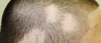
Microsporia of the scalp.
At the initial stage of the disease, a red spot appears under the hair. After a few days, the lesion becomes covered with a mass of small gray scales (“asbestos scales”). The hair becomes faded, brittle, and the hair follicle is densely filled with fungal spores. When combing, the hair easily breaks off, leaving “stumps” 5-6 mm high. Such bald spots in the hair in the form of stubble give the name “ringworm” to ringworm.
In 90% of clinical cases, microsporia of the scalp is caused by Microsporum canis.
Trichophytosis of the scalp.
This mycosis is characterized by rashes in the form of many small (up to 1.5 - 2.5 cm) isolated foci with vague contours. Their surface is covered with gray scales. Hair breaks easily at the very root, which is why dermatologists call trichophytosis “black dot” based on its appearance.
In 60% of cases, trichophytosis is caused by Trichophyton violaceum.
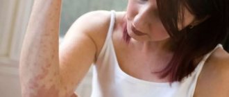
Microsporia and trichophytosis of smooth skin.
The classic clinical picture of the source of infection looks like a ring-shaped skin lesion with an inflammatory ridge along the border. The rashes have clear boundaries, tend to grow peripherally, and regression of inflammatory changes in the center of the lesion is characteristic. The surface peels off and sometimes contains blisters and pustules.
Favus (scab)
The pathognomonic sign of favus is the presence of scutulae. Scutula (from Latin scutulo - shield) is a crust of fungus, epidermal scales, dead leukocytes. The surface has a dirty yellow or gray color. The scutulae can merge, covering the head with a continuous crust. The hair does not break off, but grows through it, looking like tow. Outwardly, it resembles a picture of a honeycomb (from the Latin favus - honeycomb cell). If you remove the crust, a shiny bright red surface is revealed without scales; hair will no longer grow on it.
Infiltrative-suppurative form of microsporia and trichophytosis
Characterized by severe inflammation of the scalp with the formation of pustules and kerions - dense large painful infiltrates. When pressure is applied, yellow pus leaks out from the dilated mouths of the hair follicles. The patient is concerned about intoxication syndrome (high body temperature, headache, general malaise). The process involves regional lymph nodes (posterior cervical, occipital), and lymphadenitis develops.
Diagnostics
- External examination of the patient, characteristic picture of the lesion.
- Microscopy of pathological material (hair affected by the fungus, skin flakes, particles of nail plates). The diagnosis is confirmed by mycelium threads and fungal spores identified in the preparation.
- Fluorescent diagnostics with a Wood's lamp is used as an express diagnosis of microsporia using the photoluminescence method - under the rays of the lamp, the elements of the fungus glow with a light green color characteristic of dermatophytosis.
- Determination of the type of fungus by PCR diagnostics (based on the genetic material of the parasite identified in the scraping).
Treatment
- Systemic treatment of lichen is prescribed for mycoses of the scalp and extensive lesions of smooth skin. When treating microsporia, the drug griseofulvin is prescribed; itraconazole is effective against trichophytosis.
- Local treatment includes various compresses on the area of the rash, bandages with antiseptics to cleanse the lesions from crusts. Antifungal hair shampoos and ointments (microspor, lamisil, clotrimazole) are widely used.
Traditional methods of treatment
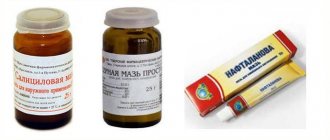
The image below shows recipes for traditional therapy (this was done so that you can keep it for yourself)
Prevention measures: mandatory hand washing after walking outside, using individual personal hygiene items (hair brushes, towels, hair accessories). After visiting swimming pools, public baths, saunas, you must shower with soap. If you have pets at home, you should regularly inspect the appearance of the animal's fur. It is advisable to detect characteristic signs of ringworm infection in time in order to disinfect the premises and treat the sick pet.
Ringworm of the skin - symptoms, treatment
29.01.2021
Ringworm is a disease caused by pathogenic fungi called dermatophytes or yeasts. The formation of these 2 types of infections is different. Dermatophytes can get onto human skin from animals (dogs, cats, hamsters, guinea pigs, often even asymptomatically infected), from the ground (farmers, small children playing in the ground) or from person to person. The last method of infection most often occurs through various objects. Factors contributing to ringworm include an individual's tendency to sweat feet and wearing windproof shoes made of artificial plastics. The second type can be described as follows: the fungi that cause them are found on the mucous membranes and skin (especially in the folds) of healthy people. They are found there in small numbers and then do not cause damage. Any factors that reduce the state of immunity ( pregnancy , diabetes , long-term treatment with antibiotics, corticosteroids or cytostatics, tumors, AIDS , etc.) cause an imbalance between the host and the yeast. They then multiply excessively and infect the skin.
Diagnosis of ringworm
The basis for diagnosing ringworm of the skin should always be a mycological examination. Hair, scales, and nail scrapings are taken for examination. Results for yeast usually take 2 days, for dermatophytes - 3 weeks. If the result of the mycological examination is negative, but is incompatible with the existing symptoms, the doctor may repeat the examination 2-3 times. The most common mistake is starting antifungal treatment before receiving the results of mycological studies. If treatment fails, it is often impossible to tell whether it was ringworm to begin with.
A common mistake (especially when treating onychomycosis of the feet or nails) is re-infection from contaminated shoes. Skin fungi can affect the hair, cuticles, or nails. The most common forms are onychomycosis of the feet and nails.
Ringworm of the legs
It is most often located in the last two interdigital spaces. The epidermis there has turned white, peels off, and sometimes small bubbles are visible. This causes itching. In other cases, skin lesions are localized on the soles. Erythema, follicles, erosions, peeling, and cracking of the epidermis may be observed.
Complications of untreated ringworm are often distant allergic rashes (on the hands, face ).
Onychomycosis
May be accompanied by ringworm of the legs or occur independently of it.
Symptoms. Usually, 1-2 nails are affected at the beginning, then move on to the next ones. Nails change color (become yellow, greenish or brown), lose transparency, and become keratosed underneath, causing them to become thicker and wither. If the cause is dermatophytes, then the nail shafts do not change inflammatoryly, when the yeast-nail shafts are red, swollen, painful, sometimes with pressure a drop of pus comes out from under them.
Oral yeast infection
This yeast infection is mistakenly called thrush . It most often occurs in newborns and infants. The infection occurs during childbirth from fungal discharge from vagina . White plaques resembling cottage cheese appear in the mouth After their collision, the mucous membrane in this place turned red, sometimes from erosion.
Published in Dermatology Premium Clinic
Lichen planus
Lichen planus in humans is a chronic dermatosis that affects the skin in the form of small, itchy and often flaky nodules.
Etiology
The specific type of pathogen has not been identified. Refers to multifactorial diseases. Pathology can be caused by various provoking circumstances: mental shock, taking certain medications (for example, tetracycline), various irritating chemicals, diseases of the gastrointestinal tract.
Clinical picture
Elements of the rash are presented in the form of itchy nodules with a diameter of 1-3 mm, in the center of the surface there is an umbilical depression. The rest of the surface of the papule is shiny. The color of the formations ranges from crimson to bluish-red with a pearlescent tint. The nodules are covered with the so-called Wickham mesh - a fine mesh pattern characteristic only of lichen planus. Favorite places for lesions are: the flexor surfaces of the limbs, symmetrically on the sides of the body, lower back, and genital area. Much less often, rashes can appear on the head, palms, and feet. With a long course, Koebner's symptom is detected - new papules appear at the sites of microtraumas of the skin.
Diagnostics
With a typical picture of the disease, characteristic clinical external signs and localization of lesions are taken into account.
Treatment
They establish a connection between the disease and stress or past infections. Conduct diagnosis and treatment of concomitant pathologies.
- Local treatment of lichen: creams and ointments with glucocorticosteroids are applied to the lesions, antihistamines and B vitamins are taken orally.
- Phototherapy methods in the form of:
- PUVA therapy (local application of a photosensitizer to the lesions, followed by irradiation with long-wave UV rays).
- Selective light therapy (combination of long-wave and mid-wave UV radiation).
Traditional methods
Versicolor (pityriasis versicolor)
Lichen versicolor is a fungal, non-inflammatory disease of the skin that affects the stratum corneum of the epidermis (hair and nails are not affected).
Etiology
Lichen versicolor is caused by fungi of the genus Malassezia Furfur, which are opportunistic microorganisms, that is, they are normally present on the human body, causing damage to the epidermis under certain conditions. Such conditions include: weakened immunity, hormonal imbalances, increased sweating, a number of concomitant pathologies (diabetes mellitus, oncology, HIV infection, chronic kidney disease, tuberculosis, systemic diseases). The fungus affects skin rich in sebaceous glands: scalp, face, surface of the upper third of the body, groin area. The rash does not cause itching.
Clinical picture
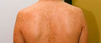
The lesions of the affected epidermis look like spots with clear edges, which often merge, forming scalloped figures. The color of the rash ranges from yellow-pink to brown. During the life of the fungus, the affected areas of the skin lose the ability to produce melanin, a coloring pigment, under the influence of ultraviolet radiation. Therefore, in light-skinned people the surface of the spots is dark, while in dark-skinned or tanned people the surface is light (no pigment). The lesions are flaky, the exfoliated scales resemble bran (hence the second name lichen).
Diagnostics
- A characteristic clinical picture of epidermal damage by the Malassezia fungus.
- Fluorescent diagnostics with a Wood's lamp (mushrooms produce a yellow or brown glow characteristic of this mycosis).
- Microscopy of a scraping of the lesion for pathological fungi (skin flakes are examined, the diagnosis is confirmed if the specimen contains threads of mycelium and fungal spores).
- An additional diagnosis is the Balzer iodine test: a 5% iodine solution is applied to the surface, and the area affected by the fungus is more colored (due to “raised” skin scales) than the healthy surroundings.
Treatment
- Systemic antimycotic therapy is prescribed for generalized forms of lesions and recurrent course of the disease. Drugs such as ketoconazole, fluconazole, and itraconazole are used.
- Local therapy: imidazole antimycotics in the form of shampoo, cream or aerosol are used to treat the surface of the lesions.
Folk remedies
Read more: Pityriasis versicolor in humans: symptoms, treatment, photos
Publications in the media
Lichen planus (Wilson's lichen, true lichen) is a chronic itchy dermatosis of unknown etiology, characterized by the appearance of flat red polygonal papules with a smooth shiny surface and a slight depression in the center. Frequency. 450:100,000. The predominant age is 30–60 years.
Risk factors • Dental diseases, unsatisfactory condition of dentures contribute to the development of lichen planus on the oral mucosa • Exposure to drugs (gold preparations, aminosalicylic acid, tetracyclines) and chemicals (paraphenylenediamine compounds - reagents for color photographic and film films) • SLE • Emotional stress .
Clinical picture • Skin lesions •• Itching, often severe •• Flat, polygonal, brownish-bluish papules with a shiny surface, 1–10 mm in diameter. The rashes can merge, forming shagreen-like plaques covered with tightly packed scales. On the surface of some papules a peculiar mesh pattern is noticeable - the Wickham mesh, caused by uneven thickening of the granular layer of the epidermis (pathognomonic sign) •• Localization - the rashes are usually located on the flexor surface of the forearms, the anterior surface of the legs, the external genitalia; less often - on the dorsum of the feet, in the inguinal and sacral areas. There have been cases of generalization of dermatosis with the development of secondary erythroderma (erythematous form) •• Characterized by an isomorphic reaction at the sites of scratching (Köbner phenomenon) •• Depending on the clinical picture, the following forms are distinguished ••• Pigmented - flat papules are barely noticeable against the background of diffuse melasma ••• Erythematous - large itchy, swollen erythematous spots and papules, difficult to distinguish until the intensity of the erythema decreases; accompanied by general intoxication ••• Blistering (bullous) - blisters form against the background of papular rashes ••• Hypertrophic (warty) - large purple papules, often covered with horny layers, usually located on the anterior surface of the legs ••• Hyperkeratotic (horny) - on surfaces of papules - pronounced hyperkeratosis, localized on the legs ••• Coraloid - papules with a diameter of up to 1 cm in the form of beads of reddish-bluish color, alternating with areas of hyperpigmentation; usually located in the abdomen and neck ••• Atrophic - skin atrophy occurs in place of the papules ••• Dull (flattened) - large hemispherical, smooth, dense, low-itching papules are located on the legs, lower back and buttocks •• Depending on the location of the elements • •• Scattered single rashes ••• Linear rashes (lichen planus linear) - papules are located linearly, sometimes along the course of a nerve or along scratches ••• Ring-shaped rashes (lichen planus annulare) - papules are located in rings growing eccentrically, usually located on the trunk and mucous membranes. • Damage to the mucous membranes - in 40-60% of patients with skin lesions. In 20%, only the mucous membranes are affected. This condition is often considered as precancerous (the development of carcinoma is possible) •• The rash is painful, especially with ulceration •• Papular, often ring-shaped rashes of a whitish-pearly color in the form of a mesh, lace pattern, usually on the mucous membrane of the cheeks, less often - on the tongue, gums, the palate, on the red border of the lips - a continuous scaly stripe •• Depending on the clinical picture ••• The vesicular (bullous) form develops quite often ••• The erosive-ulcerative form with multiple erosions on the mucous membrane of the mouth and lips - the most severe form, may combined with diabetes and essential arterial hypertension (Grinshpan-Wilapol syndrome). • Damage to hair and nails •• Scalp ••• Lichen planus pilaris - rash of small papules around the mouths of follicles on the scalp ••• Skin atrophy and destruction of hair follicles, as a result of which persistent total alopecia may develop •• Nails - proximal formation - distal grooves and partial or complete destruction of the nail bed. The big toes are most often affected.
Research methods • Wickham's grid is better visible after local application of inorganic oils • Skin biopsy - inflammation with hyperkeratosis, thickening of the granular layer, uneven acanthosis, vacuolization of cells of the basal layer, hyaline bodies under the epidermis, heavy lymphocytic infiltration of the upper layers of the dermis • Serological tests to exclude syphilis.
Differential diagnosis • Secondary papular syphilides • Psoriasis • Atopic dermatitis • Leukoplakia (if localized on the mucous membrane) • Squamous cell and basal cell skin cancer • SLE • Erythema multiforme exudative • Exposure to chemicals • Rash due to drug allergies.
TREATMENT Management tactics • Clinical observation, especially of patients with the hypertrophic form of lichen planus, periodic examination of the skin • Timely detection and treatment of somatic diseases, functional changes in the nervous system, sanitation of the oral cavity, rational dental prosthetics (dental crowns must be made of the same metal) • PUVA therapy, heliocadmium laser therapy, inductothermy of the lumbar region (especially for generalized or intractable lesions). PUVA therapy is carried out periodically for 1–2 years • CBC, liver function tests, ANAT titer determination are carried out every six months • Anti-relapse therapy - repeated courses of vitamins, spa treatment • Due to the possibility of malignancy of ulcerative-erosive forms on the mucous membranes Regular monitoring by an oncologist is necessary.
General recommendations • A regimen with long sleep, rational employment with the exception of occupational factors that irritate the skin • Correction of the reaction to stress • For lichen planus of the oral mucosa, the exception of hot, hot, spicy and rough foods.
Drug therapy • For all forms of the disease •• Sedatives •• Penicillin preparations •• Vitamins (nicotinic acid, thiamine, pyridoxine, retinol) •• Histoglobulin 2 ml 2 times/week subcutaneously, per course - 8-10 injections • For skin lesions •• Locally - HA ointments (for example, triamcinolone 0.1%) •• Antihistamines (for example, diphenhydramine 25 mg every 6 hours) - to reduce itching • For lesions of the mucous membranes •• Retinol topically, tigazon 30 –75 mg/day orally (contraindicated during pregnancy) •• GC orally • For erosive-ulcerative and bullous forms - combined treatment •• GC (for example, prednisolone 20-25 mg every other day for 2-4 weeks) •• Chloroquine 0.25 g 1–2 times a day for 4–6 weeks •• Xanthinol nicotinate 150 mg 3 times a day •• Erosion of the oral mucosa is lubricated with a paste containing solcoseryl.
Complications • Malignant degeneration of long-existing hypertrophic, ulcerative-erosive lesions on the mucous membranes (1% of cases) • Alopecia • Nail destruction.
Prognosis • The disease has a benign, but long-term course, especially erosive-ulcerative forms • An individual therapeutic approach to the patient, taking into account pathogenetic factors, contributes to positive treatment results • Spontaneous resolution is possible within several weeks • A tendency to relapse is noted, especially against the background of emotional stress. A recurrent course is observed in 20% of cases, mainly in generalized forms. Prevention. Elimination of chemical factors and drugs that provoke lichenoid reactions, thorough sanitation of the oral cavity, reduction of stress.
ICD-10 • L43 Lichen planus
Shingles
Herpes zoster is an acute viral infection characterized by fever, intoxication syndrome, damage to the ganglia of the spinal cord and the appearance of herpetiform rashes along the sensory fibers of the affected nerve.
Etiology
The causative agent of herpes zoster is varicella zoster, a neurodermatotropic virus that can cause damage to the nervous system and skin. The virus is transmitted by airborne droplets and household contact. The adult population is sick; in children, the virus usually causes the development of chickenpox (this infection has a completely different clinical picture). Once in the body, the infection penetrates the spinal ganglia, where it can lie dormant for a long time. The infection is activated by weakening of the immune system against the background of other infections, cancer, injuries, taking hormonal drugs, cytostatics.
Clinical picture
The initial stage of the disease is manifested by intense burning pain along the affected nerve. The pain usually gets worse at night. Sensitivity disturbances appear (increased, decreased or completely absent). After 5-12 days, along the course of the innervated metamer, the skin swells, turns red, and after 2-3 days characteristic rashes appear on it in the form of blisters. Over time, the contents of the bubbles darken, and brown crusts form in their place. After 3 weeks they fall off, leaving temporary pigmentation. The pain syndrome lasts longer.
Diagnostics
- The presence of a characteristic clinical picture of infection, stages of herpetic manifestations.
- Serological research methods (PCR - diagnostics, enzyme immunoassay).
- Isolation of the virus in cell cultures (the infection is grown on chicken embryo tissue).
Treatment
- Antiviral therapy. They use drugs such as Zovirax, famciclovir, valacyclovir. If antiviral therapy is started within the first 3 days from the onset of the rash, the intensity of pain decreases significantly. In addition, the duration of the disease is reduced and treatment generally has a rapid effect.
- Immunostimulating therapy. In immunosuppressive conditions, the administration of immunoglobulins is indicated. Drugs that suppress the immune system are discontinued.
- Symptomatic therapy. Antibacterial drugs are prescribed in case of secondary bacterial infection. For pain relief, non-narcotic analgesics and non-steroidal anti-inflammatory drugs are used.
Folk remedies
There is a specific prevention of herpes infection - the live vaccine Zostavax. The vaccine rarely causes side effects, but is contraindicated in patients with immunodeficiency.
Ringworm in children
Ringworm in children
Ringworm is the most common fungal disease among children, affecting the skin, hair, and nails.
From the moment of infection with the fungus to the appearance of symptoms of lichen in children, it can take from 5 days to 6 weeks. When the skin is affected, delimited round and oval spots of a reddish color are formed. The skin in these areas is covered with crusts and scales, and is very flaky; sometimes there is itching and burning. If ringworm in children affects the scalp, this is accompanied by the formation of a large rounded area of baldness, within which the hair is broken off (as if cut) at a level of 4-8 mm from the scalp. Around the main focus there may be small, sometimes numerous similar lesions.
In weakened children, ringworm can occur with lymphadenitis, fever, loss of appetite, headache, pyoderma, folliculitis and perifolliculitis of the head.
Pityriasis versicolor (varicolored) in children
The favorite localization of pityriasis versicolor in children is the “seborrheic zone” - the scalp and the upper half of the body. At the onset of the disease, yellowish dots appear around the mouths of the hair follicles, which then transform into a pink-yellow (brown-yellow) spot covered with pityriasis-like scales. The elements gradually grow along the periphery, merging into larger foci. When the scales are scraped, noticeable peeling occurs.
The color of the affected areas can vary from light cream to dark brown, which gave rise to the double name of lichen in children - pityriasis versicolor or multicolored. Areas affected by lichen tend not to darken from tanning, which explains the appearance of hypopigmented areas on the skin of children.
Seborrheic or atopic dermatitis in children, associated with the causative agent of lichen - P. orbiculare (ovale), is a risk factor for the formation of complicated forms that are resistant to traditional therapy.
Pityriasis rosea in children
In the typical form of pityriasis rosea, a primary lesion first forms on the child’s body—a single maternal plaque. It looks like a bright pink oval spot ranging in size from 2 to 5 cm in diameter. After about 7–10 days, multiple secondary rashes appear, smaller in size (1-2 cm), oval in shape. The rash is characterized by the presence of peeling in the center of the spot and a red border, free of scales, along the periphery, which is why they resemble a medallion. As a rule, spots are located in natural folds of the skin (along Langer's lines).
When affected by pityriasis rosea, children may experience slight itching. The period of rash lasts 4-6 weeks, then the elements disappear on their own without a trace. With constant irritation of the affected areas of the skin (washing, rubbing against clothing, ultraviolet irradiation), the rashes can become infected, leading to purulent complications - folliculitis, impetigo, hidradenitis.
Lichen planus in children
This type of lichen in children is extremely rare. The disease affects the skin, mucous membranes, and rarely the nails. Dermatosis is characterized by a monomorphic rash in the form of flat nodules of bright red or bluish color with a shiny surface, 2–3 mm in diameter. Lichen planus is accompanied by intense itching, depriving children of sleep. Merging, the nodules form small plaques with small scales on their surface.
The typical localization of rashes with lichen planus in children is the flexor surfaces of the forearms, wrist joints, inner thighs, inguinal and axillary areas, and mucous membranes of the mouth.
Shingles in children
Shingles (herpes) develops in children over 10 years of age and adults who have had chickenpox in the past. The appearance of skin rashes with shingles in children is preceded by a flu-like condition - malaise, chills, fever, burning sensation, numbness or tingling along the sensory nerves, in the area of future rashes.
After 1-2 days, groups of 0.3-0.5 cm vesicles filled with transparent contents appear on an erythematous-edematous background. The rash is located linearly, along large nerve trunks and nerve branches. During the period of active rashes, high fever, radiating pain along the intercostal and trigeminal nerves, and lymphadenitis are noted. After a few days, the contents of the bubbles become cloudy and dry out; In their place, crusts form, which then fall off, leaving behind light pigmentation. Recovery usually occurs within 15 days to 1 month.
With herpes zoster, children may develop stomatitis, conjunctivitis, keratitis, iridocyclitis, optic and oculomotor neuritis, and neuralgia. In weakened children, herpes zoster can be complicated by serous meningitis, encephalitis, and myelitis.
Pityriasis rosea
Lichen Zhibera (pink) is a skin disease from the category of infectious erythema.
Etiology
The pathogen has not been definitively identified. Presumably, the pathology is caused by enterovirus Coxsackie A, human herpesvirus type 7. The disease develops against the background of reduced immunity, usually in the spring-autumn period, after colds.
Clinical picture
The initial stage of the disease is manifested by mild symptoms of intoxication: arthromyalgia, weakness, headache. The primary lesion appears on the skin of the back or abdomen - an itchy large pink spot of a round shape - the maternal plaque. The center of the plaque soon turns yellow and begins to peel off (white scales). The edges remain free of scales. After 4-5 days, similar rashes of a smaller diameter affect the skin of the torso and limbs. The patient may experience itching. After 4-6 weeks, the disease usually heals on its own.
Diagnostics
Diagnosis is difficult due to the absence of a specific pathogen. The characteristic pattern of the location of the rash is taken into account - along the physiological skin folds (Langer's lines).
Treatment
- There is no specific treatment.
- Local therapy. To relieve inflammation, creams with glucocorticosteroids are prescribed.
- Symptomatic therapy. For severe itching, take antihistamines orally.
Folk remedies
The patient should refrain from visiting swimming pools and replace baths with quick showers, as water aggravates the course of lichen.
Scaly lichen (psoriasis).
Psoriasis (squamosal lichen in humans) refers to nodular-scaly non-contagious dermatoses. The cause of psoriasis has not yet been identified. However, its development is caused by immunogenetic failures. So scientists identified the psoriasis gene - PSORS1, as well as a special population of T-lymphocytes that cause inflammatory tissue damage. In psoriasis, there are disorders of fat metabolism, manifested by high levels of blood lipids and early development of atherosclerosis. In 30-60% of patients, exacerbations of psoriasis are caused by traumatic situations.
Clinical picture
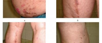
Psoriasis rashes look like dense plaques of pink-red color. The surface peels off with silvery scales and is dry, which causes itching. When scratching the papules, the scales easily come off (symptom of stearin stain), after removing the scales the surface begins to shine (symptom of terminal film), if you scratch further, a few drops of blood will be released (symptom of “blood dew”).
These symptoms are combined into the “psoriatic triad,” a characteristic feature of psoriasis. The rashes are localized on the extensor surfaces of the extremities, lower back, sacral area, and scalp (psoriatic crown). Plaques can merge, and in advanced cases, warty growths form. The scales on their surface turn yellow, stick together, and purulent bacterial flora joins.
Diagnostics
- External examination, identification of the “psoriatic triad” characteristic of psoriasis.
- Biopsy diagnosis of skin (for psoriasis, a histological specimen reveals, along with inflammation, disturbances in keratinization processes).
- Blood test (confirmation of inflammation, diagnosis of hyperlipidemia).
Treatment
- Immunosuppressive therapy for severe psoriasis (cytostatics, glucocorticosteroids, retinoids).
- Prescription of drugs that improve the rheological properties of blood (trental, rheopolyglucin, hemodez, heparin and others).
- Detoxification therapy.
- Local therapy in the form of ointments, creams containing keratoplasty drugs and/or glucocorticosteroids.
- Phototherapy methods: PUVA therapy (local application of a photosensitizer to the lesion followed by irradiation with long-wave UV rays).
Selective light therapy (combination of long-wave and mid-wave UV radiation).
- Diet contributes to the clinical cure of psoriasis. The patient limits the consumption of nightshade vegetables and spices. Refuses smoking and drinking alcohol.
Traditional methods

