A mole or nevus is a pigmented formation on the skin. It contains melanin and melanocyte cells. Moles appear on the body as a person grows older and depend on solar activity. Nevi can develop on any part of the body, become more voluminous or increase in size, causing some aesthetic inconvenience. In addition to their external beauty, nevi can degenerate into malignant melanoma through the process of metastasis. Can this be avoided? Yes, it is enough to remove the mole surgically in a trusted clinic. How exactly does the removal take place, what are the risks/consequences, and how often should I visit a dermatologist?
General characteristics of formations
A mole is a pigment formation of one of the shades of the brown palette. The base contains melanin and the melanocyte cells that produce it. Melanins are high molecular weight pigments. They are characterized by a heterogeneous structure and complex chemical composition. Pigments are found not only in moles, but also in all skin, hair, iris, inner ear and even some parts of the brain.
Content:
- General characteristics of formations
- Indications/contraindications for the procedure
- What you need to know about melanoma?
- How to properly prepare for mole removal
- Technique of the operation
- Possible complications and side effects
- Features of rehabilitation
It is important not to confuse nevi and birthmarks. Moles do not form in the prenatal period, but as a person grows older and are closely related to his lifestyle. The cause of the formation of pigment spots can be a hereditary factor, exposure to ultraviolet radiation, or hormonal changes.
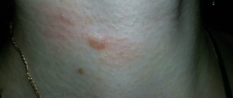
Each mole is a separate formation with a unique life cycle. At first it is a flat spot, which can increase and rise slightly above the skin as it grows and develops. The size and location of the formation depends on the level of melanocytes. If they are in the epidermis (top layer of skin), the mole will be flat. The deeper the melanocyte is immersed in the dermis, the higher the formation is located on the surface of the human body.
Convex nevi, which rise slightly above the skin, should not frighten their owners. This condition has no negative consequences and is considered a variant of the norm. The main thing is that the mole is miniature, round and uniform (in terms of color and structure) throughout its life cycle.
Can a mole disappear on its own and how safe is it? Yes, the formation can disappear without the influence of mechanical factors. This happens gradually and can alert a person. First, a white outline is formed around the formation, which resembles an orbit. This contour begins to increase and gradually covers the surface of the entire formation. The nevus disappears, and a small white spot remains in its place. Most often this occurs after severe sunburn. The disappearance of moles can also be a harbinger of the rare disease vitiligo.
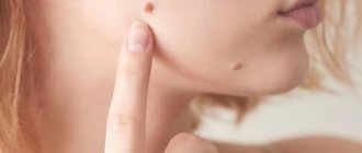
If you notice changes in a mole (increase/decrease, uneven edges, change in shade or structure), be sure to consult a specialist.
Follow-up after melanoma excision
The frequency of follow-up examinations depends on the stage of the tumor process. After excision of early stage melanoma, physical examination is performed every 6 to 12 months for several years. In the absence of alarming symptoms, the periods between visits to the doctor can be extended. Conversely, if the patient has a large number of moles, the frequency of examinations may be increased.
For thicker melanomas or those that have spread beyond the skin, a typical screening schedule may include physical examinations every 3 to 6 months for several years, after which visits to the doctor may become less frequent.
Life after melanoma removal should include careful self-monitoring. For self-examination of the skin, the ABCDE algorithm has been developed:
| A | Asymmetry. Asymmetry | Lack of symmetry of moles is a warning sign. |
| B | Border. Border | A benign birthmark, unlike melanomas, has smooth borders. The edges of melanoma are usually irregular and may be jagged or jagged. |
| C | Color. Color | Most benign formations have a uniform color, usually brown tones. A warning sign is a change in color or shade of a mole. |
| D | Diameter Diameter | Benign moles usually have a smaller diameter than malignant ones. Melanomas typically have a diameter larger than the eraser on the tip of a pencil (¼ inch or 6 mm). |
| E | Evolving. Development | Benign moles look the same over time. Any change (in size, shape, color, height or other characteristic) or the appearance of any new symptoms (bleeding, itching, crusting) is dangerous. |
Is it possible to reduce the risk of progression or recurrence of melanoma?
There are a number of recommendations that will reduce the risk of melanoma recurrence or progression:
- limit exposure to ultraviolet rays (sun, solarium);
- monthly skin examination (ABCDE);
- a complete diet;
- to give up smoking;
- physical education classes;
- maintaining normal weight.
About food supplements
There is still no scientific evidence that taking dietary supplements (BAS), including vitamins, microelements and herbal components, helps reduce the risk of relapse or prevent the appearance of metastases after treatment of melanoma. However, in some cases, these supplements may be recommended as a dietary supplement. In each case, when making a decision on this matter, it is advisable to consult a doctor.
About psychological support
Patients with skin melanoma often have questions after surgery:
- Will a relapse occur after a mole is removed?
- What are the chances that melanoma will come back?
- How will I know if the cancer comes back?
- What will I do if he comes back?
- When will he return?
In this regard, treatment after removal of melanoma should include psychological assistance from specialists and support from close relatives. Many of them should be helped to learn to live with uncertainty. Although there are no visible signs of relapse at this time, patients should understand that the disease can recur at any time.
Indications/contraindications for the procedure
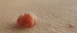
The surgical method is most often used when malignant degeneration of a nevus is suspected. Doctors recommend removing formations that are constantly in contact with clothing or located on the scalp, which increases the risk of damage. Moles in the groin, neck, chin, back or in the folds of the skin are most often damaged - consult a dermatologist to decide whether to remove them. Large formations on “legs” also pose a colossal danger. They can come off or become twisted, which can cause pain and infection.
Excision covers not only the affected area, but also healthy tissue, which prevents recurrence of skin growths. The main advantage over more modern laser removal or cryodestruction is the obtaining of material for histological examination. The excised tissue is sent for diagnostics in order to better understand the causes of the disease, understand the possible risks and create a preventive course for the patient.
Surgical procedures can only be performed in specialized medical institutions. Beauty salons or home procedures are not suitable for this and can cause irreversible consequences for the body.
| Indications | Relative contraindications |
| Increasing the size of education | Infectious diseases regardless of stage |
| Deep penetration of the mole under the skin | Acute inflammatory processes |
| Bleeding, change in color or texture | Chronic pathologies in the acute stage |
| Frequent injury to the mole, the formation causes pain or inconvenience | Pregnancy and breastfeeding period |
| Aesthetic discomfort |
There are no direct contraindications to surgical removal of nevi. In case of relative contraindications, the patient must undergo additional tests and consult a specialist.
The doctor will determine the advisability of removal, indicate possible complications and features of rehabilitation. In some cases, surgery will have to be postponed until the patient fully recovers.
Laser removal
Modern cosmetology allows using a laser to eliminate any mole without a trace on the surface of the skin. The practical benefit of the laser is that under its influence the visible area of the mole is destroyed , leaving virtually no marks on the skin. High-precision treatment of nevus does not affect the surrounding skin. As a result, the duration of the recovery course is reduced. If we talk about other advantages of the mentioned technology, they look like this:
pros
- jeweler precision of the beam;
- short duration of the operation;
- rapid regeneration of damaged skin areas;
- no bleeding;
- the patient does not need special care after completion of the procedure;
- no restrictions throughout the rehabilitation course.
Minuses
- It is impossible to obtain material for further histological examination in order to identify oncological processes, which endangers the patient’s life.
- Possible allergic reactions to ultraviolet radiation.
- If the patient suffers from other skin diseases such as herpes, dermatitis, etc., drug treatment is necessary after the procedure.
- If the tumor is large, it is impossible to remove it completely in one session.
What you need to know about melanoma?
Every mole on the human body can degenerate into a malignant melanoma. What it is? Melanoma is one of the types of malignant tumors. It also contains melanocyte cells (producing melanin), but in melanoma the cells are more aggressive.
They constantly divide, displace healthy cells, enter the vascular bed and spread throughout the body. As soon as the first aggressive cell approaches the brain or, for example, the lungs, it settles inside the organ and begins to rapidly divide. This process is called metastasis.
Melanomas are extremely dangerous because they quickly metastasize and divide at an incredible rate. The main thing is to detect the pathology in time. Surgical removal of melanoma will prevent the process from spreading throughout the body and guarantee a complete cure. Failure to see a doctor in a timely manner or complete refusal of treatment can result in death.
Life after melanoma removal
After removal of melanoma, the patient is at risk of recurrence of the disease. It is also possible for new tumors to form. Melanoma that develops after removal of a mole should be examined and removed as quickly as possible.
In some people, melanoma surgery may completely eradicate the tumor. However, there will always be a risk of relapse of the disease.
In other patients, melanoma may be inoperable, in which case you need to be prepared for immunotherapy, targeted therapy, chemotherapy and other treatments aimed at curbing tumor growth and increasing life expectancy.
Learning to live with cancer is not easy, as a person is forced to completely change his lifestyle. In this case, it is very important to develop the following plan with your doctor:
- Approximate schedule of necessary examinations and tests.
- A schedule of tests that may be needed, such as screening for other types of tumors and early detection of possible complications.
- List of possible side effects of treatment.
- What you need to pay special attention to and when to see a doctor.
- Individually selected diet and frequency of meals.
- Physical activity regimen and list of possible restrictions.
How to properly prepare for mole removal
No specific preparation is provided before removal. The patient just needs to come to the medical center and follow the necessary instructions from the specialist. Before the operation, a painkiller must be administered to make the procedure as comfortable as possible. In most cases, drugs with lidocaine are used. If there is an increased risk of bleeding, the doctor selects a medication and administers it intravenously after pain relief.
Immediately before the procedure, the doctor conducts a short consultation. He explains the essence of the method, possible complications and skin care rules.
As soon as the nuances are agreed upon, the doctor begins work. The patient is placed on a couch and the skin is treated with a special solution. It disinfects the area so that pathogenic bacteria do not enter the open wound. At this stage, the preparation ends, and the specialist proceeds directly to the surgical intervention.
Advantages of radio wave surgery for mole removal:
- The procedure is quick and virtually painless;
- There is no need to take preliminary tests;
- Minimal likelihood of complications;
- Rapid tissue healing;
- The material is sent for histological examination;
- Impeccable cosmetic effect;
- No stitches required;
- The only restriction after the procedure is that you should not take a bath or shower for 1-2 days.
Are you looking for a clinic where you can quickly and without complications remove a mole? Make an appointment with a professional oncodermatologist. If there are indications, we will remove the nevus using radio wave surgery - without complications and unnecessary hassle!
Technique of the operation
The removal technique depends on the location and size of the formation. The mole must be excised within healthy tissue. The area of excision is determined by the doctor, warning the patient about it. For example, if the nevus is located on the face, the specialist must remove a minimum amount of epidermis. On other parts of the body, the result of the intervention will be less noticeable, so the doctor can capture more healthy tissue.
There are two main methods of surgical removal. One of them is cutting off the mole without suturing. The doctor uses a scalpel to cut off the growth slightly below the skin level. If bleeding occurs during the procedure, the wound is cauterized and a local antibiotic is applied. A bandage is applied to the affected area and skin care rules are explained. Most often, the surgical area is prohibited from getting wet or subjected to mechanical or ultraviolet influence. Based on the individual characteristics of the patient, the doctor draws up a list of necessary medications, care products and sets a date for the next visit to assess the body’s reaction.
The second method is removal with sutures. Most often, the method is used in the treatment of dark or flat moles. The doctor cleanses the skin, injects an anesthetic, and excises the formation and its surrounding surface.
During the operation, the deep layers of the skin are affected, so suturing is a mandatory procedure.
The healing rate of the area is 7-14 days and depends on the individual characteristics of the patient. The operated area should be cleaned periodically, protected from damage, and ointments or oral medications should be regularly applied.
The main thing is not to self-medicate, but strictly follow the doctor’s instructions. After some time, you need to make an appointment with a dermatologist again. He will evaluate the condition of your skin, possible side effects/complications, and your overall body response to surgery.
Possible complications after surgical excision of nevi
After surgical removal of a mole, the following side effects may occur:
- Itching, pain, discomfort at the surgical site;
- Allergic reaction to a drug for local anesthesia;
- Prolonged bleeding;
- The appearance of keloid scars;
- Swelling in the area of intervention.
An increase in body temperature may indicate the presence of an inflammatory process, so in this case you should consult a doctor.
Surgical removal of moles is one of the effective methods of getting rid of nevi, which helps prevent the possible development of cancer pathologies. Our clinic employs qualified and experienced doctors who will help you efficiently and quickly get rid of not only nevi, but also other neoplasms.
Attention!
This article is posted for informational purposes only and under no circumstances constitutes scientific material or medical advice and should not serve as a substitute for an in-person consultation with a professional physician.
For diagnostics, diagnosis and treatment, contact qualified doctors! Number of reads: 4933 Date of publication: 08/06/2018
Dermatologists - search service and appointment with dermatologists in Moscow
Possible complications and side effects
The main disadvantage of surgical removal is the postoperative scar. It will be noticeable even with a cosmetic stitch, despite the fact that over time the skin will lighten and restore its structure. The surgeon’s task is to make the scar as invisible as possible, so choose a medical institution and doctor responsibly.
There is a risk of developing a keloid scar. This is a tumor-like growth of rough fibrous connective tissue. It is formed in people with a corresponding predisposition. Most often occurs when suturing large wounds.
Another risk is relapse. A repeat process is possible provided that the doctor has partially removed the mole, which is a gross violation of the surgical technique.
Immediately after excision, redness of the skin, itching, and a feeling of discomfort are possible. All these symptoms go away on their own after a few days. If something worries you, be sure to tell your doctor about it.