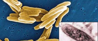
Syphilitic chancre is an ulcerative or erosive formation that appears in the primary stage of syphilis infection and is its main symptom. There are 13 types of chancre due to syphilis: ordinary and atypical. Syphilis is treated with medications and a special regimen.
Syphilitic leucoderma
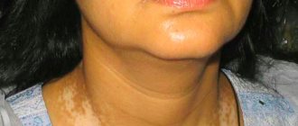
Syphilitic leukoderma or pigmentary syphilide was first described in 1854 by Hardy, and in 1883 the disease received its modern name. Leukoderma is one of the most characteristic symptoms of the disease. It occurs in both fresh and recurrent secondary syphilis and is characterized by the appearance of colorless spots on the side, back and front surfaces of the neck (“Venus necklace”).
Syphilitic leukoderma appears 4 to 6 months after infection. Its causes are profound neurophysiological changes, which are manifested by disorders of pigment formation. In 50 - 60% of patients with leukoderma, cerebrospinal fluid pathology is noted.
Most often, pigmentary syphilide is localized on the skin around the neck, but sometimes its location is noted on the anterior walls of the armpits, the area of the shoulder joints and the upper back. The chest, abdomen, limbs and lumbar region are rare locations.
At the beginning of the development of leukoderma, spots appear in the form of pale yellow hyperpigmentation with a diameter of 3 to 10 mm. Gradually, hyperpigmentation intensifies. Against its background, areas of depigmentation with rounded outlines appear. If depigmented spots are located in isolation, they speak of a spotted form of leukoderma. When spots merge, when the hyperpigmented background decreases, changes on the skin become lace-like - “lace” leucoderma. If the pigmentation around the depigmented spots is weakly expressed, they speak of “marbled” leucoderma.
Areas of syphilitic leukoderma never peel off, and there are no acute inflammatory phenomena. The patient's general condition remains satisfactory. Resistance to specific therapy is noted. There is leucoderma from several months to 4 years. Treponema pallidum is never detected in lesions.
Syphilitic leukoderma should be distinguished from pityriasis versicolor, vitiligo, plaque parapsoriasis, cicatricial atrophy, etc.
Rice. 1. Syphilitic leukoderma.
Rice. 2. A sign of secondary syphilis is leucoderma.
Vesicular syphilide
Vesicular syphilide occurs in severe syphilis. The main places of localization of syphilides are the skin of the extremities and torso. On the surface of the formed plaque, which is red in color, many grouped small vesicles (bubbles) with transparent contents appear. The vesicles quickly burst. In their place, small erosions appear, and when they dry, crusts form on the surface of the rash. When cured, a pigment spot with many small scars remains at the site of the lesion.
The rashes are resistant to therapy. With subsequent relapses they appear again. Vesicular syphilide should be distinguished from toxicerma, simple and acute herpes.
Syphilitic alopecia
Syphilitic alopecia (pathological hair loss) occurs with secondary syphilis in 15 - 20% of cases. Some patients experience loss of eyelashes, eyebrow hair, mustache and beard. This pathology occurs in both fresh (early) and recurrent syphilis. Often combined with leucoderma.
The cause of small-focal syphilitic alopecia is a disorder of hair nutrition that develops as a result of inflammation caused by Treponema pallidum. The cause of diffuse syphilitic inflammation is considered to be intoxication, disruption of the endocrine and nervous systems resulting from exposure to a syphilitic infection. In all forms of alopecia, the hair follicle is not damaged, so 1 to 2 months after adequate treatment, the hair grows back.
With small focal alopecia, the patient has many small, round-shaped foci of baldness throughout the head, but the largest number of them is recorded at the temples and in the back of the head. Due to the fact that not all hair falls out on the affected areas, patches of baldness resemble moth-eaten fur. The skin does not become inflamed. There is no peeling or itching.
With diffuse alopecia, hair begins to fall out from the temple area and then the process spreads throughout the entire scalp, which is observed in some severe acute infectious diseases.
With mixed alopecia, a combination of the two forms of the disease described above is observed.
Hair on the eyebrows falls out in the form of small patches of baldness (omnibus syphilide).
Eyelashes fall out and grow unevenly, resulting in unequal lengths (stepped eyelashes, Pincus sign).
Syphilitic alopecia should be distinguished from alopecia areata, superficial trichophytosis, microsporia, favus, early baldness, lupus erythematosus, and lichen planus.
Rice. 3. Small focal syphilitic alopecia is a sign of secondary syphilis.
Rice. 4. Syphilitic alopecia in men.
Rice. 5. Pincus symptom - stepwise growth of eyelashes with syphilis and hair loss with syphilis on the eyebrows.
How chancroid develops
Primary syphiloma forms after the incubation period has passed: 3-4 weeks after contracting the infection. It occurs in places with skin lesions in which natural body fluid contaminated with bacteria has entered: sperm, secretion of the uterine cervix.
An ulcer does not appear immediately. Initially, a red spot appears on the infected area, which, under the influence of treponemas and cells of the immune system, thickens and turns into a nodule. The compaction is not accompanied by pain or discomfort, and therefore often goes unnoticed by the patient.
Over the next 7-10 days, the nodule develops: it increases in size, thickens and then ulcerates. Ulceration can be of two types: superficial, in the form of erosion, or deep, in the form of an ulcer. The ulcer or erosion takes on its final form: it acquires clear, pronounced boundaries, an even oval or round shape.
At the bottom of the manifested syphiloma, a liquid is released containing a large number of pale treponema and cells of the immune system. The bottom itself acquires a pronounced red tint with bluish notes.
This type of chancre persists for 1-2 months, after which the process of healing and tightening begins. This signals the transition of the disease to a secondary, more dangerous and severe stage.
3-4 days before the chancre disappears, multiple rashes appear on the patient’s body, often accompanied by burning and itching.
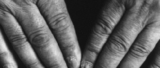
Nail damage due to syphilis
- Nails are affected in the second period of syphilis, more often in patients with pustular syphilide. This pathology is rare. The disease affects both the nail itself and the periungual fold.
- Damage to the nail fold begins with the appearance of papules or pustules. They are located isolated on the nail fold, but sometimes merge. The clinical picture resembles panaritium. Papules are red with a bluish tint. The inflammatory reaction is significantly expressed. Sometimes an abscess develops, which ulcerates over time.
- Syphilitic lesions of the nail plate develop slowly. The nail becomes dull and thickens, acquires a grayish-dirty color, and begins to crumble. Transverse and longitudinal cracks appear on it. Sometimes the nail dies completely and is rejected. Even without treatment, a normal nail plate grows back after a few months. Under the influence of specific treatment, a normal nail grows faster.
Damage to the mucous membranes (syphilis in the mouth)
On the mucous membranes of secondary syphilis, syphilitic roseola (spotted syphilide), papular and pustular syphilides are found.
Syphilitic roseola of the mucous membranes
Syphilitic roseola in the oral cavity is isolated, or the spots merge, forming continuous areas of hyperemia in the tonsils (syphilitic tonsillitis) or soft palate. The spots are red, often with a bluish tint, sharply demarcated from the surrounding tissue. The general condition of the patient rarely suffers.
When roseola is localized in the nasal passages, dryness is noted, sometimes crusts appear on the mucous membrane. On the genitals, syphilitic roseola is rare and always inconspicuous.
Papular syphilide of the mucous membranes
The most common type of syphilis is papular syphilide. Papules on the mucous membranes have a dense base and dense consistency, round in shape, smooth, flat, with clear boundaries, deep red in color, and do not bother the patient. Due to constant irritation, their central part macerates and acquires a whitish-gray or yellowish tint. Papillary growths appear on the surface. Papules are prone to hypertrophy. When they merge, fairly large plaques are formed, which have clear boundaries and scalloped edges.
The mucous membrane of the oral cavity, gums, tongue, lips, corners of the mouth, genitals, anus are the main locations of papules. Less commonly, papules are located on the mucous membrane of the pharynx, nose, eyes and vocal cords.
In some cases, in patients with secondary syphilis, erosive-ulcerative syphilide appears on the mucous membranes. Such papules are often located on the tonsils and soft palate.
Papules in the corners of the mouth often become crusty, crack, and resemble jams. Papules on the back of the tongue look like oval, devoid of papillae, bright red formations (“mown meadow symptom”).
Papules may appear on the mucous membrane of the larynx. When the vocal cords are damaged, hoarseness is noted. With a widespread process, complete loss of voice develops (aphonia).
Papular syphilide of the nasal mucosa occurs as a severe catarrhal inflammation.
Papular syphilide of the oral cavity should be distinguished from banal tonsillitis, diphtheria, lichen planus, aphthous stomatitis and planar leukoplakia.
All elements of the rash with syphilis in the oral cavity are extremely contagious. Papular syphilide of the oral cavity poses a great danger to dentists.
Rice. 6. Syphilis in the mouth - papular syphilide of the tongue.
Rice. 7. Syphilis in the mouth - papular syphilide in the corners of the mouth and on the hard palate.
Pustular syphilide of the mucous membranes
Pustular syphilide of the mucous membranes is rare. The development of the disease begins with the appearance of a diffuse infiltrate, which disintegrates over time to form a deep, painful ulcer. The bottom of such an ulcer becomes covered with pus. The process is accompanied by malaise and elevated body temperature.
All erosive and ulcerative processes localized on the mucous membranes should be examined for the presence of Treponema pallidum.
Pustular syphilide
Pustular syphilide, like vesicular syphilide, is rare, usually in weakened patients with low immunity and has a malignant course. When the disease occurs, the general condition of the patient suffers. Symptoms such as fever, headache, severe weakness, joint and muscle pain appear. Often, classical tests for syphilis give negative results.
Acne, smallpox, impetiginous, syphilitic ecthyma and rupiah are the main types of pustular syphilide. Rashes of this type are similar to dermatoses. Their distinctive feature is a copper-red infiltrate located along the periphery in the form of a roller. The occurrence of pustular syphilide is promoted by diseases such as alcoholism, toxic and drug addiction, tuberculosis, malaria, hypovitaminosis, and trauma.
Acne-like (acneiform) syphilide
The rashes are small pustules of a rounded conical shape with a dense base, located at the mouths of the follicles. After drying, a crust forms on the surface of the pustules, which falls off after a few days. A depressed scar remains in its place. The scalp, neck, forehead, and upper half of the body are the main locations for acne syphilide. Elements of the rash appear in large numbers during the period of early secondary syphilis, and scanty rashes appear during the period of recurrent syphilis. The general condition of the patient suffers little.
Acne syphilide should be distinguished from acne and papulonecrotic tuberculosis.
Rice. 14. Rash due to syphilis - acne syphilide.
Smallpox syphilide
Smallpox syphilide usually occurs in weakened patients. Pea-sized pustules are located on a dense base, surrounded by a copper-red ridge. When the pustule dries out it becomes like a smallpox element. In place of the fallen crust, brown pigmentation or an atrophic scar remains. The rashes are not abundant. Their number does not exceed 20.
Rice. 15. The photo shows manifestations of secondary syphilis - smallpox syphilide.
Impetiginous syphilide
With impetiginous syphilide, a dark red papule the size of a pea or more appears first. After a few days, the papule festeres and shrinks into a crust. However, the discharge from the pustule continues to be released to the surface and dries out again, forming a new crust. The layering can become large. The formed elements rise above the skin level. When syphilides merge, large plaques are formed. After peeling off the crusts, a juicy red bottom is exposed. Vegetative growths resemble raspberries.
Impetiginous syphilide, located on the scalp, nasolabial fold, beard and pubis, is similar to a fungal infection - deep trichophytosis. In some cases, the ulcers merge, forming large areas of damage (corrosive syphilide).
Healing of syphilide is long. Pigmentation remains at the site of the lesion, which disappears over time.
Impetiginous syphilide should be distinguished from impetiginous pyoderma.
Rice. 16. In the photo, a type of pustular syphilide is impetiginous syphilide.
Syphilitic ecthyma
Syphilitic ecthyma is a severe form of pustular syphilide. Appears 5 months after infection, earlier in weakened patients. Deep pustules are covered with thick crusts up to 3 or more centimeters in diameter; they are thick, dense, and layered. Elements of the rash rise above the surface of the skin. They have a round shape, sometimes irregular oval. After the crusts are rejected, ulcers with dense edges and a bluish rim are exposed. The number of ecthymas is small (no more than five). The main places of localization are the limbs (usually the lower legs). Healing occurs slowly, over 2 or more weeks. Ecthymas can be superficial or deep. Serological tests sometimes give negative results. Syphilitic ecthyma should be distinguished from vulgar ecthyma.
Rice. 17. Secondary syphilis. A type of pustular syphilide is syphilitic ecthyma.
Syphilitic rupee
A type of ecthyma is syphilitic rupee. The rashes range in size from 3 to 5 centimeters in diameter. They are deep ulcers with steep, infiltrated edges, covered with dirty and bloody discharge, which dry to form a cone-shaped crust. The scar heals slowly. It is often located on the shins. It spreads both peripherally and deeply. Combines with other syphilides. It should be distinguished from rupoid pyoderma.
Rice. 18. Rash due to secondary syphilis - syphilitic rupee.
Rice. 19. In the photo, the symptoms of malignant syphilis of the secondary period are deep skin lesions: multiple papules, syphilitic ecthymas and rupees.
Syphilitic sore throat
Syphilitic sore throat is one of the manifestations of syphilis in the mouth. With syphilitic roseola, spots appear in the area of the tonsils and lymphoid ring, which can be located either isolated or merge, forming continuous areas of hyperemia (syphilitic tonsillitis). The spots are red, often with a bluish tint, sharply demarcated from the surrounding tissue. The general condition of the patient rarely suffers.
With secondary syphilis, papular tonsillitis is more common. Papular elements tend to grow peripherally and often merge, forming plaques with clear boundaries. When ulcerated, the papules become covered with a whitish coating. When the mucous membrane of the pharynx is affected, there is pain when swallowing. Ulcerated papules are always accompanied by pain. The patient's general condition worsens. An increased body temperature appears.
Rice. 8. Syphilis in the mouth - syphilitic sore throat: syphilitic roseola (photo on the left) and papular syphilide (photo on the right).
Rice. 9. Syphilis in the mouth - syphilitic sore throat.
Signs of syphilitic lesions of the mucous membranes of the nose and oral cavity, pharynx and larynx:
- the disease proceeds without pronounced inflammatory phenomena,
- painlessness,
- the course of the disease is long,
- resistance to anti-inflammatory traditional therapy is noted,
- tests for syphilis are often positive.
Atypical forms of syphilitic chancre
Atypical chancres are types of syphilomas that differ from the usual types in one or more characteristics.
These include:
- Chancroid felon:
an ulcer with jagged edges that appears on the fingers. Most often it occurs on the index finger and thumb, accompanied by shooting pain, swelling, blue discoloration and suppuration. This is an “occupational disease” of surgeons and gynecologists who violate safety regulations. - Indurative edema:
chancre in the genital area, causing severe swelling, bluish skin and swelling of the genitals. Occurs on the labia and foreskin. Not accompanied by pain or inflammation. - Amygdalitis:
unilateral, less often bilateral chancre, located on the tonsils. Enlarges and deforms the tonsil on which it is located, which can cause pain. The color of the tonsil tissue does not change, so the disease can be confused with a sore throat.
With the exception of these features, atypical forms of chancroid do not differ in any way from the usual varieties. The development of atypical syphilomas, the time of their appearance and disappearance are similar to the classical forms.
Damage to internal organs, bones, joints and nervous system
Damage to internal organs
With early secondary syphilis, any organs can be affected, but the liver and stomach most often become inflamed, myocarditis and nephrosonephritis develop. The inflammatory process often does not manifest itself clinically.
Damage to the osteoarticular apparatus
Bones and joints are affected at the end of the primary period of syphilis, but more often lesions are recorded in the secondary period. Pain is the main symptom of the disease. There is pain in the lower extremities in the area of long tubular bones, knee and shoulder joints. Hydroarthrosis, periostitis and osteoperiostitis occur.