Papilloma is a benign neoplasm of a viral nature. It is a small growth that can appear on any part of the body or mucous membranes. The formation looks like a papilla on a narrow stalk. The cause of papillomas is the human papillomavirus (HPV). Papilloma does not go away on its own, it can spread, and in most cases the tumor is removed. Papillomas located on the genitals have a high risk of malignancy. Occurs in men, women and children.
Features of papillomas
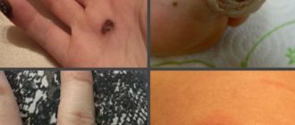
Papillomas can be of the following types: ordinary or vulgar warts, filiform (the most common type), flat, genital warts, plantar warts, juvenile warts. A dermatologist diagnoses and treats papillomas.
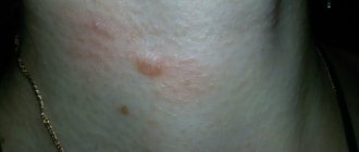
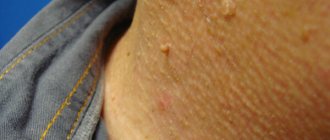
Most often, papillomas are localized in the armpits and inguinal folds, hands, on the surface of the foot, neck, under the mammary glands in women, as well as on the genitals. Papillomas located on the mucous membranes of internal organs are very dangerous; they can cause ulceration or bleeding. As a rule, papillomas that have a pronounced cosmetic defect are removed with a laser, i.e. which are located in open areas of the body, as well as those with a high risk of cancer.
When is it necessary to remove papilloma?
Benign skin tumors cannot always be removed; sometimes observation and regular examinations are sufficient. The most common indications for removal are the following:
- papillomas located on visible parts of the body and causing psychological discomfort;
- papillomas that are constantly injured by clothing, jewelry, and a comb;
- neoplasms that increase in size, tend to spread, change size, shape and color;
- papillomas located in the intimate area or soles;
- if the tumor begins to hurt, itch or bleed.
Development of the disease
HPV infection can occur through all types of sexual contact (vaginal, oral, rectal), while condoms are not a guarantee of protection, since condylomas can be localized on unprotected areas of the skin and the size of the virus is smaller than the pore size of condoms. In addition, the virus can also be transmitted to the baby during childbirth if the woman is a carrier of the virus.
The human papillomavirus (HPV) has a tropism for the skin and mucous membranes of the genitals, oral cavity, and anus. The amount of virus depends on the state of both the immune system as a whole and local immunity. The more productive the individual factors of the immune system are, the more actively the division of the virus is blocked, and the less of it is contained. That is, a person can be infected with HPV without having any external clinical symptoms.
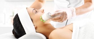
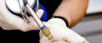
Preparation and carrying out laser removal
Laser removal is preceded by a thorough examination and examination of the tumor. The doctor must be sure of its benign quality; in case of any doubts and the borderline state of the cells, laser technology is not used, but a surgical operation is performed.
During the procedure, under the influence of a laser, heating and layer-by-layer destruction of neoplasm cells occur, so it is impossible to take biological material for subsequent research. Additionally, the doctor surveys the patient about possible contraindications to the procedure, and also conducts a number of studies (blood tests).
No special preparation is required for the procedure. At the appointed time, the patient comes to the specialist, the operation includes the following stages:
- Disinfection of the surgical field.
- Conducting anesthesia (if necessary). For this, an injection, gel or spray is used.
- Installation of the device to the required laser length.
- Layer-by-layer evaporation of the neoplasm.
- Treat with an antiseptic and apply a bandage if necessary.
To avoid relapse of the disease, it is necessary to carry out antiviral therapy, as well as take immunomodulating and immunostimulating drugs to strengthen the immune system.
Preparation for the procedure
Two weeks before laser therapy you must:
- limit exposure to the sun, use products to protect against UV rays, and do not visit the solarium;
- do not carry out cosmetic procedures in the affected area (peelings, hardware cosmetology sessions, etc.);
- if the neoplasm is located in an area where the risk of irritation is increased (for example, it constantly clings to the collar), the papilloma must be covered with a plaster
Before the sessions, the doctor will definitely tell you about contraindications and identify their presence.
Hardware diagnostic methods
Dermatoscope HEINE DELTA 20 T
Designed for dermoscopic examination for early diagnosis of malignant melanomas, for the study of non-melanocytic formations, basal cell carcinoma and dermatofibroma.
Dermatoscopy is a non-invasive method of visual diagnosis of the skin, which, thanks to the use of high technology, allows us to move from a subjective assessment of the condition of the skin to an objective one and document identified skin changes.
The essence of the method is that the surface layers of the skin are examined using a special device - a dermatoscope at 10x magnification. This allows us to more thoroughly study the symmetry of the neoplasm, its boundaries, and structure.
CLEAR VISION 3D
The most phenomenal and new development in the field of diagnostics today, absorbing all the advantages of its older brother and multiplying them thousands of times.
The evolution of cinema from 2D to 3D did not pass by other areas of activity, because until recently the Vision system was famous for its so-called two-dimensional or 2D model for diagnostics.
It is not difficult to guess that covering the treated area from several sides will give a much greater effect than “observation from one angle.”
Diagnosis of 3D in our clinic is carried out free of charge.
“3D” in our case is not a tribute to fashion, but a visual confirmation of technological progress. The installation allows for three-dimensional scanning of the client’s face using a single short image. There is no need to worry about the quality, since the device is equipped with six high-resolution cameras located at three angles, which gives us images from both sides (profile and frontal) simultaneously in one shot. If time is money, then Clear Vision 3D understands this like no other!
DUB-TPM
Ultrasound diagnostics of skin.
Ultrasound diagnostic scanning is a well-known and proven technique, which currently accounts for more than 1/3 of all diagnostic procedures in medical practice. Modern devices are already quite easy to use and are available to many clinics.
However, these studies have not previously been used in dermatology, which was due to the difficulty of technically solving this problem. In conventional devices, the sensors have a frequency of 3-10 MHz, at which it was impossible to image the structures of the epidermis, dermis and hypodermis.
The German company TPM has created unique devices with sensor frequencies of 20-100 MHz. This technique is called high-resolution digital ultrasound imaging with the ability to study the most superficial layers of the skin.
Based on research materials conducted using the DUB TPM apparatus (the list is attached separately), many articles and 2 monographs have been published.
Until now, the main method for studying skin morphology has been histological and pathomorphological examination. This technique is quite labor-intensive and expensive; in addition, a biopsy sample is examined that has already been treated with various chemical reagents.
Ultrasound diagnostics of the skin fills the gap that previously existed between external research methods and histology, since this non-invasive method allows the study of the skin in vivo.
The importance of ultrasound scanning for skin diagnosis cannot be overestimated. This method has a number of undeniable advantages - non-invasiveness, painlessness, safety and high measurement accuracy. All studies are carried out without damaging tissue and can be repeated on the same area of skin many times.
The new tool allows you to see a section of the skin and subcutaneous fat down to the muscle fascia. We can examine the skin at various time intervals, documenting all the features. The data is digitized and placed in a database. It is easy to carry out a comparative analysis of images obtained over time, the images are saved on any digital media and the data is transmitted in publicly accessible formats via the Internet.
Ultrasound examination of the skin should become the “gold standard” in skin diagnostics, as in obstetrics, gynecology and cardiology.
Distinctive features of the DUB system
- TPM was the first to develop and commercialize skin ultrasound and set the standard for skin ultrasound throughout the world.
- Only DUB devices are equipped with sensors with a maximum frequency of up to 100 MHz and a resolution of up to 8-10 microns.
- Scan modes A,B,C.
- Three-dimensional scanning.
- Cinema loop without limitation of shooting duration.
- Digitization of the signal means the image is more detailed and clear.
- Digital data processing.
- View multiple images taken at different times.
- Innovative image processing algorithms.
- Saving raw data.
- Advanced software package.
- The use of an open system with water allows you to obtain 10-20% more information than using a system with film.
- At the moment, the device has no analogues throughout the world.
Device capabilities:
- 1) Study of the condition, structure and size of all layers of skin and skin formations.
- 2) In-depth diagnosis of morphological and functional changes in acute and chronic skin diseases, including scar changes and lipodystrophies.
- 3) Assessment of the dynamics of skin condition in normal and pathological conditions.
- 4) Facilitation of diagnosis of small skin rashes.
- 5) Timely early diagnosis, since with the help of ultrasound scanning it is possible not only to identify characteristic signs of skin manifestations at the earliest stages, but also to carry out preclinical diagnosis, prevention or timely treatment.
- 6) Diagnosis of skin conditions in the case of any manifestations difficult to detect with the naked eye.
- 7) Visualization, determination of size, volume and depth of invasion, as well as assessment of skin tumors and skin metastases, selection of treatment methods, setting parameters and monitoring effectiveness.
- Preoperative measurement of the depth of spread and volume of tumors during surgical interventions, including electrosurgery, cryosurgery, laser or radiation therapy.
- 9) Study of age-related skin changes.
- 10) Determination of the depth, intensity and duration of the therapeutic effect, choice of method.
- 11) Evaluating the effectiveness and monitoring of therapeutic, physiotherapeutic and surgical treatment methods, including cosmetic procedures (for example, mesotherapy, peelings, plastic surgery, tattoo removal, hardware procedures, etc.).
- 12) Preliminary diagnosis and evaluation of the results of the introduction of fillers, hyaluronic acid, collagen, synthetic or semi-synthetic gels, etc.
- 13) Early diagnosis of osteoporosis.
- 14) Study of skin elasticity.
- 15) Study of mucous membranes.
Advantages of using this method:
- 1) A non-invasive technique for visualizing the internal structures of the skin in vivo, which allows you to obtain important information that is not available with other research methods.
- 2) The method is indispensable for assessing the dynamics of skin condition in dermatology, cosmetology and dermato-oncology. Allows you to monitor the condition of skin manifestations and use the data in the primary diagnosis, prevention and treatment of most skin diseases.
- 3) The ability to save data in computer memory and on any electronic media, print out photographs for medical records, send via the Internet for consultations with colleagues.
- 4) An objective assessment of the dynamics of the patient’s skin condition is an important legal aspect in resolving conflict situations.
- 5) Visual visualization of the state of the internal structures of the skin and its relief is a strong psychological factor in explaining to patients the need for therapeutic measures.
- 6) With the help of DUB, it is easy to prove the effectiveness of treatment in a form accessible to the client. This is a powerful marketing tool to attract new customers.
- 7) The presence of this method increases the rating of the institution and indicates high equipment and the use of advanced technologies.
- In addition to the histological picture, it increases the accuracy of the pathological diagnosis.
- 9) Conducting scientific and educational programs.
- 10) Conducting consultations and consulting and diagnostic activities.
A new aspect of using this method for manufacturers is to assess the effects on the skin of various products, including cosmetics, medications, and devices.
VIVASCOPE® 1500 MULTILASER
The most accurate diagnostic device in dermatology, dermato-oncology and cosmetology.
Confocal laser scanning microscope for in vivo histological examination of skin by fluorescence using lasers of 3 wavelengths (785 nm, 658 nm, 445 nm)
- Combining reflected laser and fluorescence detection technology
- Optical non-invasive biopsy
- Real time display
The VivaScope 1500 Multilaser system combines reflected laser acquisition technology with fluorescence confocal laser scanning microscopy. Similar to the standard VivaScope 1500, skin areas can be viewed in vivo using infrared light. The wavelengths used are 785 nm (near infrared), 658 nm (red) or 445 nm (blue). All three lasers are integrated in one device.
Before using VivaScope, a fluorescent dye (non-toxic to the body) is applied to the area of tissue that needs to be examined. The corresponding laser radiation excites the fluorophore and the resulting fluorescence allows a contrast image to be obtained, which helps to display the histological structure due to the distribution of the dye.
With the VivaScope 1500 Multilaser it is possible to image various functional aspects of tissue changes in vivo. Living tissue can be visually examined sequentially using all available laser wavelengths.
Post-procedure care
For a quick and complete recovery and to avoid possible complications, the following measures are recommended:
- avoid contact with water in the first 2-3 days;
- Do not remove the crust that has formed (it will fall off on its own);
- Treat the wound with an antiseptic 2 times a day;
- protect the manipulation site from injury;
- wounds and new skin formed must be protected from ultraviolet radiation;
- refrain from shaving and epilation if the papilloma was in this area until complete healing.
Is it possible to get rid of papillomas on your own?
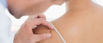
This question arises for many people. There are home remedies for acne, warts and age spots, so why can’t you remove papilloma yourself in the same way? Self-medication may be unsafe, experts warn. “Firstly, you don’t know exactly what you are going to delete,” says Maria Istomina. — Doctors have special medical diagnostic equipment that helps determine what type of tumor has appeared on the skin. Before removing any papilloma, the doctor carefully examines it with a dermatoscope. This device allows you to view the formation at significant magnification. This is necessary in order to understand whether it is really a papilloma, which can be safely removed, or something more serious. Secondly, in trying to get rid of papilloma on your own, you can injure yourself, which will cause the growth of the tumor or its transformation into a more dangerous form, even an oncological tumor.”
In the photo: Maria Istomina, dermatovenerologist at the SM-Cosmetology clinic.
Thus, it will be much safer not to experiment at home, but to seek help from a specialist. Moreover, the clinic will remove the papilloma quickly and painlessly.
Benefits of laser papillomas removal
Laser removal of papillomas has long been used to remove skin tumors, because this method has many advantages:
- Excellent result. Laser destruction removes the tumor completely, there is no risk of relapse.
- Minimum of unpleasant or painful sensations. The procedure is carried out using local anesthesia and there is no discomfort or pain.
- Speed of the procedure. Laser destruction of papillomas takes no more than 5 minutes to remove one tumor
- Minimal trauma. The laser beam has a directed effect and acts only on the cells of the formation, without affecting nearby tissues.
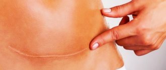
- No postoperative scars or scars. After laser removal, a small crust remains at the site of the papilloma, which disappears on its own within 1.5-2 weeks. New, intact skin appears at the site of formation.
- Versatility. Due to the precision of the laser, this method can be used on any part of the body, including areas with thin and sensitive skin, mucous membranes, intimate area, eyelid skin, etc.
- No rehabilitation period. The procedure is performed on an outpatient basis, after which you can lead your normal lifestyle.
- Minimal risk of postoperative bleeding. The action of the laser coagulates blood vessels, which eliminates the risk of bleeding.
Why are papillomas dangerous?
Typically, the appearance of skin formations is preceded by a situation that causes a decrease in immunity. This could be a previous cold, severe stress, hypothermia, exacerbation of chronic ailments and much more. In some cases, the “awakening” of the papilloma virus occurs during pregnancy.
Content:
- Why are papillomas dangerous?
- Benefits of laser papillomas removal
- How to prepare for the procedure
- Features of the procedure
- Rehabilitation after the procedure
- Contraindications for the procedure
It is almost impossible to determine the moment the virus enters the body - it can live quite peacefully in a person for decades and manifest itself only when unfavorable factors coincide, which have reduced the protective functions. Today, science knows about 130 types of HPV. The danger is that 10 of them have the ability to undergo malignant degeneration. That is, a small and unnoticeable growth on the skin can cause the development of a serious illness.
In some cases, after a certain period of time, papillomas disappear on their own - when the body’s defenses are restored. Most often, this phenomenon is observed in young and healthy women who encounter a problem during pregnancy - after childbirth, the growths begin to dry out and soon disappear. But you cannot let the course of the disease take its course - it is important to contact a specialist in a timely manner, who will select the most appropriate treatment method.
Attempts to eliminate tumors on your own can have negative consequences - from infection of the wound to the rapid spread of papillomas in healthy areas of the body. Improper treatment of HPV can cause relapse of the disease.
During laser removal, not only the body of the growth itself is destroyed, but also the pathological elements - that very thin stalk. Thanks to this, the risk of relapse is reduced by 85%.
Contraindications for laser removal of papillomas
Laser procedures are not performed under the following conditions:
- oncological diseases of any localization;
- HIV, MSID, viral hepatitis, diabetes mellitus;
- pregnancy and lactation;
- exacerbation of chronic diseases;
- infectious or inflammatory diseases;
- mental disorders;
- severe damage to the heart and lungs;
- organ failure;
- bleeding disorders;
- skin photosensitivity;
- autoimmune systemic diseases;
- inflammatory skin diseases, exacerbation of herpes infection.
Laser removal of papillomas and other skin formations can be performed in medical settings. The operations are carried out at a high professional level using modern laser technology of the latest generation, which eliminates the risk of complications or relapse. Removal of papillomas with a laser is carried out at any convenient time on an outpatient basis. After the intervention, you can go about your daily routine; it will not affect your usual lifestyle in any way. You can make an appointment by calling the center or leaving a request on the website.
Why PRESIDENTMED?
- This is the leading medical institution in the Russian Federation, using only certified and safe treatment methods!
- This is a center of excellence for training specialists in hardware methods!
- These are highly qualified specialists, professors, candidates of science, world-famous doctors!
- this is the introduction of new methods of diagnosis, treatment and the use of “last word” technologies in medicine!
Our clinic employs only experienced specialists who are constantly trained in the latest methods of hardware cosmetology. The clinic is also the largest specialist training center in the Russian Federation.
Removal of papillomas using a laser at the PRESIDENTMED clinic will give you aesthetic comfort and youth for a long time.