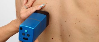
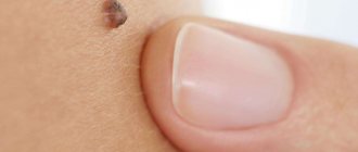
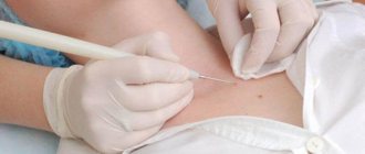
Moles (nevi) are pigmented formations that form on the human body as it grows and interacts with the environment. Some of them can jeopardize a person's health or aesthetic appearance. Such moles can be removed using a laser - evaporated or cut out with a beam. How exactly is this possible, how safe is laser removal of nevi and how to properly care for your skin after the procedure?
General characteristics of formations
Moles are brown pigmented growths. They are formed as the human body grows and develops. Additional factors for the appearance of nevi are hormonal levels, exposure to ultraviolet radiation, and genetics. Each mole contains melanin, a complex pigment that is responsible not only for color, but also for protecting the skin from sunlight.
Content:
- General characteristics of formations
- Types of nevi
- In what cases is it necessary to remove a mole?
- Contraindications to the procedure
- What you need to know about laser surgery?
- How is the removal done?
- Recovery period
- Possible complications and side effects
Most moles on the human body are not dangerous. The main thing is not to injure them, use sun filters and regularly conduct self-examination. But some pigmented formations can degenerate into melanoma. This is a malignant tumor that develops from melanocyte cells.
Melanoma is characterized by a rapid growth rate (cells rapidly divide and launch metastases throughout the body). The result is damage to organs and systems, which leads to irreversible consequences and death.
Is it possible to avoid melanoma? Yes, it is enough to conduct a self-examination and visit a dermatologist if one of the moles is suspicious. The only caveat is that melanoma cannot be removed using a laser. For this purpose, only surgical excision is used. If you notice that one of your moles has changed in color, size, or structure, contact your dermatologist immediately.
Possible consequences of the operation
Sometimes the healing process occurs with various deviations from the norm, which can occur regardless of how carefully the preventive requirements are followed. This may include the following:
- Nevus recurrence (re-formation of a mole). This situation is possible if during the operation the mole was not completely removed, and a small amount of nevus particles remained on the skin. Gradually, the cellular tissue grows, and the mole forms again. This does not pose a health risk, but in the future you will have to undergo the removal procedure again.
- Hypopigmentation is the formation of a white spot at the site of removal. This cosmetic defect occurs after removal of a deep-lying tumor. Hypopigmentation also occurs when exposed to sunlight. A white spot on the skin usually goes away on its own, but it takes at least a year.
- A hypotrophic scar is a scar at the site of a removed mole, the cause of which is the low rate of tissue restoration. This is usually a shallow defect, and over time the scar tissue smoothes out on its own.
- A hypertrophic scar is a small bump at the site of a removed mole, usually darker in color than the surrounding skin. The defect may go away on its own, or the doctor will prescribe additional cosmetic procedures that smooth the skin.
Types of nevi
Benign skin formations can be congenital or acquired. Nevi are also distinguished by shade, size, shape and surface texture. Congenital moles are conventionally divided into 4 categories based on size: small, medium, large and giant. Giant moles are extremely rare and occupy the entire anatomical area (for example, the chest or face).
Giant moles carry a hidden threat. The risk of nevus degeneration into melanoma reaches 50%. Patients with giant nevi should be regularly examined by a dermatologist to monitor the process and prevent degeneration in time.
Acquired formations are formed in early childhood. At this time, there is an intensive movement of melanocyte cells from the depths to the surface of the skin.
The number and location of moles is determined by genetic factors, exposure to ultraviolet radiation and general health.
There are only three types of acquired nevi.
The first of them is epidermal. This is the accumulation of a large number of melanocytes in the epidermis (the upper layer of the skin), which triggers the process of development of a flat miniature mole.
The second type is intradermal. Indicates an accumulation of pigment cells in the dermis (deep layers of the skin), which form convex, voluminous nevi. The third variety is borderline. This is a cluster of melanocyte cells at the border of the dermis and epidermis.
When is it necessary to remove moles?
- The mole begins to increase sharply in size;
- its forms and boundaries change;
- unpleasant sensations appear at the site of formation: irritation, pain or itching;
- its color changes;
- the mole begins to be injured by the touch of clothing or accessories.
The aesthetic aspect also plays an important role. Sometimes removing moles can be the only opportunity for women to feel much more confident.
Individual consultation
In what cases is it necessary to remove a mole?
Surgery is necessary if a mole is damaged frequently. If the formation is located on the scalp, chin, neck, groin, back or folds of skin, it constantly comes into contact with clothing/accessories, which leads to injury. Moles on “pedicles” should also be removed to avoid twisting or tearing off the stem.
Another common reason for removal is aesthetic. Volumetric nevi on the body or face make adjustments to a person’s appearance. Some people consider this their highlight, others try to hide the age spot by any means. Laser excision or vaporization is perfect for this.
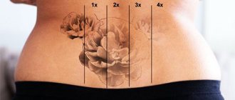
For aesthetic purposes, a mole can not only be removed, but also created. This is possible with the help of tattooing.
The only caveat is that it will not be possible to remove the tattoo without a trace, so the mole will remain with the person for life.
Cosmetic indications for birthmark removal
The mere presence of a mole is not a medical indication for its removal - but if the mole becomes a cause of complexes due to its poor aesthetic appearance, the doctor can remove it.
The “it’s better not to touch” principle, which many doctors used to adhere to, is already outdated. It is important to meet the following conditions:
- Carry out the removal procedure in a clinic, such as ours, with a certified experienced specialist using professional equipment;
- Make sure that the mole does not contain malignant cells. And for this, before removal, it is necessary to conduct a study - DERMATOSCOPE. In our clinic, this is a mandatory free study before removing any skin tumor!!!!
Contraindications to the procedure
The list of contraindications to laser removal includes:
- malignant degeneration of nevus;
- inflammatory lesions on the skin;
- exacerbation of the disease, general deterioration of the patient’s health;
- pregnancy (the appropriateness of the intervention is determined by the doctor);
- menstruation in women;
- predisposition to the formation of keloids.
Do not self-medicate and be sure to consult a dermatologist before any manipulation. He will examine the mole, determine its nature and select the optimal removal method.
What you need to know about laser surgery?
Laser surgery is one of the methods of performing surgical procedures. It involves removing/cauterizing small areas on the human body. The advantage of the method is minimal damage to surrounding healthy tissue and rapid restoration of the epidermis. For different types of operations, a specific type and power of equipment is used.
Laser surgery is used to correct vision, remove tumors, remove blockages in arteries, and excise pigmented formations.
Advantages:
- Small vessels become thrombosed.
- Bloodlessness of the operation.
- The surrounding tissues are not injured.
- The possibility of metastases is excluded,
- No traces are left.
- Painless.
- No contact with tools.
- Fast recovery period.
The only drawback is that this method does not allow us to conduct a histological examination. Therefore, it must be done in a medical facility.
Removing birthmarks and port-wine stains
How is the removal done?
With laser intervention, the nevus is literally evaporated from the skin. The beam, aimed at pigment formation, evaporates cells layer by layer, partially affecting healthy tissue. Additionally, the laser acts on blood vessels, neutralizing bleeding.
The skin reacts normally to the intervention, cells begin to actively divide, filling the affected area with a layer of healthy epidermis.
Before the intervention, the patient is given local anesthesia. If a person is confident in his abilities, and the size of the formation is minimal, then he can do without pain relief. The operation itself lasts on average 5-10 minutes, depending on the size and specifics of the formation.
Recovery period
Best materials of the month
- Coronaviruses: SARS-CoV-2 (COVID-19)
- Antibiotics for the prevention and treatment of COVID-19: how effective are they?
- The most common "office" diseases
- Does vodka kill coronavirus?
- How to stay alive on our roads?
After laser exposure, a dark crust forms at the site of the mole. This is a normal skin reaction to intervention. The crust forms a kind of protective layer that blocks damage/infection of the epidermis and helps to quickly return to normal. The main thing is not to tear off the crust, as an unaesthetic scar may form in its place.
The area should not be wetted for the first 5-7 days, but this does not negate general body hygiene. Try to protect your skin from mechanical damage, contact with cosmetics and ultraviolet rays. All these factors can slow down the recovery process and damage the structure of the skin. Treat the wound daily and lubricate it with medications prescribed by the dermatologist. After applying medications, you should use sunscreen or a spray that will prevent ultraviolet radiation from affecting the health of your skin.
Applying bandages or burning the surface with alcohol is also prohibited. If the condition of the wound bothers you (pain, redness, rash), be sure to inform your dermatologist.
Removal and treatment of moles with laser
After laser removal of a mole and to achieve the best cosmetic result until the wound heals, it is recommended to follow these recommendations:
- Avoid UV rays (and when unavoidable, use sunscreen with at least SPF 30, preferably SPF50). If the patient tans during healing, there is a high likelihood of post-inflammatory pigmentation, which can make the area where the procedure was performed more visible;
- Do not go to solariums, saunas, swimming pools, or engage in vigorous physical exercise (to avoid infections and injuries).
- As the wound heals, dry scabs form and act as a natural dressing to prevent infection. These scabs should not be scratched or otherwise torn off - wait until they fall off on their own.
During the procedures prescribed by the doctor - disinfection and dressing - the wound heals well and no infectious (or other complications) arise. Once the wound has healed, redness persists and sometimes it takes 1 to 2 months for the area of skin to reach its final appearance after the procedure. If the initial scabs have fallen off and there is an increased risk of scarring, a special scar treatment cream can be applied.
The main measures that are recommended for the treatment of scars formed after laser mole removal are as follows:
- Special purpose creams with silicone and so on.
- Special patches with silicone.
Silicone patch for scar treatment


