Pregnancy is a period when many changes occur in a woman’s body, and chronic diseases also worsen. This includes atopic dermatitis. Considering that this disease is more common in women, there is a risk of recurrence during pregnancy. Moreover, it is quite possible that the disease had not manifested itself in any way before.
Why does atopic dermatitis develop during pregnancy?
The diagnosis of “atopic dermatitis” is typical not only for pregnant women. Anyone can experience it, but more often the disease manifests itself in childhood. The reason is heredity - if at least one of the parents is atopic, then the risk of encountering dermatitis in the child increases. The first signs of the disease may appear in adulthood.
Atopic dermatitis often occurs for the first time in pregnant women. Before this, the woman may not have encountered such a disease. The reason is associated with increased production of cortisol, a hormone that plays an important role in fetal development and is also responsible for the formation of allergic reactions.
The exacerbation is also due to a decrease in immunity, which is necessary to ensure that the mother’s body does not reject the fetus. This explains the development of atopic dermatitis during pregnancy. The risk is especially high if a woman had such a diagnosis or a tendency to allergies before conception. Provoking factors also include:
- excessive psycho-emotional stress;
- consumption of allergenic foods;
- unfavorable environmental conditions;
- chronic diseases of the digestive system;
- endocrine disorders;
- hormonal changes in the body of pregnant women.
How does atopic dermatitis manifest and how is it diagnosed?
Atopic dermatitis is a chronic, often relapsing inflammatory skin disease.
There are 3 types of inflammation in AD, which can coexist in the same patient.
- Acute - erythematous papules and spots, which are combined with scratching, erosions and serous discharge.
- Subacute - erythematous, excoriated and scaly papules.
- Chronic - thickening and intensification of the skin pattern, excoriation, fibrous papules.
The classic classification of atopic dermatitis is based on three age groups.
Infant form - develops in infants under the age of 2 years (most often, the first manifestations occur at the age of 5–6 months).
In 70% of children, the weeping form with pronounced inflammation predominates. In 30% of children suffering from AD, areas of inflammation are recorded with the formation of scales and inflammatory crusts (without weeping).
Typical localization of elements at this age are the skin of the cheeks, forehead, scalp, neck, chest, elbows and knees. Sometimes the skin of the entire body is affected, with the exception of the area in the area where the diaper is worn, because there is increased humidity due to the occlusive effect of the diaper.
Child form - Occurs between 2 and 12 years of age and follows the infant form.
With this form, areas without weeping are more often recorded, but with pronounced inflammation, against which papules with scales are visualized.
It is worth noting that the older the child, the more pronounced the dry skin is and the more often a more emphasized pattern is observed.
Typical localization characteristic of the child's form is the skin of the extremities, wrists, forearms, folds, as well as in the area of folds and even feet.
Adult or adolescent form - occurs in people 12 years of age and older.
This form is characterized by pronounced lichenification against the background of areas of hyperpigmentation and bluish lesions. Most often, the elements are localized on the skin of the face, behind the ear area, the upper half of the body and on the elbows and knees.
It is important to note that each form of atopic dermatitis is characterized by a symptom such as itching.
The severity of skin itching, as well as the frequency of exacerbations, the affected area, and the morphological picture determine the severity of atopic dermatitis.
There is a mild degree of severity, in which there is less than 10% damage to the skin, mild itching and mild erythema of the skin, and the frequency of exacerbations usually does not exceed two times a year.
Moderate severity has a more widespread nature of the lesion (10–50% of the skin), moderate itching without disturbing night sleep, and the frequency of exacerbations is 3–4 times a year with short remissions.
The severe course of atopic dermatitis includes constant severe itching that disrupts night sleep, diffuse damage to more than 50% of the skin, as well as an almost continuous relapsing course.
How does the disease manifest during pregnancy?
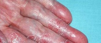
The disease is quite easy to recognize by severe itching and red rashes that appear on the stomach, knees, elbows, chest and neck. When active, the rash looks like small blisters with liquid contents. When they burst, areas of weeping appear on the skin, which then dry out and peel.
Scratching causes the skin to thicken and become rough. This phenomenon is called lichenification. The effect on the fetus of atopic dermatitis during pregnancy is minimal. The disease brings more discomfort to the woman herself, without interfering with the development of the child. Consequences can occur after birth, since the tendency to atopy is inherited. In a child, it can manifest itself not only in the form of dermatitis, but also in the form of bronchial asthma or hay fever.
What is atopy?
The content of the article
Atopy is an abnormal response of the body to an allergen, resulting in overproduction of IgE antibodies responsible for the allergic reaction. Atopic dermatitis belongs to the group of atopic diseases, like bronchial asthma, urticaria and hay fever.
Patients prone to atopy usually have several allergic diseases at the same time. The body of a person with AD responds to very low doses of allergen (molecules that can trigger an immune response) in the environment. The disease can be caused by many factors that surround us: dust mites, pollen from flowers, animals and food.
Forms and stages of atopic dermatitis
The critical periods of pregnancy are considered to be the 1st and 3rd trimester. During this period, atopic dermatitis can be severe. In other cases, everything goes smoothly.
Treatment of atopic dermatitis during pregnancy depends on the severity:
- Light form. The rashes are mild, accompanied by swelling, and have a pinkish tint. There is no peeling; skin itching occurs periodically.
- Medium shape. All symptoms intensify, the rash spreads throughout the body: on the face, back, stomach, inner thighs. Another characteristic sign is darkening of the skin around the eyes.
- Severe form. Due to severe itching, there is a possibility of nervous breakdowns and sleep problems. The skin at the site of the rash becomes swollen, peels, and becomes covered with erosions and pustules.
What should you start doing after diagnosis?
- Normalize the humidity and temperature in the room where a child with atopic dermatitis lives (the skin does not like dryness and frost, as well as heat, so the air humidity should be 50–70% according to the hygrometer, and the temperature should be 18–21 ° C).
- It is recommended to choose cotton and muslin as fabrics for clothing, bed linen, etc. Natural six, synthetics and other materials can cause exacerbation of AD.
- Replace all household chemicals with “NOT chemicals”. Pay attention to the composition of the products, and not to the labels (hypoallergenic, suitable for children, etc.). Powders, dishwashing detergents, etc. must be WITHOUT chemicals and other undesirable components.
- Use the correct care products. Bathing products and body moisturizers should also be specialized, designed specifically for atopic skin.
- To establish the presence or absence of a connection between exacerbations of atopic dermatitis and food allergic reactions. To do this, it is recommended to keep a diet diary, as well as track contact allergens, because... Sometimes rashes on the skin of the cheeks in infants and young children are associated with the fact that they smear food on the face. In this case, the product causes an aggravation due to contact, and not because the child has a food allergic reaction to it.
Even at the slightest suspicion of a connection between exacerbation of atopic dermatitis and allergic manifestations, it is necessary to visit an allergist. Only he can refute or confirm your fears and recommend adjusting your child’s diet.
It is important to note that atopic dermatitis is not always associated with nutrition (and according to statistics, only 30% of children suffering from atopic dermatitis are associated with food allergies), therefore strict restrictive diets are most often unfounded and do not lead to remission and elimination of symptoms.
Treatment tactics are determined by the attending physician and depend on the severity of the disease. It is worth considering that for atopic dermatitis there is maintenance therapy, as well as exacerbation therapy.
⠀
For mild cases, therapy includes skin care with moisturizers, bathing with mild cleansers, and avoiding trigger factors. This is called basic therapy, and most often it is enough to reduce inflammation and achieve remission.
Moisturizers for atopic skin are called emollients. They are an integral, mandatory part of care for atopic dermatitis of any course at any age - both in infants and in older children and adults.
EMOLENTS are a group of drugs that have an effective moisturizing and regenerating effect on the skin due to the presence of fats and fat-like substances. Emollients are not a medicine - they are medicinal cosmetics that have:
- moisturizing and softening effect;
- antipruritic effect;
- regenerative abilities;
- restoring skin microbiome and skin barrier effect.
⠀
In the treatment of AD, moisturizers are used to:
- maintaining skin barrier function;
- clinical improvements by reducing the severity of signs and symptoms;
- suppression of inflammation;
- prevention of exacerbations;
- steroid-sparing effect.
In moderate cases, anti-inflammatory drugs are added to basic therapy. Weak topical hormonal agents are used 1–2 times a day or maintenance therapy with topical calcineurin inhibitors 1–2 times a day.
Severe cases are most often treated in a hospital setting, using phototherapy, systemic immunosuppressants and interleukin inhibitors.
Summary: if a child has been diagnosed with atopic dermatitis, the most important thing is to start properly caring for the skin using emollients and specialized bathing products, adjust the humidity and air temperature, and try to find the trigger factor that affects the exacerbation.
Do not engage in self-diagnosis and self-medication, do not try to put your child on a diet. At the first symptoms, you need to consult a doctor to establish the correct diagnosis and select quality therapy!
Correct and safe treatment
Treatment of atopic dermatitis in pregnant women is often difficult due to the ban on the use of medications. Even many ointments that can be used in adults without consequences are prohibited during pregnancy. But the PsorMak clinic uses an absolutely harmless and safe ointment. It contains only natural ingredients, without hormones that can affect the fetus.
Only experienced doctors know what to do if atopic dermatitis worsens during pregnancy. During this period, it is especially important not to self-medicate, since the health of the child depends on the health of the woman. The PsorMak clinic has developed a treatment regimen that takes into account all the features and risks that arise during pregnancy. You can make an appointment by filling out the form on the website, or call us at +7 (800) 500-49-16.
How to diagnose atopic dermatitis and, most importantly, how to treat it?
Unfortunately, in the diagnosis of AD there are no specific histological signs, characteristic laboratory data or special skin tests that can clearly distinguish it from allergic reactions and other diseases.
⠀ With the first appearance of a skin rash, it is recommended to consult a doctor, pediatrician or dermatologist.
The doctor, in turn, will collect anamnesis, the presence of developmental risk factors, find out genetic predisposition and, of course, fully examine the child.
There are criteria in the presence of which the diagnosis is established clinically:
- itching;
- typical morphology and age-related localization features in an infant, child or adult;
- chronic relapsing course;
- personal or family history of atopy (asthma, allergic rhinitis, atopic dermatitis).
After establishing the diagnosis, the main goal of the doctor and the patient will be to prolong remission and reduce the frequency of exacerbations, because Atopic dermatitis is a chronic disease and can last for years. But, according to statistics, with proper care and treatment, AD resolves by 3–4 years.
HOW TO MAKE AN APPOINTMENT at the PsorMak Institute for Healthy Skin
1. Click the button you see below -
Make an appointment
2. Fill in the fields in the form that appears. Be sure to check the correct phone number so that our specialist can reach you. After filling out, click on the “Submit” button.
3. Wait for our specialist to call. He will answer any of your questions and agree on the date and time of your visit to PsorMak.
The initial appointment includes:
- Visual examination , which will allow the specialist to get a general understanding of the condition of your skin and the pathology itself.
- Collecting anamnesis - finding out information about the development of the disease, living conditions, previous diseases, operations, injuries, chronic pathologies, allergic reactions, heredity, etc. Together with a general examination, this allows you to make a fairly accurate diagnosis and choose a method of treatment and/or prevention.
What are the symptoms of atopic dermatitis (AD)?
The most common symptom of atopic dermatitis is eczema, an inflammation that appears as papules on reddened skin. Skin lesions usually appear in groups and in irregular shapes.
Scratching causes damage to the skin. Chronic lesions lead to lichenification (thickening) of the skin and its hyperkeratosis, that is, ichthyosis (keratosis pilaris, ichthyosis). In very severe conditions, erythrodemia may occur, that is, general inflammation of the skin, enlarged lymph nodes and fever.
Depending on the age of the patient, the course of the disease can be divided into three phases: childhood, adolescence, and adulthood. The location of changes differs from time to time. In children, symptoms of atopic dermatitis are often visible on the cheeks and forehead, while in adults it is mainly visible in the flexures of the lower and upper limbs. In addition, adults may develop other symptoms of AD, such as whitish dandruff, which does not occur in children.
Atopic patches (ATP)
Atopic patches are a type of epidermal patches in which specially prepared allergens are applied to the uninjured skin of the patient's back and sealed. They are used to detect delayed allergenic reactions that cannot be detected by skin prick tests.
Atopic patches are a very good addition to the diagnosis of AD with substances that the patient must eliminate from life in order to reduce the symptoms of the disease.
Atopic patches
Atopy is not only a disease of the body
Atopic dermatitis is a disease that can have a negative impact on the patient's psyche. Children and adolescents diagnosed with AD are more likely to have problems being accepted in their community. It is difficult to explain to your peers where the red spots and scars come from after an exacerbation of the disease.
Fortunately, only about 30% of patients continue to have symptoms into adulthood. In the remaining 70%, the symptoms disappear completely or appear in a milder form, which the patient can control with the help of a good dermatologist.
Skin application with SAFT nutrition test
SAFT is a food allergy testing method primarily used for young children. The technique involves eating the food that is suspected of causing the child's allergy and monitoring symptoms.
If erythema, itching and swelling of the skin occurs during use, this indicates a positive result, that is, an allergy. Most often, this test is carried out on allergenic fruits, vegetables, milk, chicken eggs and nuts.
Determination of total concentration of IgE antibodies
The simplest test performed to diagnose AD is to determine the total concentration of IgE antibodies in the blood. This concentration is indicated in IU, and the standard is 100 IU/ml. In patients with atopy, the total concentration of IgE antibodies increases by 80%. There is often a relationship between IgE concentrations 10 times normal and the clinical condition of the patient's skin. The higher the antibody concentration, the worse and more severe the skin changes.
Therefore, patients with atopic dermatitis can be divided into two groups: those with elevated antibody levels and those with normal antibody levels (about 20% of patients).
- In the first case, we talk about extrinsic atopic dermatitis (extrinsic atopic dermatitis - EAD) because the symptoms are caused by allergens coming from the environment.
- Less commonly, we deal with intrinsic AD (intrinsic atopic dermatitis - IAD), where symptoms arise from non-immune causes, such as a skin defect or a nonspecific inflammatory response. This means that an IgE result within the normal range does not necessarily rule out atopic dermatitis.
Introduction
There are itchy rashes that are characteristic of pregnancy and the postpartum period.
In 1983, Holmes and Black proposed a classification of skin diseases of pregnancy, which included pemphigoid gravidarum, multiform dermatosis of gravidarum, prurigo of gestation, and pruritic folliculitis of gestation. In 1998, Shornick proposed adding cholestasis of pregnancy to this list. The latest classification was proposed by Ambros-Rudolph et al in 2006, based on a large retrospective study of 505 patients, and includes four dermatoses: pemphigoid gravidarum, multiform dermatosis gravidarum, atopic dermatitis of pregnancy (including prurigo gravidarum and pruritic folliculitis of gestation), and intrahepatic cholestasis of gestation. .
The main reason for skin changes during pregnancy is considered to be changes in the pregnant woman's immune system. To prevent fetal rejection, an imbalance develops between the cellular and humoral immunity. The production of cytokines by T helper type 2 (Th2) cells predominates over Th1, an increase in humoral immunity and a delay in the growth of cellular immunity are observed. The influence of changes in the level of maternal hormones is assumed, because many skin conditions develop during the third trimester. This article is devoted to skin diseases characteristic of pregnancy, with an emphasis on clinical manifestations, possible complications for the fetus, pathogenesis, diagnosis and treatment.
Discussion
Pemphigoid gravidarum (PG)
Pemphigoid gravidarum, formerly known as herpes gravidarum, is the rarest of the skin disorders of pregnancy, with an incidence of 1:2000 to 1:60,000. PG initially appears as papules and plaques that transform into vesiculobullous elements. PG is characterized by the appearance of rashes in the navel area, spreading to the chest, back and limbs. The palms and soles may be involved, but usually the mucous membranes rather than the face. The rash most often develops in the third trimester. Dermatosis exists throughout pregnancy and in 75% of patients, exacerbation occurs during childbirth. PG usually resolves spontaneously within a few months after delivery. Typically, there is a recurrence of dermatosis during subsequent pregnancies, with earlier onset of dermatosis and greater severity compared to the previous pregnancy. There are also reports of exacerbations during menstruation or when using oral contraceptives. There is an increase in the incidence of prematurity, especially with more severe cases, blistering and the onset of dermatosis before the third trimester.
Approximately 10% of children develop a temporary, bullous rash due to the transfer of antibodies across the placenta. PG is an autoimmune disease in which there are antibodies against the NC16A region of collagen XVII (BPAG2, BP180), which is present in amniotic, placental, and umbilical tissue, in addition to the basement membrane of the skin. Antibodies activate the complement cascade with inflammation and blistering.
IgG4 antibodies are detected. Women with this disorder are at high risk of autoimmune diseases, especially Graves' disease. An association with HLA-DR3 and HLA-DR4 is observed. Histologically, bullous PG is characterized by cutaneous edema and perivascular inflammation with lymphocytes, histiocytes, and eosinophils. Subepidermal blisters are observed in vesiculobullous lesions with a predominance of eosinophilic infiltrates.
Direct immunofluorescence shows linear deposition of complement 3 (C3) along the basement membrane zone in all patients. Some patients also have IgG deposits along the basement membrane. Enzyme-linked immunosorbent assay (ELISA) can detect specific antibodies against collagen XVII, which correlates with disease activity and can be used to monitor the effectiveness of treatment. Treatment of PG should be aimed at reducing itching and blistering. In mild cases, topical corticosteroids and antihistamines are effective. In severe cases of bullous PG, it is advisable to use systemic corticosteroids. The dose can be reduced once adequate control is achieved, however, it is often not reduced due to the high risk of exacerbation. The use of systemic corticosteroids does not increase the risk to the fetus.
Polymorphic dermatosis of pregnancy (PEP)
PEP is a benign, pruritic, inflammatory condition with an incidence of 1 in 160 pregnancies. It usually occurs in the late third trimester or immediately after delivery in the first pregnancy, with an increased risk in multiple pregnancies and rapid weight gain. Urticarial papules and plaques appear first on the abdomen and, unlike PG, do not affect the umbilicus area. The rash usually extends to the thighs and buttocks and is rarely generalized. Blisters with a diameter of 1-2 mm may develop, but unlike PG, bubbles are not observed. Rashes with clear boundaries regress spontaneously within 4-6 weeks without connection with treatment.
PEP does not pose a risk to the fetus. The histological picture is similar to PG. At the onset of PEP, superficial to mid-skin perivascular infiltrates of lymphocytes, histiocytes, and eosinophils are observed with dermal edema. Late stages of PEP are characterized by epidermal spongiosis. In contrast to PG, the immunofluorescence assay is negative. Treatment of PEP is based on symptomatic relief using topical corticosteroids and antihistamines. If the rash becomes generalized, a short course of systemic corticosteroids may be used.
Atopic dermatitis of pregnancy (AEP)
AEP is the most common skin disease in pregnant women, accounting for almost 50% of all dermatoses. It has also been called by other names, including prurigo gravidarum, prurigo gravidarum, Spangler's papular dermatitis of gestation, pruritic folliculitis of gestation, and eczema of gestation. AEP is a benign condition characterized by a pruritic, eczematous or papular rash. Develops until the third trimester, unlike other dermatoses of pregnancy. Two thirds of AEP cases are characterized by eczematous skin changes localized to atopic areas of the body such as the neck and flexor surfaces of the extremities. The remaining cases are characterized by a papular rash in the abdomen and limbs. The lesions usually respond well to treatment and resolve spontaneously after delivery. However, AEP is likely to recur in subsequent pregnancies. Dermatosis does not significantly affect the fetus, but there is an increased risk of developing atopic dermatitis in the infant.
It is believed that the development of AEP is initiated by pregnancy-related immune changes in the body. In this case, there is a shift towards humoral immunity with increased activation of Th2. Pregnant women with AEP are predisposed to atopic dermatitis, but 80% of these pregnant women develop these skin changes for the first time during pregnancy. There is often a family history of atopic dermatitis in relatives.
AEP is usually a diagnosis of exclusion because Diagnostic testing is nonspecific. Serum IgE levels are elevated in 20-70% of patients. AEP is differentiated from ICP, scabies and drug allergies. The mainstay of treatment is topical corticosteroids. In severe cases, a short course of systemic corticosteroids and antihistamines may be used. Phototherapy may also be used.
Intrahepatic cholestasis of pregnancy (ICP)
ICP, formerly known as obstetric cholestasis, cholestasis of pregnancy, and jaundice of pregnancy, is a reversible cholestasis that is likely due to hormonal changes late in pregnancy in predisposed women. ICP is characterized by acute-onset itching that often begins on the palms and soles and then generalizes. The skin contains mainly secondary lesions, such as excoriations, but there may also be papules. In 10%, jaundice develops due to concomitant extrahepatic cholestasis. After childbirth, the itching goes away within a few weeks. There is a risk of recurrence in subsequent pregnancies and when using oral contraceptives.
Diagnosis of ICP is important because... may be associated with serious consequences. Possible fetal complications include preterm birth, intrauterine fetal distress, and intrauterine fetal death. The incidence of complications in the fetus correlates with the total level of bile acids in the maternal blood serum. In cases of severe ICP complicated by jaundice, there is a risk of bleeding in the mother or fetus due to impaired absorption of vitamin K. Severe itching in ICP is associated with increased levels of bile acids in the blood caused by impaired secretion, a multifactorial process resulting from genetic disorders, the influence of environmental factors environment and endogenous hormones. There is a high incidence of ICP in multiple pregnancies. ICP is diagnosed based on the detection of elevated bile acid levels. Hyperbilirubinemia is seen only in the most severe cases, about 10-20%, and liver tests may be normal in 30%. Histology is nonspecific and immunofluorescence is negative.
Treatment is aimed at normalizing serum bile acid levels to reduce risk to the fetus and to control maternal symptoms. Treatment with ursodeoxycholic acid (UDCA) is recommended. Other drugs that reduce itching, such as antihistamines, S-adenosyl-L-methionine, and dexamethasone, may be used. Anion exchange resins such as cholestyramine may cause vitamin K deficiency regardless of the presence of ICP and should therefore be avoided.
Conclusion
The four skin diseases characteristic of pregnancy, pemphigoid gravidarum, polymorphic dermatosis of pregnancy, atopic dermatitis of pregnancy, and cholestasis of pregnancy can be distinguished by clinical presentation, histopathology, pathogenesis, and risk of fetal complications. Only pemphigus of pregnancy and intrahepatic cholestasis of pregnancy are associated with a significant risk to the fetus. Because all of these dermatoses are pruritic, careful evaluation of any pruritic pregnancy is necessary. Adequate treatment of the pregnant woman and prevention of any potential risk to the fetus is imperative.
Skin testing (STP)
Skin prick tests test for a specific allergen. This test is based on the fact that IgE antibodies are present on the surface of immune system cells. The purpose of the test is to bring such cells into contact with the allergen.
A solution containing the allergen is applied to the patient's skin, and then it is pricked with a special lancet. Typically, this test tests several substances at the same time, such as dust, hair or pollen.
Skin tests
This test is no different from a test that determines an allergy to any substance. Therefore, the presence of a positive allergic reaction does not automatically mean atopic dermatitis. In addition to the allergic reaction, other symptoms such as dry and itchy skin should also appear. Additionally, a negative test result does not mean that the disease is not AD; it only means that this set of substances does not cause allergic reactions.