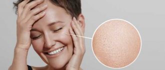
Atopic dermatitis of the facial skin is the most problematic form of the disease, since all signs are visible to the naked eye. Because of this, the patient experiences not only physiological, but also psychological discomfort. In most cases, the disease manifests itself in childhood, but sometimes the first symptoms appear in adulthood. Therefore, the causes and treatment of atopic dermatitis can be completely different. To achieve good results, it is necessary to select therapy individually.
What is atopic dermatitis (AD)?
The term “atopic dermatitis” was proposed back in 1935, but was introduced into medical practice much later – in 1972.[1]
Officially, “atopic dermatitis” was adopted only in 1992 and introduced into the ICD-10 classification of diseases. This explains the existence of a large list of synonyms that are still used to designate the manifestations of this specific skin disease by doctors from different countries. Among the medical definitions of this disease one can find such as exudative diathesis, eczema, neurodermatitis, atopic eczema, childhood eczema, atopic dermatitis. So, atopic dermatitis is a chronic inflammatory skin disease that develops in individuals with a genetic predisposition to allergies and is characterized by rashes, severe itching and increased sensitivity to allergens.
The prevalence of atopic dermatitis among children reaches 15–30%, among adults – 2–10% [5]. Atopic dermatitis can be the debut of the so-called “allergic march”, when in the future such patients develop other atopic diseases, for example, food allergies, bronchial asthma, allergic rhinitis.
In recent decades, there has been a significant increase in the incidence of AD. The disease brings quite a lot of problems to patients and significantly worsens their quality of life.
Dermatomycosis
Mycoses of the scalp
They appear as bright red infiltrated plaques, which are covered with gray scales on top. The elements are mainly formed around the hair in the form of a muff. Occasionally, deep inflammatory foci are possible, which are large in size and covered with massive grayish-yellow crusts. At the site of a fungal infection, hair breakage is observed at a height of 5-8 mm or at the very root. A person complains of severe itching and rapid contamination of hair after washing.
Mycoses of skin folds
The main representative of this group is dermatophytosis inguinalis. The disease affects the inguinal folds and adjacent areas of the skin: the inner surface of the thighs, the perianal area, and the perineum. Due to self-infection, the process may spread to the armpits, elbows, popliteal fossae, and in severe cases to any area of smooth skin. The frequency of inguinal dermatophytosis in the structure of all dermatomycosis is up to 10%.
At the initial stage, the pathology is represented by pink swollen spots that have a round shape, clear contours and a smooth surface. If treatment is not carried out, the lesions merge to form large polygonal spots, the marginal zone of which is covered with polymorphic elements: bubbles, erosions, crusts. Most of all, patients are worried about the painful itching, which interferes with sleep and daily activities, forcing them to scratch the affected area until it bleeds.
Mycoses of the feet
The clinical picture of epidermophytosis of the distal extremities depends on the form of the lesion. As a rule, the pathology begins with an erased form, in which there is slight peeling in the interdigital spaces, which does not cause any concern to the patient. Sometimes, against the background of peeling, superficial skin cracks form, which do not become inflamed or bleed.
In the squamous-hyperkeratotic form, abundant peeling is accompanied by reddish-bluish plaques and yellowish-gray calluses, which arise due to excessive keratinization of the skin. With the dyshidrotic form, multiple blisters with a thick tire appear, when opened, bright pink weeping erosions are formed. For mycoses of the feet, secondary allergic rashes, called dermatophytids, are typical.
Onychomycosis
Fungal infection of nails has different symptoms, which depend on the shape and depth of the lesion. The pathology is mainly manifested by grayish-yellow stripes on the nail plate, its increased fragility, tendency to deformation, transverse striations and cracks. At an advanced stage of the disease without treatment, significant destruction of the nail is observed, in addition to a cosmetic defect, itching and painful sensations occur.
The normotrophic form is characterized by decreased transparency of the nail and thickening of its edges due to subungual hyperkeratosis. In the hypertrophic variant, the thickening of the nail plate is more pronounced; in severe cases, the nail acquires a curved beak-like shape (onychogryphosis) and a dirty gray tint. For atrophic onychomycosis, total destruction and detachment of the nail plates is typical, and onycholysis is considered the extreme degree of this process.
Mycoses of smooth skin
Classic dermatomycosis is characterized by the appearance of flat, scaly spots of pink or red color with a raised border. There may be inflammatory papules or vesicles along the edges of the lesions. Over time, the central part of the elements becomes brown due to hyperpigmentation, and the edges continue to grow, so that the lesions merge into large polygonal spots. Subjectively, patients experience itching, burning, and pain in the affected area.
Causes of development of atopic dermatitis
Atopic dermatitis is being actively studied by dermatologists and allergists, but its causes have not yet been definitively established. It is a generally accepted fact that atopic dermatitis is based on a hereditarily determined excessive immune response to allergens, manifested by skin inflammation.
However, there is also a significant group of patients - about 20-30% - in whom hypersensitivity to any allergens has not been identified, but manifestations of atopic dermatitis are present [2].
Factors that provoke the development of the disease include:
- Genetic predisposition to the development of allergic reactions;
- Violation of the integrity of the skin barrier, facilitating the penetration of allergens and microorganisms into the deeper layers of the skin and the development of an allergic immune reaction;
- Excessive intake of allergens in early childhood from food (cow's milk protein, citrus fruits, chocolate, etc.);
- Impaired immunity after infectious diseases or against the background of a chronic illness.
Perioral dermatitis: causes
Experts have not fully identified the causes of perioral dermatitis. However, it is generally accepted that this is a complex, multifactorial disease that has a wave nature (with stages of exacerbation and remission, respectively).
It is usually customary to associate the appearance of a rash around the mouth with hormonal changes that occur:
- when taking various medications, primarily when using ointments based on corticosteroids. Most often, corticosteroids are prescribed as one of the main means for the treatment of acne, rosacea, blackheads, eczema, etc. It is in the case of using corticosteroid ointment that perioral dermatitis can be detected in teenage boys;
- during pregnancy: this type of dermatitis is often diagnosed in women during pregnancy, which makes it possible to assume a hormonal origin of the disease, since pregnancy most often entails serious hormonal changes in the female body. For the same reasons, dermatitis most often worsens before the start of the menstrual cycle. Hormonal changes often occur in newborns, which fully explains the possibility of dermatitis in young children.
Among other things, such dermatitis can occur due to:
- abuse of cosmetics: most often we are talking about both decorative cosmetics (foundation and powder in the first place), and moisturizing creams, scrubs, tonics;
- using fluoride-containing pastes that can irritate the skin around the mouth;
- severe chapping of the skin, especially in the cold, as well as severe insolation (absorption of large amounts of sunlight);
- infections: often during the diagnostic process various infectious agents are discovered (streptococcus, staphylococcus, etc.), but specialists have not yet been able to identify a single agent that can cause the development of this type of dermatitis;
- failure of the immune system, which most often occurs due to a lack of vitamins in the body, especially in adolescents, who during the period of active growth experience a special need for vitamins. In this regard, it is also customary to include an imbalance of intestinal microflora, which occurs due to problems with the digestive organs, as provoking factors for dermatitis.
Perioral dermatitis: consequences
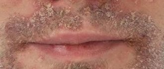
Dermatitis of this type is a very unpleasant dermatological problem, which, in the absence of timely professional intervention, can spread further over the skin of the face. In addition, usually the papules are destroyed, and in their place crusts form, the premature removal of which can cause hyperpigmentation (more intense coloring) of certain areas of the skin.
In case of large skin lesions, especially against the background of weakened immunity, including in children, purulent, weeping ulcers can form in place of papules (pimples), causing patients a lot of discomfort.
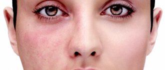
In addition, sometimes, in relatively rare cases, oral dermatitis can affect not only the area around the mouth, but also be found on the skin around the eyes, on the lower and upper eyelids. At the same time, dermatitis poses a potential danger to the patient’s vision (primarily if diagnosed in small children), and therefore requires timely consultation with an ophthalmologist
.
What is the cause of atopic dermatitis on the face?
The appearance of rashes on the face is one of the manifestations of atopy. The term itself is interpreted as a person’s high sensitivity to certain external stimuli. The mechanisms of atopic dermatitis are still being studied.
A significant role in the occurrence of atopic dermatitis on the skin of the face is played by such a factor as hereditary predisposition. If the disease is diagnosed in one or both parents, then the likelihood of a similar problem in the child increases to 30-80%.
Atopic manifestations on the skin of the face can also occur for other reasons. Provoking factors for exacerbation of atopic dermatitis include:
- regular skin contact with aggressive chemical compounds (they can be contained in care products and decorative cosmetics);
- exposure to household chemicals (for example, aggressive components of washing powders, cleaning products and detergents);
- consumption of foods that are potentially allergens (these include honey, nuts, some fruits and berries, etc.);
- taking certain medications;
- adverse environmental impacts (living in places with poor ecology, in industrial areas, near waste sites, etc.);
- stress associated with increased physical and mental stress.
An important role in the occurrence of atopic dermatitis on the face is played by disruption of the skin microbiome. Recent research in this area suggests that cutaneous manifestations of atopy may be exacerbated in response to increased activity of fungi of the genus Malassezia spp.
Colonies of Malassezia spp. for most healthy people there is no danger. But in patients prone to atopic dermatitis, sensitivity to these fungi increases. As a result of the interaction of lipophilic yeast with the human immune system, the production of specific IgE is activated. Therefore, in people with atopic skin, in addition to rashes, other manifestations of allergies are possible. A similar increased response was found to the bacteria Staphylococcus aureus (Staphylococcus aureus).
Treatment of candidal dermatitis
Treatment for candida skin infections is usually quite simple. If the immune system works well and the infection does not spread to internal organs, hospitalization is not required. The doctor will usually prescribe a drying agent along with antifungal creams, ointments, or lotions to apply to the affected skin. Sometimes medications in tablets are needed.
Treatment of candidal dermatitis with medications
- The topical antifungals most often prescribed to treat candidal dermatitis are ketoconazole or clotrimazole. They are used topically and have very few side effects.
- Fluconazole is an oral tablet that is used when topical treatments are ineffective. It is quite safe, there are few adverse reactions to it, and sometimes one dose is enough to fight the infection.
- Nystatin and amphotericin B are more powerful drugs that are used intravenously for a common infection in a hospital setting.
Most medications are used twice daily. Some medications (such as miconazole and clotrimazole) are safe in the first trimester of pregnancy.
Side effects include:
- Itching at the application site,
- Redness and burning sensation in the area of application,
- Headache,
- Upset stomach and indigestion,
- Skin rash.
Intravenous antifungals cause more severe effects:
- Loss of appetite,
- General weakness
- Stool disorder
- Pain in muscles and joints,
- Various types of rashes.
In rare cases, these drugs cause dangerous allergic reactions or severe skin damage with the death of the epithelium and the formation of blisters like a burn.
Patients with liver disease should not use antifungal agents without medical supervision. They cause liver damage and, if used uncontrolled, can lead to liver failure.
Medicines that, when used simultaneously, can react with antifungals and change their activity:
- Rifampin, an antibiotic
- Benzodiazepines, which are prescribed to improve sleep and reduce anxiety,
- Estrogens and progesterones, which are contained in oral contraceptives and hormone replacement therapy,
- Phenytoin, which is sometimes prescribed for epilepsy.
- Among drying agents, the most effective and inexpensive is zinc oxide, which has virtually no contraindications or adverse effects. This remedy can even be used in infants with diaper dermatitis caused by fungi.
- Fukortsin is another drug that has a drying, anti-inflammatory and antiseptic effect. This is an aqueous solution of fuchsin, it has a bright pink color, it is recommended for use in acute cases of the disease. It is not advisable to use it in children due to some toxicity.
- A weak solution of apple cider vinegar may be useful as an additional remedy for acute dermatitis in adults.
- Benzoyl peroxide has shown effectiveness in subacute cutaneous candidiasis.
- It is also recommended to cover the affected area with bandages as little as possible, try to keep it open, and ensure that there is no weeping or rubbing of the skin.
Symptoms and types of atopic dermatitis
The first manifestations of the disease in children are usually noted at the age of 6 months in 45%, up to 1 year in 60%, and up to 5 years in 85% of cases. [5]
Depending on age, the following forms of atopic dermatitis are distinguished:
- infant (from 1 month to 2 years);
- children's (from 2 to 12 years);
- teenage (over 12 years old).
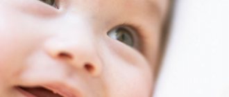
A triggering factor for the development of AD in an infant can be a food allergy and it manifests itself in the form of rashes on the baby’s cheeks, thighs, legs and arms. In severe cases, large areas of the skin may be affected. The rashes are moist, weeping, and are accompanied by severe itching of the skin, which significantly disrupts the quality of life of both the baby and his mother. As a result of scratching, wounds appear on the baby’s skin, which are entry points for fungi, viruses and bacteria. Therefore, in the absence of proper care and timely treatment, children with atopic dermatitis have a high risk of secondary infection. This form of atopic dermatitis is often called exudative diathesis.
As children grow older, the disease may go away on its own. But in approximately 50% of children, atopic dermatitis persists [3]. At the same time, the manifestations of the disease also change - wet rashes disappear, and instead of them, itchy, dry areas of skin appear, sometimes with a rash in the form of nodules and microbubbles. The location of the rashes also changes - now they are in the area of the folds - knees, elbows, wrists; on the back of the neck. Doctors often differentiate these manifestations of the disease from neurodermatitis.
After 12 years, with the onset of adolescence, the clinical picture of the disease changes again. In adolescents and adults, it manifests itself as rashes on the backs of the hands, feet, and fingers, and is often accompanied by severe, unbearable itching. In severe forms, the disease can affect large areas of skin throughout the body.
Candidal dermatitis in an adult
Candidal dermatitis in adults, unlike children, develops more often in the armpits, inguinal folds, under the mammary glands, less often between the fingers and in the folds of the skin in obesity (as shown in the photo), and in this case, treatment also consists of eliminating provoking factors and the use of antifungal drugs.
In bedridden patients, dermatitis caused by these fungi develops on the back.
Cases have been described in which candidiasis developed under the ring on the finger in patients with reduced immune system function.
In young mothers who are breastfeeding, an inflamed nipple with very severe soreness can also be caused by candidiasis. This makes feeding the baby much more difficult. The infection is most often transmitted from the baby's mouth, especially if he has candidiasis of the oral mucosa. Some researchers believe that cutaneous candidiasis is the main cause of nipple pain associated with breastfeeding.
What does atopic dermatitis look like on the face?
The key symptom of atopic skin is severe itching. Along with it, redness forms on the surface of the face, often accompanied by weeping and the formation of crusts, since damage to the facial skin is characteristic of the infant and child form of atopic dermatitis. Frequent “companions” of atopic dermatitis on a child’s face are other manifestations of atopy:
- bronchial asthma;
- allergic rhinitis;
- allergic lacrimation from the eyes.
Ignoring the problem and expecting the rash to go away on its own is a big mistake. It is much more correct and effective to contact a specialist to prescribe adequate therapy and personal selection of medications.
Types and stages of the disease
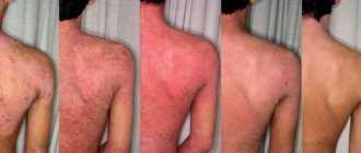
Facial skin dermatitis occurs in several stages, which differ in symptoms. It all starts with an acute form, accompanied by profuse rashes, severe swelling and redness of the skin. All symptoms are pronounced.
The disease then progresses to the subacute stage. At this stage, the bubbles burst and dry out, leaving flaky areas. A person may experience a headache and a fever. After the scales disappear, hyperpigmented spots remain in their place. When the symptoms subside, the disease progresses to the chronic stage. It is characterized by alternating periods of exacerbation and remission.
In addition to stages, there are different types of dermatitis on the face:
- exudative, characteristic of childhood;
- erythematous-squamous, often found in adults;
- pruriginous, combining both forms.
Treatment of atopic dermatitis
Treatment of atopic dermatitis in patients of different ages still remains a pressing problem in pediatrics, allergology and dermatology. Modern principles of treatment for this disease include:
- Alleviation of the patient’s general condition;
- Relief of skin manifestations of the disease (itching, rashes, weeping, dry skin, etc.);
- Prevention or control of secondary infection;
- Prevention of repeated exacerbations (relapses)
- Extension of the period of temporary recovery (remission);
- Creation of an anti-allergenic environment, compliance with skin care rules;
- Compliance with the work and rest regime, taking care of the psycho-emotional state.
Perioral dermatitis: symptoms
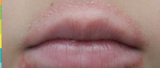
Symptoms of this disease are small round or oval-shaped papules located primarily around the mouth and on the chin. In some cases, papules, as mentioned above, can be found on the wings of the nose, on the bridge of the nose, on the temples and cheeks, as well as around the eyes. In some cases, papules may be filled with clear fluid.
The color of the papules, depending on the stage of the disease - exacerbation or remission - can vary from pale pink with a bluish tint to bright red. In some cases, papules are accompanied by hyperemia (general redness) of the skin, or may not cause a change in skin color. A distinctive feature of this type of dermatitis is considered to be a characteristic (light) strip of healthy skin around the mouth, which can be up to 4 mm wide. In turn, the localization of papules can be different - from single formations to groups of several pimples with clearly defined edges. In the case of a large number of papules located nearby, the skin becomes rough, which is felt well when touched.
Also, symptoms of perioral dermatitis, especially during an exacerbation of the disease, may include:
- dryness and tightness of the skin;
- skin itching;
- burning.
However, in a large number of cases, patients do not observe or experience any additional concern with dermatitis.
The sooner you start treating oral dermatitis on the face, the easier and faster it will be cured. That is why, when you detect the first signs of a possible disease, you must seek advice from a dermatologist.
.
Initial appointment
An initial appointment with a specialist involves a thorough examination of the patient, as well as drawing up a preliminary medical history (recording complaints, symptoms, concomitant diseases), which allows making preliminary assumptions regarding the cause of the disease. Additionally, tests and other diagnostic measures may be prescribed to clarify the clinical picture and understand how to treat oral dermatitis on the face.
Diagnostics
Diagnosis of this type of dermatitis is necessary as a way to differentiate from diseases with similar manifestations - eczema, herpes simplex, rosacea, etc.
Diagnostic measures in this case include:
- dermatoscopy: allows you to assess the general condition of the skin, the degree of development of pathology through a thorough examination of neoplasms;
- scrapings of skin material from the affected areas: bacterial culture of the test material makes it possible to determine the presence and type of infection (for example, candida);
- Allergy tests for staphylococcus and streptococcus: also allow you to determine the sensitivity of the skin to these agents and identify their presence.
Treatment plan
If the tests confirm the initially suspected diagnosis of oral dermatitis, the treatment prescribed by a specialist will be comprehensive (using ointments and other medications, as well as diet) aimed at eliminating the manifestations of the disease on the face.
The impact on perioral dermatitis on the face when developing a treatment regimen involves two successive stages leading to the gradual disappearance of the rash:
- At the first stage, it is necessary to completely abandon cosmetics (primarily decorative cosmetics) and ointments with corticosteroids. The latter are most often recommended to be discontinued gradually, reducing the dose and quantity, otherwise the so-called “withdrawal syndrome” may occur, which leads to an exacerbation of all existing symptoms - redness, swelling of the face, burning, itching. At this stage, it is most often assumed to use antihistamines (which relieve itching and burning), as well as herbal medications to relieve local inflammation. Additionally, reflexology can be used, which is an effect on active points on the skin of the face, either by simple pressure or with the help of special needles, to reduce the unpleasant manifestations of dermatitis.
- Second stage: after the acute phase of the disease passes, the patient is prescribed antibiotics, which are designed to destroy the infection and are selected taking into account the infectious agent. Antibiotics are taken only under the supervision of a doctor, who has the ability to adjust their composition and dose taking into account the patient’s body’s reaction to them. Particular care should be taken when treating oral dermatitis during pregnancy: in this case, taking antibiotics is contraindicated, and full treatment is possible only after the birth of the child (normalization of hormonal levels is also important). During pregnancy, only partial relief of the symptoms of the disease - burning, itching, etc. - is possible with the use of medications that are safe for the fetus. The same applies to therapeutic measures for dermatitis in children: treatment in this case can only be carried out under the supervision of a doctor, otherwise it can harm the child’s health.
In addition to the described methods of influence, the patient may be recommended:
- strengthen the immune system and take vitamins;
- establish intestinal microflora;
- adhere to a diet: a diet for this type of dermatitis involves avoiding fried, fatty and spicy foods, increasing the amount of vegetables and fruits consumed, and preferring boiled rather than fried foods. During the period of taking antibiotics, you must completely abstain from alcohol.
Treatment result
Most often, if you follow medical recommendations, it is possible to cure perioral dermatitis. In some cases, a course of treatment is assumed rather than a one-time treatment. In this case, taking antibiotics can usually last quite a long time (several weeks), and the overall course of treatment can last from several months to a couple of years.
Prevention
Because dermatitis is a multifactorial disease with an unidentified underlying cause, preventive measures include the following:
- careful use of cosmetics, primarily foundations, as well as ointments based on corticosteroids;
- compliance with personal hygiene measures;
- strengthening the immune system (taking vitamins, reducing psychophysical stress, etc.);
- proper nutrition with plenty of fruits and vegetables;
- monitoring the state of the digestive system;
- control of hormone levels, especially during pregnancy.
When the first signs of the disease appear, you should not wait long, you must seek advice from a specialist, undergo an appropriate examination and begin therapy, since in order to achieve the best results, treatment of oral dermatitis must begin as early as possible - in this case it can be cured in a relatively short time .
You can make an appointment with a specialist at the Energo clinic either by phone or by using a special form for registering patients on the clinic’s website.
Take care of your beauty and health!
How to treat atopic dermatitis on the face?
Therapy for atopic dermatitis is always selected individually and is usually complex. Today, there are tested and proven effective practices on how and how to treat atopic dermatitis on the face in adults:
- Stick to a hypoallergenic diet. A common cause of exacerbations of facial rashes is the consumption of allergenic foods. If rashes appear, you should review your diet and remove allergenic foods from it (for example, honey, citrus fruits, strawberries, nuts, seafood, etc.).
- Eliminate other sources of a possible allergic reaction - remove feather pillows, limit contact with pets, and perform regular wet cleaning, since dust often provokes aggravation.
- Use external therapy products in accordance with your doctor’s recommendations.
- Use hormonal ointments. Hormonal medications are used only in cases where the disease progresses and becomes severe. Since this group of products has a fairly serious effect, they should not be used for long and strictly under the supervision of a specialist.
Hormonal ointment for the treatment of atopic dermatitis on the face can only be prescribed for severe lesions for a short period of time, since the skin on the face is especially sensitive and thin, which increases the risk of unwanted reactions. Preference is given to non-hormonal drugs for the treatment of atopic dermatitis on the face.
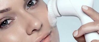
In addition to treatment, atopic facial skin needs special care. An important role here is played by the use of special products (emollients) to soften, moisturize and restore the natural barrier of the skin.
If you encounter such a problem, do not try to ignore it. Today there are effective ways to help not just control atopic dermatitis, but improve the quality of life and instill confidence in the future.
Allergy to cosmetics
Allergies to cosmetics are common. In most cases, allergic reactions occur on the mucous membranes of the eyes, eyelids, skin of the face and shoulders. There are several causes of cosmetic allergies. At the same time, the body’s reaction to the use of cosmetics can be expressed in completely different ways. The most common intolerance to certain ingredients in makeup, creams, masks and lotions.
Allergy to facial cosmetics
No one is immune from allergic reactions to cosmetics. This may occur only once with a new therapy or as an ongoing occurrence.
The average number of beauty products a person uses per day is increasing. The number of allergic reactions to them logically increases. People at risk include people with allergies and people with dry, thin and allergic skin.
Cosmetic allergies can develop on any part of the body, but local reactions to cosmetics are more common. Usually this is the skin of the face. After all, she tries every day, especially for those women who often wear makeup:
- blush;
- ink;
- Contour pencil
- Eyeliner and lip liner;
- Eye shadow;
- Pomade.
Consumers generally reject cosmetic recommendations.
For example, a jar of cream or a tube of ink bears the name in the form of a round box with a half-open lid. This box always contains a number (usually 6m or 12m) that indicates the maximum shelf life of the product when the package is opened. The shelf life can be several years, but after opening such cosmetics can only be used for 6 or 12 months. Failure to comply with deadlines often causes skin irritation.
The body's negative reaction to cosmetics varies from person to person. This happens to some people even when used correctly. However, allergies do not always occur immediately. Using mascara or cream incorrectly can take hours or days. In this case, it is difficult to immediately understand what happened to the reaction.
Main allergy symptoms:
- Redness of the skin (may be accompanied by itching);
- Tingling or burning (both symptoms may occur at the same time).
- Swelling (usually in the eye area);
- Tears, conjunctivitis (the white eyes become covered with a dense network of red blood vessels and purulent discharge occurs).
Allergic manifestations are divided into two types - simple and allergic dermatitis. The simpler form is more common.
Simple dermatitis is more common. Its symptoms:
- Increased skin sensitivity with minor burns;
- The affected area may include slight swelling, dry and flaky skin, a small red rash, and blisters.
The listed manifestations can be displayed separately, simultaneously or alternately.
Allergic dermatitis does not depend on shelf life, but is the result of a special reaction of the body to certain ingredients. The symptoms are similar to simple dermatitis, but with allergic rhinitis, watery eyes and dark circles under the eyes. And performance doesn't stop at the part where cosmetics are used: pimples, rashes and swelling are visible all over the body.
Some substances most often cause allergies:
preservative
Preservatives are aggressive allergens. They are used to extend shelf life. In most cases, manufacturers choose salicylic acid or benzoic acid.
paint
Without them there can be no cosmetics. The brighter the color of lipstick, eye shadow or blush, the stronger the allergen. People with dry, sensitive and allergic skin should choose cosmetics based on natural ingredients. In this case, the tone of lipstick, eye shadow or blush should be neutral. The lighter the color of the product, the less risk of staining and irritation 1 .
Whitening agent
Lightening and whitening elements can be found in special creams and lotions for the face. Cosmetics manufacturers use hydrogen peroxide and hydroquinone as whitening agents. Due to increased toxicity, the use of hydrogen peroxide and hydroquinone has been banned in some parts of the world. When choosing a whitening cream, pay special attention to the composition of the product.
Perfume
The smell gives the product a pleasant aroma. They are natural and artificial. The first one is usually hypoallergenic, but there are also substances that can cause allergies.
Artificial components are more aggressive. Professionals recommend taking your choice of cosmetics seriously. The likelihood that a perfume is a synthetic product increases as the cost of the product decreases.
Supplements
Food additives added to cosmetics can cause allergies, even if they are natural. The principle of producing food additives is to concentrate substances in plants, seeds and fruits. Housing and animal source. Anti-wrinkle creams made from cosmetic industry additives to activate the natural regeneration process of the subcutaneous layer. This is not a bad thing, but be aware that hyaluronic acid and collagen can be very irritating for people with allergies.
What is atopy
The term “atopy” refers to an abnormal reaction of the body to an allergen, which generally explains the nature of the disease.
With the development of atopy, there is an overproduction of IgE antibodies, which are responsible for the appearance of an allergic reaction. AD belongs to the same group of diseases as urticaria, hay fever and bronchial asthma. The pathology is immune-dependent, since the main factor in its development is a mutation in genes. Specifically, we are talking about the genes encoding filaggrin. This structural protein is located in the skin and is involved in the formation of the skin barrier, preventing water loss and the entry of allergens and microorganisms. For this reason, the pathology is inherited from one of the parents, most often from the mother.
Interestingly, atopic dermatitis was essentially known back in ancient times. In those days, this disease was called “idiosyncrasy.” That is, the name reflected the mechanism of development of the pathology - as an increased reaction to an allergen - but did not explain its etiology.
The term “atopy” was first used in 1922 to define the body’s increased sensitivity to external factors.