The causative agent of herpes is one of those viruses that, once introduced into the body, remain in it forever. How can you protect yourself from a dangerous disease? And how to resist infection if infection does occur?
Herpes (from the Greek herpete, meaning “to crawl”) is a widespread disease caused by viruses of the Herpesviridae family, which has about 100 microorganisms and is characterized by a variety of clinical manifestations, usually a chronic course, as well as various routes of transmission of infectious agents. Of the large family of herpes viruses, only 8 cause human diseases.
Simple recurrent (recurring) herpes is a disease caused by the herpes simplex virus (HSV), manifested by itchy rashes in the form of blisters (vesicles), which can occur on any part of the skin and mucous membranes. The infection rate of the world's population with HSV is extremely high, more than 95%.
There are two main groups of HSV: types 1 and 2 (HSV-1. HSV-11). HSV-1 most often causes lesions on the skin of the face (popularly called “fever” or “cold”) and upper extremities, while HSV-11 causes lesions on the genitals.
The journey of the virus in the body
The source of HSV infection is a patient or a virus carrier (there are no external manifestations of the disease, but the virus is excreted in saliva, urine, and discharge from the genitourinary system). The virus is transmitted by contact, airborne droplets, blood transfusions and organ transplants. During pregnancy, infection of the fetus can occur transplacentally (through the placenta) and during childbirth.
It has been established that in 40% of cases, primary HSV infection occurs by airborne droplets in early childhood, and the source of infection, as a rule, is family members who have active signs of herpes infection (usually recurrent herpes of the lips). The herpes simplex virus enters the body through injured skin or mucous membranes (red border of the lips, mucous membranes of the mouth, genitals, conjunctiva) - where typical blistering rashes appear (the result of the activity of the virus), and penetrates the bloodstream and lymphatic system. Viral particles reach the nerve nodes (ganglia) of the central nervous system, where they remain inactive for life. For example, with herpes of the face, the virus is stored in the ganglia of the trigeminal nerve, and with herpes of the genitals - in the ganglia of the lumbosacral spine. From the nerve ganglia, viral particles begin to move to the periphery - the skin, mucous membranes, and a focus of infection develops during a relapse.
Under certain conditions, HSV multiplies in lymphocytes (blood cells), which leads to their damage and disruption of the genetic mechanisms that control immune responses. Clinically, this manifests itself in frequent colds, decreased performance, weakness, increased body temperature, and enlarged lymph nodes.
A recurrent course of the disease is observed in 17-50% of the population infected with HSV. Exacerbations of herpes occur after various provoking factors: hypothermia, mental or physical trauma (dental, gynecological medical procedures), alcohol intake, against the background of hormonal surges (“menstrual herpes”). “Solar herpes” is known, which appears under the influence of ultraviolet rays.
However, it is worth noting that the herpes virus may never manifest itself as a disease during a person’s life.
Causes
Herpes often occurs due to direct contact with a person whose disease is in the third stage, when an infection is released with the contents of the blisters, but pathology more often develops if the virus living inside the body begins to act too actively.
Herpes in the head area is dangerous due to possible complications on the organ of vision.
As a rule, several doctors treat herpes in this area. An ophthalmologist and a dermatologist primarily treat the infection.
Dermatovenerologist, cosmetologist
Zhikhoreva Inna Viktorovna
6 years experience
Lead to this phenomenon:
- overvoltage and stress:
- severe hypothermia;
- polluted external environment;
- the presence of a cancerous tumor;
- decreased immunity.
Blood test for herpes
Herpes simplex virus (HSV) types 1 and 2 cause genital infections and are the most common cause of genital ulcers.
People who move little, have undergone complex surgery, or have diabetes suffer from herpes.
Shingles is caused by smoking, drinking alcohol, and a deficiency of microelements and vitamins in the body, which is caused by a lack of adequate nutrition. The pathology sometimes develops due to injury, a course of chemotherapy, long-term use of hormonal drugs, or after heavy physical exertion.
Herpes, which affects the scalp, is dangerous because it can lead to inflammation of the ears, affect the eyes, and this can lead to loss of vision and hearing. The pathogen, when activated in the brain, causes paralysis of the limbs.
If the infected fluid released from the bursting bubbles enters the respiratory tract, the lungs become inflamed.
The activity of the virus provokes the development of:
- infertility;
- liver pathologies;
- radiculitis:
- joint destruction.
Many-sided, insidious
The properties of HSV are such that almost all human organs can be involved in the infectious process, which allows us to speak of herpes as a “herpetic disease” with a predominant lesion of one or another organ system. For example, the eyes, ENT organs (pharynx, larynx, ear), lungs, genitals, gastrointestinal tract, central nervous system (meninges, nerves), skin and mucous membranes (face, lips, oral mucosa) may be affected. ) etc.
The frequency and intensity of exacerbations of recurrent herpes depend on the activity (aggressiveness) of the pathogen, as well as on the resistance of the human body.
Exacerbations of herpes infection are not always accompanied by the appearance of typical rashes in the form of blisters. The insidiousness of herpes lies in the fact that very often a person, without knowing it, becomes a source of infection for others. This applies to both facial herpes and genital herpes, which often manifests itself only in severe pain (in the facial area or in the pelvis, respectively).
When herpes worsens (when rashes appear), the patient becomes acutely contagious. Kissing a loved one or relative during an exacerbation can lead to the appearance of herpes in a previously healthy person. In addition, under unfavorable circumstances and failure to comply with personal hygiene rules, conditions for self-infection may arise. Thus, the virus can be carried from a lesion on the lips by hands into the eyes and genitals. In the presence of microtraumas in these areas, new foci of the disease arise: ocular herpes or genital herpes.
Genital herpes
Genital herpes (GG) is one of the most common infections of the genitourinary system, most often caused by HSV-11. Infection occurs through close physical contact with a patient or virus carrier during genital, oral-genital, genitorectal and oral-anal contact. There are primary and recurrent genital herpes.
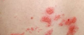
With primary genital herpes, the incubation period (the period from infection to the appearance of the first symptoms of infection) is 1-10 days and differs from subsequent relapses in a more severe and prolonged course. Clinical symptoms of primary HH develop in only 10% of infected people and are characterized by the appearance on the mucous membranes of the genital organs and adjacent areas of the skin of single erosions or grouped small blisters filled with liquid, with redness around them. After 2-4 days, the contents of the blisters become cloudy, and they burst, forming weeping ulcers, which then heal. Subjectively, patients are bothered by itching, burning, and pain in the affected area. Some patients experience an increase in body temperature up to 38°C and a painful enlargement of the inguinal lymph nodes. If the course of the disease is favorable, after 5-7 days the crusts disappear and a stain remains in their place. Even without treatment, symptoms usually go away on their own within 2-3 weeks. Subsequently, for many, the disease recurs, and the time until the next relapse can range from several weeks to several years.
Recurrent genital herpes is characterized by a chronic course, disruption of the patient’s sexual and reproductive functions. The disease is difficult to treat. Herpetic rashes can appear on the labia majora and minora, vaginal mucosa, cervix, and perineum. The duration of the rash does not exceed 3-5 days. In some cases, during a relapse, visible rashes are not detected at all, but swelling, itching, and a feeling of discomfort in the genital area appear. The disease may be accompanied by fever, general weakness and malaise, enlargement and tenderness of the inguinal lymph nodes (usually on one side).
A feature of genital herpes is multifocality: the pathological process often involves the lower part of the urethra (urethra, bladder, which is manifested by pain and pain at the beginning of urination, frequent urination) and the mucous membrane of the rectum (recurrent cracks occur), as well as the upper parts of the genital tract (uterus, ovaries and fallopian tubes). In the latter case, the appearance of mucous discharge from the vagina, periodic pain in the pelvis, in the area of the projection of the uterus, ovaries (symptoms of irritation of the pelvic nerve plexus) are possible. Moreover, these symptoms are often associated with a certain phase of the menstrual cycle (with ovulation or the perimenstrual period). It is not uncommon that the viral nature of the disease is not recognized, and patients are treated for a long time by gynecologists with antibacterial and antifungal drugs to no avail.
Shingles
Shingles (herpes zoster) is a common human disease that is characterized by general infectious symptoms, skin manifestations and neurological disorders of the central and peripheral nervous system.
The disease is caused by the Varicella zoster virus, which is also the causative agent of chickenpox. The virus contains DNA, being neurodermotropic, it affects the skin, cells of the central and peripheral nervous system. The virus is unstable in the external environment: it quickly dies when heated, under the influence of ultraviolet rays and disinfectants. It lasts for a long time at low temperatures.
Initially or after chickenpox, the virus penetrates through the skin and mucous membranes, then through the lymphogenous and hematogenous route into the intervertebral nodes and dorsal roots of the spinal cord, where it can persist for a long time in a latent state. With a decrease in immunological reactivity under the influence of various factors, such as exacerbation of chronic diseases, taking immunosuppressants, intoxication, latent infection, it can become more active. Herpes zoster is most severe in patients with cancer, HIV-infected people, as well as in people who have received corticosteroids or radiotherapy. Activation of the virus is accompanied by the development of ganglioneuritis with damage to the intervertebral ganglia or ganglia of the cranial nerves, as well as the dorsal roots (E. S. Belozerov, Yu. I. Bulankov, 2005). In severe cases, the anterior and posterior horns, white matter of the spinal cord, and brain may be involved in the process. The virus can also infect the autonomic ganglia, causing dysfunction of internal organs.
Pathomorphological changes in the brain with lesions of the central nervous system can be varied. In mild cases, changes occur only in the spinal cord and root ganglia; swelling is recorded in the brain. In severe cases, pronounced infiltration of the subarachnoid space, cerebral edema, hemorrhage in the white matter, basal ganglia and brain stem are noted.
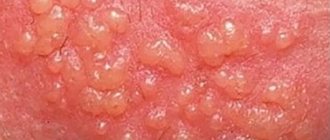
The incubation period for herpes zoster can be several years from the moment of infection. In the clinical course, the main ones are: the prodromal period, the period of clinical manifestations and the period of residual effects. It all starts with an increase in temperature, a tingling sensation, burning, itching at the site of the rash, and a headache. Along the nerve trunks of the trunk, limbs or head, limited pink spots up to five centimeters in diameter appear. On the second day, bubbles 2–3 mm in diameter appear, filled with transparent contents. The number of lesions can vary from one to several, closely adjacent to each other, forming a continuous line. Over time, the contents of the bubbles become cloudy. At about 8–10 days, the blisters dry out and crusts form, which disappear after 3–4 weeks. In many patients, neurological manifestations can last for several months (up to a year).
Typical clinical manifestations of herpes zoster are characterized by a certain sequence of skin rashes. The rashes are segmental, unilateral and do not spread to the other side of the body, unlike chickenpox.
Lesions of the nervous system with herpes zoster occupy first place among the complications of this disease. In the structure of neurological disorders, the leading place is occupied by lesions of the peripheral nervous system [10]. The most common disorders are neuralgia, neuropathy of the cranial and peripheral nerves, serous meningitis, etc. The most common manifestation is pain in the area of skin rashes. The pain is paroxysmal in nature, intensifying at night. In the future, the pain may intensify and bother you for several months and even years. Herpes zoster can also occur only with the symptoms of radicular pain, which is not preceded by a period of rash.
Most often, the rashes are located on the skin of the torso and limbs. The localization of pain and the appearance of a specific rash correspond to the affected nerves, most often intercostal nerves and are encircling in nature. The intensity of the pain increases with the slightest touch to the skin, with movement, or temperature changes. After the blisters disappear, the erosions epithelialize, and temporary red or red-brown pigmentation may remain on the skin. Some patients may have no pain. And sometimes herpes zoster can manifest itself only as neurological symptoms without the absence of skin manifestations.
Very often the localization of the disease is the skin of the face and head, especially the branches of the trigeminal nerve. Manifestations of the disease begin acutely, with general symptoms of intoxication and fever. Some patients may experience facial paralysis and trigeminal neuralgia lasting up to several weeks.
There may be manifestations of motor functions that occur not only when herpes zoster is localized in areas of the skin innervated by cranial nerves, but also when the cervical, thoracic and lumbar spinal cord, roots and ganglia are involved. Almost 5% of patients with rashes of various locations experience paresis of the upper and, more often, lower extremities, which indicate the phenomenon of focal myelitis.
To understand the pathogenesis of herpes zoster, data from pathological studies are important, indicating a connection between areas of the rash and damage to the corresponding ganglia. Head and Campbell (1900), based on pathohistological studies, came to the conclusion that both the neurological phenomena of herpes zoster and the areas of skin rashes that characterize them arise as a result of the development of a pathological process in the spinal nodes and their homologues (Gasserian node, etc.). But Volville (1924), having studied the nervous system of patients who died from a generalized form of herpes zoster, came to the conclusion that damage to the intervertebral ganglia in herpes zoster is not necessary. The inflammatory process often involves the spinal cord, and not only the posterior horns are affected, but also the anterior ones. Volville and Shubak (1924) described cases in which herpetic rashes were the first manifestations of a polyneurotic process occurring like Landry's paralysis. Volville believes that the inflammatory process first affected sensory neurons, and then spread to the spinal segments and peripheral nerves. In the case described by Shubak, a pathological examination revealed nests of inflammatory infiltration in the sciatic nerves, cervical sympathetic ganglia and the corresponding spinal ganglia, and the dorsal horns of the spinal cord.
Thus, the process involves not only the spinal and cerebral ganglia, which are most often affected, but also the substance of the spinal (anterior and especially posterior horns, white matter) and brain (oblongata, pons, hypothalamic region) brain, as well as the meninges.
Pathological and virological studies indicate that the herpes zoster virus widely disseminates throughout the body. During illness, it can be isolated from the contents of vesicles, saliva, tear fluid, etc. This gives reason to believe that herpetic eruptions can be caused not only by the settling of the virus in the sensory ganglia and damage to the parasympathetic effector cells located in them, but also by direct penetration it into the skin. Penetrating into the nervous system, it is localized not only within the peripheral sensory neuron (spinal ganglia, etc.), but also spreads to other parts of the central nervous system. When it is introduced into motor cells and roots, a picture of amyotrophic radiculoplexitis arises; in the gray matter of the spinal cord - myelitic syndrome; into the cerebrospinal fluid system - meningoradiculoneuritis or serous meningitis, etc.
The clinical picture of herpes zoster consists of skin manifestations and neurological disorders. Along with this, most patients experience general infectious symptoms: fever, enlargement of hormonal lymph nodes, changes (in the form of lymphocytosis and monocytosis) and cerebrospinal fluid. Typically, erythematous spots of round or irregular shape, raised, swollen, are found on the skin; when running a finger over them, a certain shagreen-like appearance of the skin is felt (tiny papules). Then, in these areas, groups of bubbles appear successively, often of varying sizes. The bubbles can merge with each other, but most often they are located in isolation, although close to one another - the vesicular form of herpes zoster. Sometimes they look like a small bubble surrounded on the periphery by a red rim. Since the rash occurs simultaneously, the elements of the rash are at the same stage of their development. However, the rash may appear in separate clusters over 1 to 2 weeks. In the latter case, when examining the patient, rashes can be detected at various stages of evolution. In typical cases, the bubbles at first have transparent contents, which soon turn cloudy, and then dry out in the form of a crust. A deviation from the described type is a milder abortive form of herpes zoster. With this form, papules also develop in the foci of hyperemia, which, however, do not transform into vesicles, which is how this form differs from the vesicular one. Another type is the hemorrhagic form of herpes zoster. The blisters have bloody contents, the process spreads deeply into the dermis, the crusts become dark brown. In severe cases, the bottom of the vesicles becomes necrotic - a gangrenous form of herpes zoster, after which scarring changes remain. The intensity of the rashes in this disease is very variable: from confluent forms, leaving almost no healthy skin on the affected side, to individual blisters, although in the latter case the pain can be sharply expressed. Such cases have given rise to the assumption that herpes zoster may exist without a skin rash.
Skin manifestations correspond to the level of damage to certain vegetative formations. By localization, lesions of the following ganglia are distinguished: gasserian, geniculate, cervical, thoracic, lumbosacral. One of the leading symptoms of the disease are neurological disorders, usually in the form of pain. Most often it occurs 1–2 days before the rash appears. The pain, as a rule, is intense, burning in nature, and the area of its distribution corresponds to the roots of the affected ganglion. It should be noted that the pain syndrome intensifies at night and under the influence of a variety of stimuli (cold, tactile, kinesthetic, barometric) and is often accompanied by vegetative-vascular dystonia of the hypertensive type. In addition, patients experience objective sensitivity disorders: hyperesthesia - the patient can hardly tolerate the touch of linen, hypoesthesia and anesthesia, and hyperalgesia may exist simultaneously with tactile anesthesia. Objective sensitivity disorders vary in form and intensity, they are usually limited to temporary sensitivity disorders in the area of rash or scars. Anesthesia affects all types of sensitivity, but in some cases a dissociated type of disorder is observed; sometimes within the same type of sensitivity, such as hot and cold. Occasionally, hyperesthesia takes on the character of irritation in the form of causalgia. Not in all cases, the intensity of the pain syndrome corresponds to the severity of skin manifestations. In some patients, despite the severe gangrenous form of the disease, the pain is minor and short-lived. In contrast, a number of patients experience prolonged intense pain with minimal skin manifestations.
Some patients in the acute phase have diffuse cephalgia, which intensifies with changes in head position, which may be associated with a meningeal reaction to herpes zoster infection. According to a number of authors [11, 12], herpetic ganglionitis of the Gasserian ganglion is more common than ganglionitis of the intervertebral ganglion. Most patients with this localization of the process experience increased temperature and swelling of the face on the affected side, as well as pain at the exit points of the trigeminal nerve.
The cornea is often affected in the form of keratitis of various types. In addition, other parts of the eyeball are affected - episcleritis, iridocyclitis, iris zoster. The retina is very rarely involved (hemorrhages, embolisms), more often the changes affect the optic nerve - optic neuritis resulting in atrophy, possibly due to the transition of the meningeal process to the optic nerve. With ophthalmoherpes (iritis), glaucoma may develop; Usually, with zoster, hypotension of the eyeball is observed, which is apparently caused by damage to the ciliary nerves. Complications of zoster from the motor nerves are quite common and are located in the following order: III, IV, VI nerves. Of the branches of the oculomotor nerve, both external and internal branches are affected. Ptosis is often observed. Skin rashes with ophthalmic zoster are often more severe than on other parts of the body, possibly depending on the structure of the skin in the eye area. Quite often, necrosis of the vesicles and severe neuralgia, accompanied by lacrimation, are observed. Bubbles appear not only on the skin, but also on the mucous membranes of the eye. As a result of the process in the cornea with ophthalmic zoster, optic nerve atrophy and complete blindness can develop. In addition, some patients experience loss of eyebrows and eyelashes on the affected side. The maxillary branches of the trigeminal nerve are affected both in the skin and in the mucous membranes (half of the hard and soft palate, velum, upper gum, inner surface of the buccal mucosa, while the nasal mucosa may remain unaffected). The branches supplying the mucous membranes may be more affected than the cutaneous branches, and vice versa. Lesions of the nerves of the upper and lower jaws do not always remain strictly localized, since pain sometimes radiates to the area of the ophthalmic and other branches.
Herpes zoster usually affects the autonomic nervous system. However, clinical observations have shown that the animal nervous system may also be involved in the pathological process. Evidence of this is that some patients, simultaneously with damage to the Gasserian ganglion, had peripheral paresis of the facial nerve on the side of the herpetic rash. With ophthalmic zoster, both the external and internal muscles of the eye are paralyzed. IV paralysis is rare. Oculomotor paralysis is more often partial than complete; More often than other muscles, m. is affected. levator palpebrae. There are cases of ophthalmic zoster with isolated changes in the shape and size of the pupil; unilateral Argil-Robertson (Guillén) sign. These paralysis sometimes partially or completely resolve spontaneously without special treatment.
Simultaneous damage to the facial, auditory and trigeminal nerves was first described by Frankl-Hochwart in 1895. Hunt (1907) described in detail the four clinical forms of this disease, which later became known as Hunt's syndrome, or herpes zoster oticus. The defeat of the geniculate ganglion in this form of herpes zoster was first pointed out by Nordahl (1969). Usually, herpetic rashes appear on or around the ear, and sometimes in the ear canal and even on the eardrum. There is sharp pain in the circumference of the auricle. Dysfunction of the facial, cochlear, and vestibular nerves occurs in the first days of the rash or precedes it. Pain in such cases is localized in the depths of the ear canal and the auricle with irradiation to the mastoid region, auricular and temporoparietal areas.
Objective sensitivity disorders are found behind the ear, in the fold between the auricle and the mastoid process. This skin area is supplied by the auricular branch of the X pair, which innervates the posterior walls of the auditory canal. Finally, in cases of very common ear zoster, the latter affects not only the external auditory canal, the auricle, the mastoid process, but also the eardrum, which is sometimes extremely seriously affected. In such cases, the area innervated by pairs V, VII and X is affected, and damage to these nerves is accompanied by damage to the ganglia, corresponding cranial nerves, or anastomoses connecting the terminal branches of all of the above nerves.
Often, simultaneously with paralysis of the VII pair, there is paralysis of the soft palate, anesthesia and paresthesia in the tongue, and often a taste disorder in the anterior two-thirds of the tongue due to the lesion. The defeat of the VIII pair usually begins with tinnitus, which sometimes lasts a long time after the disappearance of other phenomena. Hyperacusis with damage to the VIII pair is caused by paresis n. stapeblii, although this symptom can also occur with isolated and previous damage to the auditory nerve and in such cases represents a symptom of irritation. Hypoacusis can occur regardless of damage to the auditory nerve due to local lesions of the middle ear, eruption of bubbles on the eardrum, blockage of the external auditory canal, due to swelling of the mucous membrane due to eruption of zoster.
Vestibulatory phenomena, in contrast to cochlear phenomena, usually develop extremely slowly and are expressed differently: from mild subjective symptoms of dizziness to significant static disorders.
Neuralgia with auricular zoster, as opposed to ophthalmic zoster, is rare. Long-term results are not always favorable, as persistent paresis of the facial nerve and deafness may occur.
Volville emphasizes that the combination of paralysis of the VII and VIII pairs, although it occurs especially often with zoster, nevertheless the same combination occurs when the Gasserian node, II, III, cervical ganglia are affected, and, finally, all of the above areas can be affected simultaneously.
Zoster rashes are also described in the area of innervation of the IX pair; the posterior part of the soft palate, the arch, the posterolateral parts of the tongue, part of the posterior wall of the pharynx; In addition to IX, this same area is also innervated by branches of the X pair: the root of the tongue, larynx, epiglottis, basal and posterior parts of the pharyngeal wall. Although zoster predominantly and even selectively affects the sensory systems, movement disorders are sometimes observed with it, especially when the rash is localized in the head, neck, and limbs. Paralysis with zoster is radicular in nature, and damage to the posterior roots in these cases is accompanied by phenomena from the corresponding anterior roots.
Damage to the cervical sympathetic nodes is often accompanied by rashes on the skin of the neck and scalp. Pain in this case is observed not only in places of rashes, but also in the area of paravertebral points. Sometimes attacks that mimic facial sympathalgia may occur.
With ganglionitis of the lower cervical and upper thoracic localization, along with the usual symptoms of this disease, Steinbrocker syndrome can be observed. The dominant picture of this syndrome is pain of a sympathetic nature in the form of burning or pressure, occurring initially in the hand, and then in the entire arm. Soon swelling of the hand appears and quickly increases, spreading to the entire arm. Trophic disorders are added in the form of cyanosis and thinning of the skin, hyperhidrosis, and brittle nails. Movement of the fingers is limited and painful. Often pain and other autonomic disorders persist even after the rash disappears. Thoracic ganglionitis often simulates the clinical picture of myocardial infarction, which leads to diagnostic errors.
With herpetic lesions of the ganglia of the lumbosacral region, the rashes are most often localized on the skin of the lower back, buttocks and lower extremities; Along with pain in the areas of rashes, pain syndromes simulating pancreatitis, cholecystitis, renal colic, and appendicitis may occur. Herpetic lesions of the lumbosacral ganglia are sometimes accompanied by involvement of the animal nervous system in the process, giving a picture of ganglioradiculitis (radicular syndrome of Pori, Matskevich, Wasserman).
Sometimes, along with rashes along the nerve trunk, vesicular rashes appear throughout the skin - a generalized form of herpes zoster. Usually the disease does not recur. However, it is known from the literature that recurrent forms of the disease occur against the background of somatic complications: HIV infection, cancer, diabetes mellitus, lymphogranulomatosis, etc.
Treatment . When treating herpes zoster of varying localization and severity, early administration of antiviral drugs is necessary. It is known that the virus contains proteins that form its shell and carry enzymatic functions, as well as nucleic acid, the carrier of its genetic properties. Penetrating into cells, viruses are freed from their protective protein shell. It has been shown that at this moment it is possible to inhibit their reproduction using nucleases. These enzymes hydrolyze the nucleic acids of viruses without damaging the nucleic acids of the cell itself. It was found that pancreatic deoxyribonuclease sharply inhibits the synthesis of DNA-containing viruses, such as herpes virus, vaccinia, and adenoviruses. Considering the above, it is recommended that patients with herpes zoster be prescribed deoxyribonuclease intramuscularly 1-2 times a day, 30-50 mg for 7 days. In addition, in patients with rashes on the oral mucosa, conjunctiva and cornea, the drug is used topically in the form of an aqueous solution. The administration of deoxyribonuclease promotes rapid regression of skin rashes and a reduction in pain.
The drug Isoprinosine has a good effect in the treatment of herpes zoster. This is an immunostimulating agent with an antiviral effect. Isoprinosine blocks the reproduction of viral particles by damaging its genetic apparatus, stimulates the activity of macrophages, the proliferation of lymphocytes and the formation of cytokines. The second component increases the availability of Isoprinosine for lymphocytes. Reduces the clinical manifestations of viral diseases, accelerates convalescence, and increases the body's resistance.
Indications: viral infections in patients with normal and weakened immune systems (diseases caused by herpes simplex viruses types 1 and 2, Varicella zoster, including chickenpox, measles, mumps, cytomegalovirus (CMV), Epstein-Barr virus); viral bronchitis; acute and chronic viral hepatitis B and C; diseases caused by the human papillomavirus; subacute sclerosing panencephalitis. Chronic infectious diseases of the urinary and respiratory systems; prevention of infections in stressful situations; the period of convalescence in postoperative patients and persons who have suffered serious illnesses; immunodeficiency states. Isoprinosine is taken orally, for adults - 50 mg/kg/day in 3-4 doses; for children - 50–100 mg/kg/day in 3–4 doses. Duration of treatment is 5–10 days, in severe cases up to 15 days. For diseases caused by herpes simplex viruses types 1 and 2, treatment is continued until the symptoms of the disease disappear and for another two days. For subacute sclerosing panencephalitis for adults and children - 50–100 mg/kg/day in 6 divided doses. For acute viral encephalitis for adults and children - 100 mg/kg/day in 4–6 divided doses for 7–10 days. This is followed by a break for 8 days, then a repeat course for 7–10 days. If necessary, the dose and duration of the continuous course can be increased, subject to a mandatory break in taking the drug for 8 days. Long-term treatment is carried out under medical supervision. For genital warts in complex therapy with a CO2 laser - 50 mg/kg/day in 3 divided doses for 5 days, then with a 3-fold repetition of the specified course at intervals of one month.
In recent years, antiviral chemotherapy drugs from the group of synthetic acyclic nucleosides have been used to treat herpes zoster. The most well studied drug at present is acyclovir. The mechanism of action of acyclovir is based on the interaction of synthetic nucleosides with the replication enzymes of herpes viruses. Thymidine kinase of the herpes virus binds to acyclovir thousands of times faster than cellular ones, so the drug accumulates almost only in infected cells. This explains the complete absence of cytotoxic, teratogenic and mutagenic properties in acyclovir. The synthetic nucleoside is arranged in a chain of DNA being built for the “daughter” viral particles, and this process ends in this way: the reproduction of the virus stops. The daily dose of acyclovir for herpes zoster is 4 g, which should be divided into 5 single doses of 800 mg. The course of treatment is 7–10 days. The best therapeutic effect is achieved with early administration of the drug; The duration of rashes is reduced, crusts form quickly, intoxication and pain are reduced. Second generation acyclovir - valacyclovir, retaining all the positive aspects of acyclovir, due to increased bioavailability, allows you to reduce the dose to 3 g per day, and the number of doses - up to three times. The course of treatment is 7–10 days. Famciclovir has been used since 1994. The mechanism of action is the same as that of acyclovir. The high affinity of the virus thymidine kinase for famciclovir (100 times higher than the affinity for acyclovir) makes the drug more effective in the treatment of herpes zoster. The drug is prescribed 250 mg 3 times a day for 7 days.
Along with antiviral drugs, ganglion blockers, such as Gangleron, are used to reduce pain. Gangleron is used intramuscularly in the form of a 1.5% solution, 1 ml once a day for 10–12 days or 0.04 g in capsules 2 times a day for 10–15 days, depending on the severity of the pain syndrome. In addition, the use of carbamazepine gives good results, especially in cases of damage to the Gasserian node; the drug is prescribed with 0.1 g 2 times a day, increasing the dose by 0.1 g per day, if necessary, up to 0.6 g daily dose (in 3– 4 doses). After the pain decreases or disappears, the dose is gradually reduced. Usually the effect occurs 3-5 days after the start of treatment.
In cases of severe pain, analgesics and, in the form of injections, reflexology are prescribed. In reflexology, both points of general action and points corresponding to the affected ganglion are usually used. The course consists of 10–12 sessions. It is also recommended to prescribe multivitamins, in particular B vitamins. Local irrigation with interferon or ointments with interferon, aniline dyes, Eridin aerosol, Florenal, Helepin, Alpizarin ointments can be used. For gangrenous forms of herpes zoster, pastes and ointments containing an antibiotic, as well as Solcoseryl, are used.
Good results are obtained by irrigation with Epigen spray 4–5 times a day for 7–10 days from the first days of the disease. When combined with oral acyclovir therapy, a decrease in pain is noted.
After the skin rash resolves, treatment is carried out by neurologists until the neurological symptoms disappear.
Thus, treatment of herpes zoster should be comprehensive and include both etiological and pathogenetic agents.
Literature
- Batkaev E. A., Kitsak V. Ya., Korsunskaya I. M., Lipova E. V. Viral diseases of the skin and mucous membranes. Textbook manual, RMAPO. M.: Pulse, 2001.
- Butov Yu. S. Skin diseases and sexually transmitted infections.
- Kartamyshev A.I. Skin and venereal diseases. Medgiz, 1954.
- Skin and venereal diseases: Directory. Ed. O. L. Ivanova. M.: Medicine, 1997.
- Paltsev M. A., Potekaev N. N., Kazantseva I. A. et al. Clinical and morphological diagnosis of skin diseases (atlas). M.: Med., 2004.
- Pospelov A.I. A short textbook of skin diseases. M., 1907.
- Skripkin Yu. K., Kubanova A. A., Prokhorenkov V. I. et al. Dermatological syndromology. M. - Krasnoyarsk, 1998.
- Sukolin G.I. Clinical dermatology. St. Petersburg, 1997.
- Lezvinskaya E. M., Piven A. L. Laboratory diagnostics: skin diseases and sexually transmitted infections. M.: Practical Medicine, 2005
- Yushchuk N. D., Stepanchenko A. V., Dekonenko E. P. 2005.
- Kalamkaryan A. A., Kochetkov V. D. 1973.
- Zucker M. B. 1976.
I. M. Shakov, Candidate of Medical Sciences
GOU DPO RMAPO, Moscow
Contact information about the author for correspondence
Complications of genital herpes
Complications of genital herpes include dryness and the formation of painful bleeding cracks on the mucous membranes of the external genitalia, which occurs due to mechanical stress (for example, during sexual intercourse).
A special place among other complications is occupied by pain syndrome caused by specific herpetic neuralgia of the pelvic nerve plexus. In this case, women complain of periodically occurring nagging pain in the lower abdomen, in the area of the projection of the ovaries, radiating to the lumbar region and rectum, pain in the perineum. Pain with recurrent herpes can occur regardless of the presence of skin rashes, which greatly complicates diagnosis. Recurrent genital herpes, disrupting the normal sex life of patients, often causes neuropsychiatric disorders and leads to conflicts in the family.
Genital herpes in some women causes miscarriage and infertility.
Herpes in newborns
A child becomes infected with the pathogen from the mother in utero or by passing through an infected genital tract during childbirth. In newborns, the mucous membranes of the eyes and mouth, skin, and genitals are affected. When the pathogen enters the child’s brain, meningoencephalitis occurs, causing death or severe disability.
A generalized form is possible, in which all organs and systems of the newborn are affected. The child experiences jaundice, respiratory distress, and urinary retention. Children are restless, do not latch on to the breast, and are vomiting. Death occurs from shock, bleeding, dehydration, intoxication, and organ failure.
The likelihood of infection increases with primary infection of the mother in late pregnancy.
Diagnostics
If you suspect the presence of herpes simplex, you should not self-medicate. It is urgent, without delay or masking the rash, to come to an appointment with a dermatovenerologist. A carefully collected anamnesis (questioning of the patient) is important for establishing the correct diagnosis. Herpes, regardless of the location of the pathological process, is characterized by a wave-like course, when painful states are replaced by periods of well-being, even without treatment. The presence of blistering rashes on the skin and mucous membranes, severe subjective symptoms (itching, burning) allows doctors to visually diagnose herpes simplex, prescribe treatment in a timely manner and inform the patient about the danger of infecting a sexual partner.
Only laboratory research methods, which are fundamentally divided into two groups, can reliably confirm the herpetic nature of the disease in the absence of typical manifestations on the skin and mucous membranes:
- isolation and identification of HSV from infected material (material for analysis are scrapings from the lesion, blood, urine, saliva, tear fluid, cerebrospinal fluid, discharge of the cervical canal, vagina, urethra, rectum);
- detection of specific antibodies (protective proteins) to the herpes virus in blood serum - serodiagnosis.
What is herpes on the head
Children who have had chickenpox usually develop immunity to the virus, but it does not leave the body, but finds a place in the nerve ganglia. When the protective forces are weakened, the herpes pathogen begins to act, provoking the development of the pathological process.
A herpetic rash on the scalp is usually a manifestation of chickenpox.
Itchy red blisters appear on the scalp that leave crusts; characterized by fever and poor health.
Diagnostics: examination, PCR, ELISA, culture isolation.
Treatment: antiseptics and antiviral ointments; general antiviral, antihistamine, immunomodulators; Physiotherapy UFO.
Bubbles form on the forehead and scalp; when they open, the liquid upon contact is transferred to a crack or microtrauma that is located on the skin of a healthy person.
A rash on the head, accompanied by unbearable itching, is formed if the virus is activated in the trigeminal nerve, which is located on top of the visual organ or near the ear.
Isolation and identification of HSV
In specialized virology laboratories, HSV is isolated using the culture method. Its essence lies in the fact that the material for research (the contents of herpetic eruptions, human secretions) is placed on specially grown cells in which the virus begins to multiply. Then, after 5 days, the presence of HSV is determined by characteristic changes. Thus, we can say for sure that this disease is of a herpetic nature.
To identify the causative agent of HSV, the polymerase chain reaction (PCR) method is widely used, which makes it possible to find out what type of herpes virus is present in the body. The material for research is taken with a special brush from the rash sites. The PNR method is highly sensitive and is performed within 24–48 hours.
Scalp diseases: names and causes
Common skin diseases are considered to be:
- diseases of a dermatological nature (including seborrhea, eczema and hyperkeratosis);
- autoimmune diseases of the scalp (including psoriasis and scleroderma);
- infectious (formation of pustules, appearance of lice, etc.);
- fungal rashes.
Whatever the cause of the disease, it must be eliminated in time. It is important to start treatment as early as possible, because wasted time can have serious consequences.
Serodiagnosis
Antibodies to the herpes virus appear in the blood serum by 4-7 days after the initial infection, reach a peak after 2-3 weeks and can persist throughout life. The diagnosis is made by the characteristic increase in antibodies and determination of their class. For example, the detection of a certain level of immunoglobulin M (antibodies) indicates a primary infection of a person or an exacerbation of herpes. The detection of a certain level of immunoglobulin G indicates that the human body has encountered the virus and has developed antibodies - the person is infected, but not infectious to others. The result is known the next day. The analysis can be repeated at intervals of several days.
How can you get infected with herpes
The content of the article
The disease is caused by the herpes simplex virus (HSV, Herpes simplex virus). Of the eight types of this pathogen, genital lesions are the result of two infectious agents. HSV-2 causes 80% of cases of the disease, and HSV-1 - 20%. There are also combined infections, in which both viral types are to blame.
There is a misconception that herpes on the lips and genitals are completely different diseases. In fact, both types of pathogens often “swap places” during domestic infection and oral sex. And both infections are equally dangerous.
Both types of virus are transmitted:
- For all types of sexual contact - vaginal, oral, anal, during which the virus enters the body through microdamage to the mucous membrane. You can also become infected from a partner who does not have rashes on the genitals or other manifestations of the disease. This condition is possible with strong immunity, when the virus lies low and waits in the wings. A person becomes a carrier of a herpes infection, but does not experience any health problems.
- Through common objects - washcloths, sponges, bed linen, towels. A person with a cold sore caused by the type 1 virus can become a source of genital herpes infection.
- During autoinoculation (self-infection), when the patient transfers the pathogen from the face to the genitals.
- A child becomes infected from a sick mother when the virus penetrates from the vagina into the uterus or transplacentally through the placenta. During childbirth, infection occurs as the newborn passes through the birth canal.
Statistics of hidden and overt forms of herpes
The body cannot overcome the pathogen on its own. The virus settles in the roots of the spinal cord, and the infected person continues to live with the disease throughout his life.
The pathogen can remain in the body for many years without causing symptoms. Therefore, it is impossible to know without testing for herpes whether a person is infected with the virus. According to WHO, type 1 herpes, which causes a rash on the lips and genitals, affects 67% of the world's inhabitants, and type 2 affects 11% of people. Only 20% of the disease occurs in the classical form, which is beyond doubt. In the rest, the course of the disease is latent or asymptomatic.
Principles of treatment
Herpes can and should be treated. Currently, medicine has a whole arsenal of drugs that, in most cases, make it possible to obtain a lasting clinical effect in those who suffer from recurrent herpes and to successfully control periods of activation of the disease in asymptomatic forms. It is impossible to achieve complete removal of the virus from the body using currently existing treatment methods; it is possible to maintain the body in a state where the virus does not have a chance to activate. Currently, there are two main directions of treatment for herpes simplex:
- The use of antiviral drugs, the main place among which is occupied by acyclovir-containing (ACV) drugs. In case of exacerbation of herpes, the first aid remedy is medications containing acyclovir: ZOVIRAX, VAPTREX, ACICLOVIR-ACRI, VIROLEX. They are available in the form of tablets and creams. The same drugs are used locally in the form of cream and ointment. Taking these medications should begin in the first hours of an exacerbation.
- A complex treatment method that includes immunotherapy in combination with antiviral therapy. As a rule, the disease occurs against the background of suppressed immune reactions, so immunotherapy plays an important role in the treatment of herpes. If relapses of the disease are seasonal (autumn, spring) and they are accompanied by symptoms of ARVI, then to antiviral treatment with drugs containing acyclovir, it is advisable to add a course of drugs that stimulate the synthesis of interferon (a substance produced by the body and providing an antiviral effect): herbal immunostimulants (for example, IMMUNAL) and synthetic drugs such as ARBIDOL, AMEXIN, VIFERON, KIPFERON. Moreover, they can be used for preventive purposes about a month before the onset of autumn slush and spring hypovitaminosis). If relapses of herpes (at any location) occur more than 3-4 times a YEAR, this is a reason to consult a doctor. This means that the body's defenses (immunity) cannot cope with the infection. In such cases, after studying the immune status, the patient is prescribed complex treatment, including antiviral and immune drugs. In patients with recurrent herpes, the drugs TACTIVIN, TIMALIN, THIMOGEN, MYELOPID, etc. are successfully used (treatment must be carried out after an immunological study).
Stages and symptoms
The pathology occurs in 4 stages, each of which corresponds to certain symptoms:
- Initially, the scalp begins to itch, tingle, and painful sensations appear. Later the scalp turns red. The general condition of the patient is similar to the onset of a cold; the temperature may be elevated. Possible headache.
- After 12 hours, acute inflammation of the scalp begins. Bubbles filled with liquid (vesicles) appear, and over time the number of rashes steadily increases.
- The vesicles burst. The pain subsides.
- In place of the blisters, scabs form, which fall off on their own after a few days. This symptom indicates recovery.
After a month of the disease, the pathological process subsides. But if the patient’s immunity is weak, painful sensations along the affected nerves can persist for 1–2 months.