Herpesvirus infections are a group of infectious diseases that are caused by viruses from the Herpesviridae family, can occur in the form of localized, generalized, recurrent forms of the disease, and have the ability to persist (the virus is constantly present) in the human body.
Herpesvirus infections (HVI) are among the most common human viral diseases. Their infection and morbidity rates are increasing every year. In all countries of the world, 60-90% of the population is infected with one or another herpes virus.
How can you get infected with herpes
The content of the article
The disease is caused by the herpes simplex virus (HSV, Herpes simplex virus). Of the eight types of this pathogen, genital lesions are the result of two infectious agents. HSV-2 causes 80% of cases of the disease, and HSV-1 - 20%. There are also combined infections, in which both viral types are to blame.
There is a misconception that herpes on the lips and genitals are completely different diseases. In fact, both types of pathogens often “swap places” during domestic infection and oral sex. And both infections are equally dangerous.
Both types of virus are transmitted:
- For all types of sexual contact - vaginal, oral, anal, during which the virus enters the body through microdamage to the mucous membrane. You can also become infected from a partner who does not have rashes on the genitals or other manifestations of the disease. This condition is possible with strong immunity, when the virus lies low and waits in the wings. A person becomes a carrier of a herpes infection, but does not experience any health problems.
- Through common objects - washcloths, sponges, bed linen, towels. A person with a cold sore caused by the type 1 virus can become a source of genital herpes infection.
- During autoinoculation (self-infection), when the patient transfers the pathogen from the face to the genitals.
- A child becomes infected from a sick mother when the virus penetrates from the vagina into the uterus or transplacentally through the placenta. During childbirth, infection occurs as the newborn passes through the birth canal.
Statistics of hidden and overt forms of herpes
The body cannot overcome the pathogen on its own. The virus settles in the roots of the spinal cord, and the infected person continues to live with the disease throughout his life.
The pathogen can remain in the body for many years without causing symptoms. Therefore, it is impossible to know without testing for herpes whether a person is infected with the virus. According to WHO, type 1 herpes, which causes a rash on the lips and genitals, affects 67% of the world's inhabitants, and type 2 affects 11% of people. Only 20% of the disease occurs in the classical form, which is beyond doubt. In the rest, the course of the disease is latent or asymptomatic.
Cytomegalovirus infection
An infectious disease that is caused by cytomegalovirus (CMV) and is characterized by a variety of clinical forms (from asymptomatic to severe generalized with damage to many organs) and course (acute or chronic). CMV transmission factors can be almost all biological substrates and human secretions that contain the virus: blood, saliva, urine, cerebrospinal fluid, vaginal secretions, sperm, amniotic fluid, breast milk. Potential sources of infection are organs and tissues in transplantology, as well as blood and its products in transfusiology. Routes of transmission of CMV infection: airborne, sexual, vertical and parenteral.
There are congenital and acquired forms of CMV infection. Congenital CMV infection. During antenatal infection of the fetus, infection occurs predominantly transplacentally. During intrapartum infection, CMV enters the body through aspiration of infected amniotic fluid or secretions from the mother's birth canal.
In older children, acquired CMV infection occurs in a subclinical form in 99% of cases. The most common manifestation of this form of CMV infection in children over one year of age is mononucleosis-like syndrome. As a rule, a clinical picture of acute respiratory disease is observed in the form of pharyngitis, laryngitis, and bronchitis.
Infections caused by the sixth, seventh and eighth types of herpes viruses Type six herpes viruses (HHV-6) can cause erythematous and roseolous rashes (sudden exanthema), lesions of the central nervous system and bone marrow in immunocompromised children. Herpesvirus type seven (HHV-7) causes neonatal exanthema
For the diagnosis of herpes infection, cytological, immunofluorescent, serological and PCR methods are valuable. Virological testing for herpes infection reveals complement-fixing antibodies to HSV-1 or -2 in the mother's blood, fetal cord blood and amniotic fluid. PCR method. The material for testing for herpes is blood, throat swabs, the contents of blisters, ulcers, and urine.
The study of specific antibodies of various subclasses: IgM, IgG1-2, IgG3 and IgG4 to herpes viruses is important. The detection in the blood serum of children of specific immunoglobulins M, IgG3, IgG1-2 in a titer > 1:20, viral antigen and specific immune complexes with antigen indicates the severity of the infectious process (active phase), and the determination of only specific IgG4 is regarded as the latent phase of infection or carriage of maternal antibodies.
Primary herpes
There are three periods of illness:
- Prodromal
(precursor period), during which the temperature rises, the inguinal lymph nodes become inflamed, weakness, fatigue, and headache appear. A person experiences a condition similar to the symptoms of flu and colds. If you take an antiviral drug during this period, the disease may not develop. If the symptoms are ignored and treatment is not started on time, the second period of the disease begins. - A rash
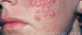
that occurs three to four days after the onset of a flu-like condition. Common symptoms include pain, heat, swelling, itching in the genital area, anus, buttocks, pubis and perineum. Soon, numerous blisters filled with clear liquid appear on the inflamed skin and mucous membranes, reminiscent of a “cold” on the lips. The rash causes pain and itching, worse at night and leading to insomnia. - Ulcerations.
After some time, the blisters burst and ulcers (erosions) form in their place, surrounded by a focus of inflammation. Contact with stool and urine on the eroded surface increases pain and complicates tissue regeneration. - Healing,
during which the ulcers gradually heal. If the genital organs are poorly cleaned, the immune system is weak, and infection occurs, the healing process is delayed.
After the ulcers disappear, the disease goes into remission. During this period, despite the presence of the virus in the body, there are no rashes on the genitals.
Prevention
Following simple rules of prevention will help to significantly reduce the risk of a child becoming infected with herpes. Basic rules include:
- preventing child contact with sick children and adults;
- strict adherence to hygiene rules;
- balanced diet;
- immunostimulating therapy;
- specific vaccination.
In our multidisciplinary medical center, experienced doctors provide effective treatment for children with any manifestation of herpetic infection. Timely consultation with a doctor will help avoid the development of infection and dangerous complications.
Recurrent herpes
In 50%-70% of people the disease becomes chronic. This is facilitated by:
- untimely or incorrect treatment;
- decreased immunity, including that caused by taking medications that suppress the immune system;
- presence of other STDs;
- strict diets, vitamin deficiencies;
- state of chronic stress.
Why do patients develop relapses, during which the disease affects the same parts of the body? There are several reasons for this:
- Having penetrated the body, the virus settles in the tissues of the genital organs. As it multiplies, it affects cells deeper and deeper until it reaches nerve cells - neurons connected to each other by processes - axons. Through them, like bridges, the pathogen reaches the cells of the spinal cord, into which it introduces its DNA.
- An infected brain cell becomes an “incubator” for viruses, which periodically return via axon bridges to the mucous membrane or skin, causing new rashes. Schematically, the process is similar to the migration of birds, constantly returning to their “native places.”
- The immune system cannot kill viruses hidden in the spinal cord, but it deals with those that have left the “shelter”. Therefore, with high immunity, relapses occur rarely or not at all. But as soon as the body “loses its vigilance” - weakens, gets sick, catches a cold, is exposed to stress - the pathogens begin to overcome the immune defense. As a result, they reach their target - the skin and mucous membranes of the genital organs. Here, the increased reproduction of viral particles inside the “captured” cells begins. Cellular structures, dying, release myriads of viruses that cause inflammation, redness, swelling and the appearance of blisters. Herpes is causing another aggravation.
- Gradually, the immune system fights back the virus, and within 10 days the ulcers at the site of the rash dry out and heal. Everything returns to normal, so that at the slightest weakening of the immune system it can start again.
From the mechanism of exacerbations, it becomes clear why herpetic eruptions appear in the same places. It’s just that the virus cells return back only through the nerve cell from which they penetrated deep into the body.
Types of recurrent herpes
Depending on the number of exacerbations, several types of the disease are distinguished:
- mild, occurring up to 3 times a year;
- moderate severity, in which exacerbations occur 4-6 times a year;
- severe, accompanied by frequent exacerbations, intervals between which do not exceed a month;
- arrhythmic, in which after a period of imaginary well-being, lasting from a month to five, rashes appear. This form of herpes is characterized by a pattern - the longer the remission, the more severe the disease;
- menstrual, manifested by rashes during menstrual periods. This type of disease is severe and difficult to treat;
- subsiding, in which the manifestations of the disease constantly become weaker, and the inter-relapse periods become longer. The subsidence of the manifestations of the disease indicates the positive dynamics of treatment and the restoration of immune defense that suppresses the virus.
Herpesvirus infection is currently one of the leading medical and social problems not only in Russia, but throughout the world. The variety of clinical manifestations, characteristics of pathogens, almost 100% infection of the population with certain types of herpes, as well as the possibility of spreading the herpes virus through almost all known routes of transmission allowed the WHO Regional Office for Europe to classify herpesvirus infection as a group of diseases that determine the future of infectious pathology.
Herpesvirus infections, or herpetic infections, include infectious diseases caused by a group of human herpes viruses, which are characterized by a variety of clinical forms and tend to be chronic. Predisposing factors for the development of these diseases are often immunodeficiency states [1, 2].
Herpesvirus diseases are an opportunistic infection that develops against the background of immunodeficiency, and their long, recurrent and severe course is regarded by many dermatologists as para-oncological dermatosis or as an AIDS-marker disease [1, 3, 4].
However, difficulties in solving this problem are associated not only with the peculiarities of the course of herpetic infections themselves, but also with the lack of continuity in providing care to patients. Thus, herpetic lesions of the skin and mucous membrane of the oral cavity are treated by dermatologists and dentists, diseases in childhood are observed by infectious disease specialists and pediatricians, genital herpes is dealt with by urologists, gynecologists and venereologists, ophthalmoherpes - by ophthalmologists, damage to the nervous system - by neurologists, damage to the lymphatic system - by hematologists, oncologists, immunologists.
Herpetic infection is one of the very first viral infections that a small child encounters. Unfortunately, it is not possible to prevent children from becoming infected with viruses at such a high level of infection in the population. A large share in the structure of infectious diseases is accounted for by herpes simplex virus (HSV) infection, the clinical manifestations of which in childhood can have significant differences and an unequal prognosis. In most cases, diseases caused by HSV are characterized only by damage to the skin and mucous membranes (dermatological syndrome) and are mild. However, we should not forget that the course and prognosis of herpes infection in children can be different and depend primarily on the state of antiviral immunity. The presence of passive immunity protects most children under 6 months of age from HSV infection and explains the rarity of herpetic diseases in newborns and infants [3].
In childhood, primary infection with HSV is observed, which can manifest not only with skin rashes and damage to the mucous membranes of the oral cavity, genitals, eyes, but also with a pronounced intoxication syndrome, involvement of the central nervous system (CNS) and other organs. The most severe types of HSV infection are combined forms that develop against the background of immunological and/or allergic disorders. In immunodeficiency states (primary or secondary origin), the development of generalized forms of herpetic infection with the risk of death is possible.
Epidemiology of herpes simplex virus
HSV is ubiquitous, highly pathogenic, and causes a wide variety of clinical conditions. There are two types of viruses: herpes simplex virus type 1 (HSV-1) and type 2 (HSV-2). About 80% of initial contacts with HSV (onset of the disease) are asymptomatic. Lesions accompanied by clinical manifestations may differ in severity and variety of symptoms, as well as a high frequency of recurrence. In immunocompromised people, the infection can cause life-threatening conditions [3].
Given the high contagiousness of HSV, infection with the type 1 virus in the vast majority of people occurs in early childhood, after the infant’s passive immunity disappears—maternal antibodies to various infectious agents transmitted transplacentally are destroyed [4]. Most often, children become infected as a result of contact with the saliva of sick parents containing the virus (Fig. 1).
Rice. 1. Residual appearance of labial herpes in the mother and primary infection (Kaposi's eczema) in the child.
A surge in the incidence of HSV-2 is observed in adolescence and adulthood, and detection of this virus in children can be interpreted as an indirect sign of sexual abuse.
HSV can be transmitted directly through close contact with a sick person, as well as sexually and transplacentally. Less commonly observed are airborne, transplantation (during organ transplantation) and transfusion routes of transmission. The incubation period ranges from 3 to 6 days. HSV infection occurs when the virus is inoculated onto the surface of a susceptible mucous membrane (eg, oropharynx, cervix, conjunctiva) or through small skin lesions [4, 5].
The incidence of HSV increases with age and correlates with socioeconomic status, race, and culture. By age 30, 50% of people with high socioeconomic status and 80% with low socioeconomic status are seropositive [6]. A national health survey conducted in the United States [6] revealed HSV-2 seropositivity in 45% of African Americans, 20% of Mexican Americans, and 17% of fair-skinned individuals. It was also noted that seropositivity for HSV-2 is more common in women (25%) than in men (17%). When studying the effect of HSV of various types on the human body, it was noted that antibodies to HSV-1 in patients alleviate the course of the disease caused by HSV-2, and the primary orolabial localization of HSV-1 manifestations protects against genital infection with HSV-1, but not HSV-1. 2 [6, 7].
Clinical manifestations of herpes virus infection in children
Acute herpetic gingivostomatitis
is the most typical manifestation of primary HSV-1 infection, which occurs in children aged 6 months to 5 years. The onset of the disease is acute, the clinical picture is characterized by a violation of the general condition of the child: fever (38-39 ° C), regional lymphadenopathy, anorexia and lethargy. Considering the short incubation period characteristic of HSV, upon examination one can note the presence of signs of the disease in the baby, and residual manifestations of herpes simplex in the parents (hemorrhagic crusts on the lips, wings of the nose, etc.) [8].
Herpetic gingivitis is characterized by swollen, hyperemic, sharply painful “fragile” gums. The vesicles are localized on the mucous membrane of the mouth, tongue and lips, later they quickly open and merge, forming painful erosions covered with a whitish coating. A characteristic sign of herpetic gingivostomatitis is the spread of vesicular-erosive rashes to the mucous membrane of the red border of the lips (Fig. 2).
Rice. 2. Herpetic gingivostomatitis.
The duration of acute herpetic gingivostomatitis is 5-7 days. Virus shedding from saliva can last up to 3 weeks or more. Often, parents “miss” the debut of a herpetic infection, explaining the symptoms of dysphagia and the child’s poor health with teething.
Acute herpetic pharyngotonsillitis
(or herpangina) also refers to frequent clinical manifestations of primary HSV infection. Vesicular elements form erosion-aphthae on the tonsils and posterior wall of the pharynx, covered with a white coating. In school-age children, herpetic pharyngitis and tonsillitis are more common than gingivostomatitis. Characteristic is a combined lesion of the oropharynx and the red border of the lips. You feel unwell: fever, weakness, sore throat. Differential diagnosis is carried out with chronic aphthous stomatitis and damage to the oral mucosa with viral pemphigus caused by Coxsackie enterovirus.
Labial herpes
- the most typical manifestation of a recurrent HSV-1 infection, rashes can be localized not only on the lips, but also on the chin, wings of the nose, periorbital, periauricular, etc. Often manifestations of HSV-1 infection are observed on the skin of the fingers and palms ( especially when saliva gets on injured hands, for example when a child sucks fingers) (Fig. 3) [9].
Rice.
3. Herpetic rashes on the hands of a child. With relapses of herpetic infection in “favorite” places, patients note prodromal sensations of pain, burning and tingling, which are stereotypically felt at the site of further formation of typical herpetic eruptions. The typical form of herpes simplex is characterized by the dynamic development of the elements of the rash: hyperemic papules quickly turn into small thin-walled intraepidermal vesicles, leaving behind erosions. With a long course of the disease (2-3 weeks), the formation of superficial cicatricial atrophy is possible as a result of the disease. A pathognomonic sign of the evolution of herpetic elements is the formation of hemorrhagic crusts, often massive and confluent (Fig. 4).
Rice. 4. Massive hemorrhagic crusts are residual manifestations of labial herpes.
Less commonly, atypical forms of herpes simplex are observed in adolescents. Hemorrhagic-necrotic form
characterized by a large number of grouped vesicles on an edematous hyperemic background.
Some of the vesicles have hemorrhagic contents. When these elements resolve, necrosis and scar formation are observed. Regression of rashes is quite rapid (14-16 days). The edematous form
of herpes simplex often develops in areas of the skin with loose subcutaneous fat (lips, periorbital area, vulva, foreskin).
This form of herpes simplex can lead to the development of persistent swelling, and frequent relapses can lead to the development of elephantiasis-like herpes
.
An impetigo-like form
has been described , reminiscent of streptococcal impetigo in its clinical picture, while rashes in the form of conflicts tend to grow peripherally, and after opening they form massive layered crusts.
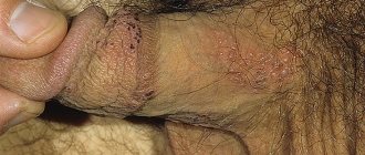
Genital herpes
develops in adolescents and young adults soon after the onset of sexual activity. Clinical manifestations of primary genital herpes are more severe in young women. The inflammatory process is characterized by ulcerative or necrotic lesions. Herpetic vesicles are localized on the skin of the external genitalia: labia majora and minora, vestibule and vaginal walls. On the mucous membranes, the blisters quickly open, leaving behind painful ulcers. The vaginal mucosa is inflamed and swollen. The cervix is involved in the inflammatory process in 70-90% of cases. HSV-1 causes urethritis much more often than HSV-2 (Fig. 5, 6).
Rice. 5. Anogenital localization of herpetic rashes in a girl.
Rice.
6. Herpes simplex rash on the buttocks of a boy. In young men, herpetic blisters are localized on the skin of the glans and shaft of the penis, foreskin, and sometimes on the scrotum, inner thighs and buttocks [7].
A rare but extremely severe manifestation of the disease is primary herpes of newborns.
Infection occurs through direct contact of the child with herpetic rashes on the mother during the passage of the birth canal. The risk of infection of a child from a mother with a primary herpes infection is 50% [10, 11]. Primary herpes of newborns is a classic systemic viral disease, often fatal. An unfavorable prognosis is associated with HSV damage to vital internal organs (central nervous system, lungs, etc.). Primary herpes in newborns is characterized by multiple dense vesicular-hemorrhagic rashes on the child's skin; often the mucous membrane of the oral cavity and the conjunctiva of the eyes are involved in the inflammatory process.
An extremely severe form of generalized herpes is herpetic eczema,
or Kaposi's eczema
. The peak incidence in young children occurs between the ages of 8 months and 1.5 years [12]. In children, there is a sharp rise in temperature to 39-40 ° C, a violation of the general condition, and lymphadenopathy. The development of meningeal symptoms, as well as the phenomenon of generalized herpetic infection with the involvement of internal organs in the pathological process, is possible. With recurrent herpetic eczema, in contrast to the primary form, general health is slightly impaired. Rashes in the recurrent form of Kaposi's eczema are localized mainly in places of greatest damage to the skin (for example, the location of herpetic elements in atopic dermatitis on the extensor surfaces of the extremities) (Fig. 7, 8, 9, 10). Often herpetic rashes spread to the periorbital area (Fig. 11). A serious complication in this case is ophthalmoherpes (herpetic conjunctivitis, keratitis, uveitis, chorioretinitis, acute retinal necrosis), which can result in loss of vision [13, 14].
Rice. 7. Severe Kaposi's eczema in an 11-month-old child.
Rice. 8. Vesicular rashes in Kaposi's eczema with umbilical depression.
Rice. 9. Kaposi's eczema.
Rice. 10. Kaposi's eczema.
Rice.
11. Kaposi's eczema. Studies conducted in patients with herpetic eczema that developed against the background of atopic dermatitis confirmed that the greatest risk of this complication is observed in patients with an impaired IFN-γ response [15], with a high level of IgE in the blood serum and polyvalent sensitization [16] . A poor prognostic sign for the development of Kaposi's eczema is also a low level of the antimicrobial peptide cathelicidin peptide LL-37 in the blood [17].
Herpes-associated erythema multiforme exudative (HAMEE) -
a type of infectious-allergic form of exudative erythema multiforme caused by HSV. Erythema multiforme exudative (EME) of herpetic etiology usually develops in adolescents and adult patients, but is not typical for young children [18].
In 95% of patients, GAMEE is combined with orofacial (labial or nasal) localization of herpetic eruptions. The clinical picture of MEE of herpetic etiology is characterized by the appearance of a widespread polymorphic rash on the skin of the trunk and extremities on the 4-6th day from the onset of relapse of herpes simplex. In contrast to the toxic-allergic form of MEE, typical maculopapular target-shaped rashes are characterized by less pronounced exudation, solitary location, stagnant color of the rashes, and relatively small sizes - up to 4 cm in diameter (Fig. 12, 13).
Rice. 12. Residual manifestations on the lips of labial herpes in a teenager with GAMEE.
Rice. 13. Target-shaped maculopapular rash in an adolescent with GAMEE.
The duration of the disease is usually 12-20 days. In some patients, this form may recur several times a year [19].
Researchers have noted that prognostic criteria that increase the risk of developing HAMEE are the presence of the following changes in the immune status of patients with herpes simplex:
- an increase in the spontaneous production of IL-4 and IL-6 in combination with inhibition of their induced production (spontaneous/induced ratio - 1:1);
— in the humoral link there is an imbalance in the synthesis of immunoglobulins with a predominance of IgE and a decrease in IgA with an increase in the absolute content of B-lymphocytes;
- decrease in IFN-α and IFN-γ;
— no increase in NK [20, 21].
One of the hypotheses that explains the occurrence of MEE only in a small number of patients suffering from recurrent herpes is the assumption that the development of HAMEE requires hematogenous spread of HSV - viremia. Recent studies have noted that patients with HAMEE have HSV circulation in affected CD3+, CD4+ cells [22].
Another significant component of the pathogenesis of MEE of herpetic etiology is the development of autoimmune changes in the body: hypersensitivity with an immune complex component of varying severity [23].
Therapy for children suffering from HSV infection
Basic therapeutic measures should be aimed primarily at eliminating the impact of the etiological factor. Causal therapy for herpes simplex is a generally accepted approach to the treatment of acute manifestations of a viral infection and the prevention of its exacerbations and is included in all international standards and recommendations [24, 25]. The main direction of therapy is the use of highly specific antiviral drugs - acyclic nucleosides, which in their chemical structure are modified analogues of the viral DNA component. The first drug was acyclovir. To increase biological activity in the body, acyclovir undergoes three stages of phosphorylation. Acyclovir has specific inhibitory activity against viruses: Herpes simplex
Types 1 and 2,
Varicella zoster
, Epstein-Barr, cytomegalovirus and human herpes virus type 6.
The mechanism of action of acyclovir is associated with inhibition of viral DNA synthesis immediately after phosphorylation of the drug and conversion to the active form - acyclovir triphosphate. The first stage of phosphorylation takes place with the participation of virus-specific enzymes (viral thymidine kinase, which is present in cells affected by the virus). Acyclic nucleosides are most effective when warning signs and/or first symptoms of herpes infection appear. Long-term use of these drugs during the period between relapses serves preventive purposes.
In children, acyclovir is approved for use from birth. Doses of acyclovir are calculated based on 20 mg per 1 kg of body weight in 4-5 doses per day, both parenterally and in tablet form.
The modern second generation acyclic nucleoside is valacyclovir.
(L-valine ester of acyclovir).
Valacyclovir
has a higher bioavailability (54%) and half-life (10-20 hours) compared to acyclovir.
Its effectiveness, high safety profile and ease of use have been repeatedly proven in numerous studies [26, 27]. Valacyclovir
is approved for use
in children over 12 years of age
. Prescription of the drug in adolescents is especially recommended in cases of treatment and prevention of recurrent herpes simplex of various localizations, with herpetic eczema, as well as in complex therapy of GAMEE.
In adolescents, the drug valacyclovir
is prescribed at a dose of 500 mg 2 times a day for a long time (for 7-10 days). The results of treatment of a patient with Kaposi's eczema are presented in Fig. 14 and 15.
Rice. 14. Patient K., 13 years old. Kaposi's eczema that developed against the background of atopic dermatitis - before treatment.
Rice.
15. The same patient. After therapy with valacyclovir 500 mg 2 times a day for 6 days. GAMEE therapy begins with the combined use of steroid hormones and antiviral drugs [28]. All patients are prescribed infusion therapy. Glucocorticosteroids (GCS) are administered parenterally at a dose of 3-5 mg per 1 kg of body weight per day (calculated using prednisolone). The course of therapy with GCS at the indicated dose is 2-4 days, followed by withdrawal within 2-3 days. Valaciclovir
prescribed at a dose of 500 mg 2 times a day for 7-10 days. Long-term prophylactic administration of valacyclovir is recommended for patients who have undergone MEE due to recurrent herpes infection.
Atypical forms of herpes
Sometimes genital herpes is erased. These forms of the disease account for 65% of cases of the disease:
- In women,
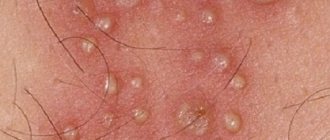
atypical herpes resembles inflammation of the vagina or vulva. There is pain, itching and swelling of the genital organs, profuse leucorrhoea and pain during sexual intercourse. External manifestations are limited to areas of redness or pinpoint rashes. - In men,
the atypical form of the disease is similar to inflammation of the head and foreskin (balanitis or balanoposthitis). A reddish rash appears on the mucous membrane of the penis, accompanied by pain and burning, which does not look like a herpetic rash. Inflammation of the prostate gland occurs, causing pain radiating to the anal area. Damage to the urethra leads to pain and burning when urinating, and the appearance of traces of blood in the urine.
There is a latent form of the disease in which there are no clinical manifestations, but despite this, the person remains a source of infection. But imaginary well-being does not last forever. With hypothermia, loss of strength, decreased immune defense, pregnancy, stress, severe concomitant diseases and other unpleasant conditions, the virus begins to multiply rapidly and the person becomes ill.
The disease is activated by concomitant sexually transmitted infections, especially ureaplasmosis. Due to the ability to rapidly “bloom” against a background of weakened immunity, herpes occurs in 90% of HIV patients. Therefore, if herpetic rashes appear, you need to be examined for other STDs.
Questions
- Which doctor treats balanoposthitis in children?
Diagnostics and medical care for balanoposthitis is provided by a pediatric urologist. - Is surgery necessary for balanoposthitis?
In 90% of cases, the disease can be managed with conservative therapy. The exception is chronic forms, when adhesions form between the head of the penis and the foreskin with the development of phimosis. In this case, circumcision is indicated. - Do folk remedies help with balanoposthitis in children?
Various decoctions of plants with an antiseptic effect can be used as baths for inflammation of the foreskin and glans penis. However, this approach is justified only after the child has been examined by a doctor. Otherwise, parents may miss the moment of active progression of the disease and allow it to become chronic and develop complications. - Is circumcision beneficial for balanoposthitis?
Sometimes, with frequent episodes of acute balanoposthitis, pediatric urologists recommend circumcision to prevent relapses of the disease. This removes the preputial sac, which is a container for the secretions of the glands in the area of the head of the penis. With the help of planned intervention, the risk of recurrent inflammation can be permanently eliminated. However, the choice in this case remains with the patient and parents.
Features of the course of genital herpes during pregnancy
The number of positive (seropositive) reactions to herpes viruses 1 and 2 in pregnant women is 50-70%. Due to the increased load on the body and decreased immune defense, herpes often relapses during this period. But only 30% of women experience the classic development of the disease. Basically, the symptoms of herpes during pregnancy are limited to the appearance of areas of redness and cracks, which women mistake for irritation.
The manifestation of a herpetic infection caused by recurrence is not dangerous for the child. The woman’s body has already built up immunity to infection by producing antibodies—substances that protect her from the virus. Some of the antibodies will pass from mother to baby, protecting him from infection.
Only a relapse that occurs immediately before childbirth is dangerous. To prevent infection of the baby and rupture of inflamed tissues, women with a herpetic rash on the genitals are advised to deliver by cesarean section.
It is much worse when the infection is primary. Herpes belongs to a group of infections, infection of which for the first time during pregnancy leads to the birth of children suffering from developmental delays and congenital defects.
Herpes tests for pregnant women: interpretation and prognosis of pathologies in the fetus
To determine the degree of risk for the baby, a woman’s blood is taken for IgM and IgG antibodies, the concentration of which determines when infection occurred. The analysis is carried out using the ELISA method (immunofluorescence), which reacts to IgM and IgG antibodies to the virus. By the presence or absence of antibodies, you can find out whether a woman is infected and when the infection occurred:
- IgM antibodies
appear after 2-3 weeks. after the onset of the disease, therefore indicating a “fresh” infection or relapse of infection. These antibodies disappear after 1-2 months. after recovery. The presence of IgM in the analysis is a bad sign. - IgG class antibodies
- appear as protection against the virus after 2 weeks. field of infection, quickly increase the titer (concentration) and persist throughout life. Their detection means that the body encountered a herpes infection and managed to cope with it.
It is bad if a fourfold increase in IgG is combined with the detection of IgM. This means that antibodies are being formed right now, i.e. the woman is sick.
Infection with the herpes virus is determined in the laboratory. It is better to undergo such an examination twice - before pregnancy and during it.
Table of interpretation of results for herpes
| IgM | IgG | What does it mean | What to do | |
| — | — | The woman is healthy (seronegative) - she has never had herpes | Before pregnancy | During pregnancy |
| In order not to become infected, you need to avoid contact with people who have manifestations of herpes, incl. on the lips. Periodic antibody monitoring is recommended | ||||
| — | + | Female carrier (seropositive) | The infection occurred a long time ago and does not pose a danger to the child, no measures are needed | |
| + | — | Recent infection | It is better to plan pregnancy after acute symptoms subside and re-examination | Consult a doctor and undergo additional diagnostics to find out if everything is okay with the baby. |
| + | + | Exacerbation of infection | It is better to plan pregnancy after acute symptoms subside | Recurrent herpes is less dangerous than primary herpes, so the likelihood of complications and intrauterine infection is only 0.02% |
Fetal pathologies in children infected with herpes during pregnancy
The most dangerous thing for a child is primary infection of the mother during pregnancy, which leads to many complications.
| Gestational age at primary infection | Possible complications |
| I trimester (up to 13 weeks) | Fading pregnancy, miscarriages, severe malformations |
| II trimester (14-27 weeks) | Child infection, internal organ defects, fetal death |
| III trimester (29-40 weeks) | Intrauterine infection, death of a child after birth, premature birth, hearing, vision, and nervous system defects of the child. Subsequently - mental retardation. |
Sources
- Vos T., Allen C., Arora M., Barber RM., Bhutta ZA., Brown A., Carter A., Casey DC., Charlson FJ., Chen AZ., Coggeshall M., Cornaby L., Dandona L ., Dicker DJ., Dilegge T., Erskine HE., Ferrari AJ., Fitzmaurice C., Fleming T., Forouzanfar MH., Fullman N., Gething PW., Goldberg EM., Graetz N., Haagsma JA., Johnson CO., Kassebaum NJ., Kawashima T., Kemmer L., Khalil IA., Kinfu Y., Kyu HH., Leung J., Liang X., Lim SS., Lopez AD., Lozano R., Marczak L ., Mensah GA., Mokdad AH., Naghavi M., Nguyen G., Nsoesie E., Olsen H., Pigott DM., Pinho C., Rankin Z., Reinig N., Salomon JA., Sandar L., Smith A., Stanaway J., Steiner C., Teeple S., Thomas BA., Troeger C., Wagner JA., Wang H., Wanga V., Whiteford HA., Zoeckler L., Abajobir AA., Abate KH ., Abbafati C., Abbas KM., Abd-Allah F., Abraham B., Abubakar I., Abu-Raddad LJ., Abu-Rmeileh NM., Ackerman IN., Adebiyi AO., Ademi Z., Adou AK ., Afanvi KA., Agardh EE., Agarwal A., Kiadaliri AA., Ahmadieh H., Ajala ON., Akinyemi RO., Akseer N., Al-Aly Z., Alam K., Alam NK., Aldhahri SF ., Alegretti M.A., Alemu ZA., Alexander LT., Alhabib S., Ali R., Alkerwi A., Alla F., Allebeck P., Al-Raddadi R., Alsharif U., Altirkawi KA., Alvis- Guzman N., Amare AT., Amberbir A., Amini H., Ammar W., Amrock SM., Andersen HH., Anderson GM., Anderson BO., Antonio CA., Aregay AF., Ärnlöv J., Artaman A ., Asayesh H., Assadi R., Atique S., Avokpaho EF., Awasthi A., Quintanilla BP., Azzopardi P., Bacha U., Badawi A., Balakrishnan K., Banerjee A., Barac A., Barker-Collo SL., Bärnighausen T., Barregard L., Barrero LH., Basu A., Bazargan-Hejazi S., Bell B., Bell ML., Bennett DA., Bensenor IM., Benzian H., Berhane A ., Bernabé E., Betsu BD., Beyene AS., Bhala N., Bhatt S., Biadgilign S., Bienhoff K., Bikbov B., Biryukov S., Bisanzio D., Bjertness E., Blore J., Borschmann R., Boufous S., Brainin M., Brazinova A., Breitborde NJ., Brown J., Buchbinder R., Buckle GC., Butt ZA., Calabria B., Campos-Nonato IR., Campuzano JC., Carabin H., Cárdenas R., Carpenter DO., Carrero JJ., Castañeda-Orjuela CA., Rivas JC., Catalá-López F., Chang JC., Chiang PP., Chibueze CE., Chisumpa VH., Choi JJ ., Chowdhury R., Christensen H., Christopher DJ., Ciobanu LG., Cirillo M., Coates MM., Colquhoun SM., Cooper C., Cortinovis M., Crump JA., Damtew SA., Dandona R., Daoud F., Dargan PI., das Neves J., Davey G., Davis AC., Leo D., Degenhardt L., Del Gobbo LC., Dellavalle RP., Deribe K., Deribew A., Derrett S., Jarlais DC., Dharmaratne SD., Dhillon PK., Diaz-Torné C., Ding EL., Driscoll TR., Duan L., Dubey M., Duncan BB., Ebrahimi H., Ellenbogen RG., Elyazar I., Endres M., Endries AY., Ermakov SP., Eshrati B., Estep K., Farid TA., Farinha CS., Faro A., Farvid MS., Farzadfar F., Feigin VL., Felson DT., Fereshtehnejad SM ., Fernandes JG., Fernandes JC., Fischer F., Fitchett JR., Foreman K., Fowkes FG., Fox J., Franklin RC., Friedman J., Frostad J., Fürst T., Futran ND., Gabbe B., Ganguly P., Gankpé FG., Gebre T., Gebrehiwot TT., Gebremedhin AT., Geleijnse JM., Gessner BD., Gibney KB., Ginawi IA., Giref AZ., Giroud M., Gishu MD ., Glaser E., Godwin WW., Gomez-Dantes H., Gona P., Goodridge A., Gopalani SV., Gotay CC., Goto A., Gouda HN., Grainger R., Greaves F., Guillemin F ., Guo Y., Gupta R., Gupta R., Gupta V., Gutiérrez RA., Haile D., Hailu AD., Hailu GB., Halasa YA., Hamadeh RR., Hamidi S., Hammami M., Hancock J., Handal AJ., Hankey GJ., Hao Y., Harb HL., Harikrishnan S., Haro JM., Havmoeller R., Hay RJ., Heredia-Pi IB., Heydarpour P., Hoek HW., Horino M., Horita N., Hosgood HD., Hoy DG., Htet AS., Huang H., Huang JJ., Huynh C., Iannarone M., Iburg KM., Innos K., Inoue M., Iyer VJ ., Jacobsen KH., Jahanmehr N., Jakovljevic MB., Javanbakht M., Jayatilleke AU., Jee SH., Jeemon P., Jensen PN., Jiang Y., Jibat T., Jimenez-Corona A., Jin Y ., Jonas JB., Kabir Z., Kalkonde Y., Kamal R., Kan H., Karch A., Karema CK., Karimkhani C., Kasaeian A., Kaul A., Kawakami N., Keiyoro PN., Kemp AH., Keren A., Kesavachandran CN., Khader YS., Khan AR., Khan EA., Khang YH., Khera S., Khoja TA., Khubchandani J., Kieling C., Kim P., Kim CI ., Kim D., Kim YJ., Kissoon N., Knibbs LD., Knudsen AK., Kokubo Y., Kolte D., Kopec JA., Kosen S., Kotsakis GA., Koul PA., Koyanagi A., Kravchenko M., Defo BK., Bicer BK., Kudom AA., Kuipers EJ., Kumar GA., Kutz M., Kwan GF., Lal A., Lalloo R., Lallukka T., Lam H., Lam JO ., Langan S.M., Larsson A., Lavados PM., Leasher J.L., Leigh J., Leung R., Levi M., Li Y., Li Y., Liang J., Liu S., Liu Y., Lloyd BK., Lo WD., Logroscino G., Looker KJ., Lotufo PA., Lunevicius R., Lyons RA., Mackay MT., Magdy M., Razek AE., Mahdavi M., Majdan M., Majeed A ., Malekzadeh R., Marcenes W., Margolis DJ., Martinez-Raga J., Masiye F., Massano J., McGarvey ST., McGrath JJ., McKee M., McMahon BJ., Meaney PA., Mehari A ., Mejia-Rodriguez F., Mekonnen AB., Melaku YA., Memiah P., Memish ZA., Mendoza W., Meretoja A., Meretoja TJ., Mhimbira FA., Miller TR., Mills EJ., Mirarefin M ., Mitchell PB., Mock CN., Mohammadi A., Mohammed S., Monasta L., Hernandez JC., Montico M., Mooney MD., Moradi-Lakeh M., Morawska L., Mueller UO., Mullany E ., Mumford JE., Murdoch ME., Nachega JB., Nagel G., Naheed A., Naldi L., Nangia V., Newton JN., Ng M., Ngalesoni FN., Nguyen QL., Nisar MI., Pete PM., Nolla JM., Norheim OF., Norman RE., Norrving B., Nunes BP., Ogbo FA., Oh IH., Ohkubo T., Olivares PR., Olusanya BO., Olusanya JO., Ortiz A ., Osman M., Ota E., Pa M., Park EK., Parsaeian M., de Azeredo Passos VM., Caicedo AJ., Patten SB., Patton GC., Pereira DM., Perez-Padilla R., Perico N., Pesudovs K., Petzold M., Phillips MR., Piel FB., Pillay JD., Pishgar F., Plass D., Platts-Mills JA., Polinder S., Pond CD., Popova S., Poulton RG., Pourmalek F., Prabhakaran D., Prasad NM., Qorbani M., Rabiee RH., Radfar A., Rafay A., Rahimi K., Rahimi-Movaghar V., Rahman M., Rahman MH., Rahman SU., Rai RK., Rajsic S., Ram U., Rao P., Refaat AH., Reitsma MB., Remuzzi G., Resnikoff S., Reynolds A., Ribeiro AL., Blancas MJ., Roba HS ., Rojas-Rueda D., Ronfani L., Roshandel G., Roth GA., Rothenbacher D., Roy A., Sagar R., Sahathevan R., Sanabria JR., Sanchez-Niño MD., Santos IS., Santos JV., Sarmiento-Suarez R., Sartorius B., Satpathy M., Savic M., Sawhney M., Schaub MP., Schmidt MI., Schneider IJ., Schöttker B., Schwebel DC., Scott JG., Seedat S., Sepanlou SG., Servan-Mori EE., Shackelford KA., Shaheen A., Shaikh MA., Sharma R., Sharma U., Shen J., Shepard DS., Sheth KN., Shibuya K., Shin MJ., Shiri R., Shiue I., Shrime MG., Sigfusdottir ID., Silva DA., Silveira DG., Singh A., Singh JA., Singh OP., Singh PK., Sivonda A., Skirbekk V ., Skogen JC., Sligar A., Sliwa K., Soljak M., Søreide K., Soriano JB., Sposato LA., Sreeramareddy CT., Stathopoulou V., Steel N., Stein DJ., Steiner TJ., Steinke S., Stovner L., Stroumpoulis K., Sunguya BF., Sur P., Swaminathan S., Sykes BL., Szoeke CE., Tabarés-Seisdedos R., Takala JS., Tandon N., Tanne D., Tavakkoli M., Taye B., Taylor HR., Ao BJ., Tedla BA., Terkawi AS., Thomson AJ., Thorne-Lyman AL., Thrift AG., Thurston GD., Tobe-Gai R., Tonelli M ., Topor-Madry R., Topouzis F., Tran BX., Dimbuene ZT., Tsilimbaris M., Tura AK., Tuzcu EM., Tyrovolas S., Ukwaja KN., Undurraga EA., Uneke CJ., Uthman OA ., van Gool CH., Varakin YY., Vasankari T., Venketasubramanian N., Verma RK., Violante FS., Vladimirov SK., Vlassov VV., Vollset SE., Wagner GR., Waller SG., Wang L. , Watkins DA., Weichenthal S., Weiderpass E., Weintraub RG., Werdecker A., Westerman R., White RA., Williams HC., Wiysonge CS., Wolfe CD., Won S., Woodbrook R., Wubshet M., Xavier D., Xu G., Yadav AK., Yan LL., Yano Y., Yaseri M., Ye P., Yebyo HG., Yip P., Yonemoto N., Yoon SJ., Younis MZ. , Yu C., Zaidi Z., Zaki ME., Zeeb H., Zhou M., Zodpey S., Zuhlke LJ., Murray CJ. Global, regional, and national incidence, prevalence, and years lived with disability for 310 diseases and injuries, 1990-2015: a systematic analysis for the Global Burden of Disease Study 2015. // Lancet - 2016 - Vol388 - N10053 - p.1545 -1602; PMID:27733282
Herpes in newborns
A child becomes infected with the pathogen from the mother in utero or by passing through an infected genital tract during childbirth. In newborns, the mucous membranes of the eyes and mouth, skin, and genitals are affected. When the pathogen enters the child’s brain, meningoencephalitis occurs, causing death or severe disability.
A generalized form is possible, in which all organs and systems of the newborn are affected. The child experiences jaundice, respiratory distress, and urinary retention. Children are restless, do not latch on to the breast, and are vomiting. Death occurs from shock, bleeding, dehydration, intoxication, and organ failure.
The likelihood of infection increases with primary infection of the mother in late pregnancy.
When to see a doctor
With a primary infection, the child almost always develops a fever. The baby has no appetite, anxiety, crying, and sleep disturbances. If one of the above symptoms is present or if there are several of them, you need to immediately show the newborn to a specialist - a pediatrician (children's doctor) or a dermatovenerologist.
If the infection develops, it can lead to complications, including death. The herpes virus in newborns should be identified as quickly as possible so that treatment can begin without delay. If there are signs of herpes in an infant, you can visit JSC “Medicine” (academician Roitberg’s clinic) in the center of Moscow. The Pediatrics Department has a staff of highly qualified specialists, new generation equipment and instruments for examination. You can make an appointment with a pediatrician or dermatovenerologist on the website or by calling +7(495)993-00-33.
Diagnosis of herpes
If genital herpes is suspected, after an external examination, scrapings from the affected areas, blood, and urine are taken for examination. This will allow you to differentiate the disease from other skin lesions that have similar symptoms.
For diagnostics the following is used:
- Cultural method - during the study, the contents of the vesicles are transferred to a growing chicken embryo; if it dies, the disease is confirmed. The method is reliable, but too time-consuming.
- PCR is a test that detects viral DNA in tissues. The method allows you to calculate the pathogen even from a small amount of viral material. Effective for diagnosing any form of disease, incl. hidden. The method can be used immediately after infection before symptoms of the disease appear. PCR not only detects the virus, but also determines its type.
- Methods aimed at determining antigens and antibodies to the pathogen: ELISA - immunofluorescence diagnostics, CFR (complement fixation reaction), various types of agglutination reactions. With their help, you can identify the type of pathogen, determine its quantity and duration of infection.
A complete diagnosis of herpes virus infection allows you to make the correct diagnosis in order to prescribe treatment.
Diagnosis of balanoposthitis in children
Diagnosis of balanoposthitis in children is based on an analysis of the patient’s complaints, collection of anamnesis and examination of the pathological area.
In 95% of cases, the urologist correctly determines the cause of the characteristic symptoms at the first consultation. To confirm the diagnosis and exclude other possible diseases, the doctor prescribes a comprehensive examination:
- general blood analysis;
- blood chemistry;
- general urine analysis;
- bacteriological examination of discharge from the preputial sac;
- blood test for infections (HIV, syphilis, viral hepatitis B, C).
If necessary, the child is examined by related specialists (pediatrician, ophthalmologist, gastroenterologist) to exclude somatic diseases.
Complications of herpes
The worst thing is that a herpes infection brings not only unpleasant symptoms. This virus causes many terrible complications. The most obvious ones are:
- Irritation and dryness of the skin and mucous membranes of the genital organs;
- Cracks in the genitals caused by metabolic disorders in tissues caused by herpes;
- Adhesive and cicatricial deformities of the labia, vaginal opening, perineum, urethra and rectum. The complication arises due to the specific feature of herpetic rashes appearing on the same areas of the skin and mucous membranes
- Cervical erosion leading to cancer of the reproductive organs.
- Lacerations during childbirth caused by poor distensibility of inflamed perineal tissue.
- Infection of ulcers due to microbes entering tissues not protected by the mucous membrane;
- cicatricial deformities of the penis, provoked by constant inflammatory rashes and ulcerations.
- Chronic herpetic lesions of the genitourinary area, spreading to the urethra, bladder and kidneys.
- Herpetic proctitis is inflammation of the rectum.
- Erectile dysfunction and male and female infertility.
- Neurological symptoms caused by a viral infection of the nervous system. Patients complain of pain in the sacrum, lower back, and groin. With recurrent lesions on the inner surface of the thighs, hypersensitivity of the skin occurs, manifested by a burning sensation, crawling “goosebumps”, tingling.
In severe cases, with a pronounced weakening of the immune system, viremia occurs - viral infection of the blood leading to damage to the brain, liver, lungs, adrenal glands, and intestines. This condition, even with adequate treatment, is often fatal.
Classification
Herpes infection is classified according to the signs of predominant tissue damage, the nature of the disease and the type of rash.
There are 8 types of herpesvirus that affect children, but 7 and 8 have not been studied enough:
| Type | Features of manifestation |
| HSV type 1 (herpes labialis) | Herpes manifests itself on the lips of a child in the form of blistering rashes. May provoke the development of herpetic stomatitis and encephalitis. |
| Type 2 | It affects the genitals with a rash (genital herpes). |
| Type 3 | The causative agent of chickenpox and recurrent herpes zoster. |
| Type 4 (Epstein-Barr virus) | Causes the development of malignant lymphoma and infectious mononucleosis - an acute viral pathology accompanied by fever, damage to the respiratory system, spleen, liver, lymph nodes and blood. |
| Type 5 (cetomegalovirus) | Affects the respiratory, nervous and urinary systems, intestines, brain and eyes. |
| Type 6 | It mainly affects young children, manifesting itself as a viral exanthema - a rash similar to rubella. |
According to the development of the disease, herpes is divided into: acquired, primary, recurrent and congenital.
According to the form of infectious prevalence, 4 stages of development are distinguished:
- latent - carriage without the manifestation of specific symptoms;
- localized - with a single lesion;
- widespread – with the presence of 2 or more lesions;
- generalized - disseminated and visceral (spreading to internal organs).
Treatment of genital herpes
It is difficult to cure such a dangerous disease. Treatment of genital and labial herpes is long and complex. Therapy includes:
- Prescription of antiviral agents that slow down the reproduction (replication) of viruses. To maintain the effectiveness of treatment during long-term therapy, it is necessary to constantly change the prescribed medications.
- The use of drugs based on interferons - proteins released in response to viral invasion. Scientists have found that a lack of this substance provokes exacerbations of the disease.
- The use of local antiviral ointments, gels and sprays that kill viruses that have penetrated the skin and mucous membranes.
- Prescribing symptomatic medications that combat the general symptoms of the disease - reducing pain, swelling, inflammation, lowering temperature, normalizing sleep.
- Local procedures - baths and irrigations that relieve pain and prevent the penetration of secondary infections.
- Taking immunoglobulins, immunomodulators and stimulators of cellular immunity, which improve the functioning of the immune system, activate the production of antiviral antibodies and prevent relapses.
Patients suffering from genital herpes are advised to avoid wearing synthetic underwear.
Prevention of infection consists of a thoughtful choice of sexual partners, compliance with hygiene rules, periodic examination by a gynecologist and the use of emergency measures at the first symptoms of the disease.
Where to get tested and cure herpes in St. Petersburg, prices
Doctors at the modern Diana Clinic treat herpes infections in St. Petersburg. Here you can take express tests for all infections - smears, blood, etc. Tests are performed using the best methods - PCR, ELISA, etc. The cost of visiting a doctor based on test results is 500 rubles. The price of tests depends on the type of examination. For example, the cost of taking blood from a vein is only 170 rubles.
Prices
| Name of service (price list incomplete) | Price |
| Appointment (examination, consultation) with a dermatovenerologist, primary, therapeutic and diagnostic, outpatient | 1950 rub. |
| Consultation (interpretation) with analyzes from third parties | 2250 rub. |
| Prescription of treatment regimen (for up to 1 month) | 1800 rub. |
| Prescription of treatment regimen (for a period of 1 month) | 2700 rub. |
| Consultation with a candidate of medical sciences | 2500 rub. |
| Dermatoscopy 1 element | 700 rub. |
| Setting up functional tests | 190 rub. |
| Excision/removal of cutaneous/subcutaneous elements and formations (1 element) | 2550 rub. |
| Removal of milia of one unit using electrocoagulation | 350 rub. |