Dermatovenerologist
Khasanova
Alina Rashidovna
9 years experience
Make an appointment
Fibroids are a group of benign neoplasms that affect various tissues of the human body. Tumors are formed from collagen fibers with varying densities and elasticity. Foci of pathology can be located on the skin, bones and walls of internal organs of children and adults. Oncologists consider fibromatosis as a precancerous condition.
General information
Single or multiple fibromas can affect the lungs, mammary glands, liver, skin, oral mucosa, etc. The proliferation of connective tissue occurs under the influence of various factors: unfavorable environmental conditions, severe injuries, bacterial or viral infections. When making a diagnosis and developing a treatment strategy for a patient, dermatologists, oncologists and surgeons take into account laboratory data. Information about the morphology of the tumor allows doctors to assess the risk of its malignant degeneration.
Removal of breast fibroadenoma
Preparation for removal of breast fibroadenoma includes a consultation with a mammologist and undergoing diagnostic tests. Methods for diagnosing breast fibroadenoma include:
- breast biopsy;
- ECG of the heart and consultation with a therapist;
- general and biochemical blood tests;
- blood test for pathogens of hepatitis B and C, HIV and syphilis;
- Ultrasound of the mammary glands.
After diagnosing fibroadenoma, the doctor prescribes surgical treatment. At the initial stage of fibroadenoma, sectoral resection (partial removal) is performed. Removal of breast fibroids is performed under local anesthesia. After removing the formation, the surgeon applies a cosmetic suture using self-absorbing threads.
For large breast fibroadenoma, total resection of the tumor and the affected gland is performed. Typically, the operation is performed under local anesthesia and lasts from one to two hours.
Reasons for the development of pathology
The causes of fibroids are varied. Doctors identify several factors that contribute to the formation of connective tissue tumors in patients’ bodies:
- systematic use of alcohol and tobacco;
- repeated injuries to the skin, muscles, bones;
- unfavorable environmental conditions in the region of residence;
- the predominance of fatty, spicy and sweet foods in the diet of children and adults;
- viral and bacterial infections;
- immunosuppression due to the use of immunosuppressants.
Doctors often diagnose fibromatosis in people working in chemical production. Benign skin tumors can develop under the influence of excessive sun exposure. Damage to internal organs is often a consequence of endocrine disorders.
Why does fibroma appear?
Fibroids can develop as a result of a hereditary predisposition. In addition, poor diet, smoking and excessive consumption of alcoholic beverages lead to the development of fibroids.
The causes of fibroids also include:
- hormonal imbalance;
- overweight;
- metabolic disease;
- frequent skin damage (scratches, bruises, cuts);
- endocrine diseases (diabetes mellitus, thyroid disease).
Abortions in late pregnancy, inflammatory diseases of the genitourinary system and ovarian dysfunction can lead to the development of uterine fibroids. Breast fibroids can be triggered by prolonged breastfeeding or refusal of lactation.
Kinds
Doctors use several typologies of fibroids. The most common classification takes into account the localization of the tumor focus: the pathological process can affect the skin (for example, desmoid fibromatosis), internal organs (fibrosis of the uterus, mammary gland, lungs), mucous membranes of body cavities (gingival fibroma), bone tissue (non-osteogenic fibromatosis).
Desmoid tumors form on the skin of the back, limbs and chest. Externally, the tumor looks like a growth with a bumpy surface up to 50 millimeters in diameter. About 5% of patients experience malignant transformation of tumors - a small fibroma on the skin of the shoulder or thigh can lead to the development of skin cancer.
Fibrous lesions of the uterus and other internal organs of the human body can affect various tissues: muscle, epithelial, glandular. Often the diseases are asymptomatic; the focus of the pathology is discovered by chance - during ultrasound, magnetic resonance imaging or radiography.
Odontogenic neoplasms form in the oral cavity. The pathological process develops slowly, the patient does not experience pain. If left untreated, the tumor can reach significant sizes and lead to jaw deformation.
Non-osteogenic tumors form in the femur and provoke the destruction of the tubular bones of the skeleton. In an advanced stage, the disease causes a pathological bone fracture. Rarely (3–5% of clinically diagnosed cases) the tumor decreases in size without surgical intervention. Other types of bone tumors grow aggressively. Thus, desmoplastic fibroma can double in volume in 2–3 weeks.
Predictions of why fibroids are dangerous
Dermatofibroma does not have serious complications.
Complications of plantar fibroids usually occur as a result of surgical interventions. These include flattening of the arch of the foot, postoperative nerve entrapment, and postoperative growth of larger and recurrent fibromas. Article sources:
- Desmoid fibroma. Staged, organ-preserving surgical treatment. Khvastunov R.A., Mozgovoy P.V., Lukovskova A.A., Yusifova A.A. Volgograd scientific and medical journal No. 2, 2022. p. 54-57
- https://www.ncbi.nlm.nih.gov/pmc/articles/PMC2927520/ Central odontogenic fibroma: a case report with long-term follow-up. Marco T Brazão-Silva, Alexandre V Fernandes, Antônio F Durighetto-Júnior, Sérgio V Cardoso, Adriano M Loyolacorresponding
- https://www.ncbi.nlm.nih.gov/pmc/articles/PMC3351722/ Literature survey on epidemiology and pathology of cardiac fibroma. SuguruTorimitsu, Tetsuo Nemoto, Megumi Wakayama, Yoichiro Okubo, Tomoyuki Yokose, Kanako Kitahara, Tsukasa Ozawa, Haruo Nakayama, Minoru Shinozaki, Daisuke Sasai, Takao Ishiwatari, Kensuke Takuma, Kazutoshi Shibuya
The information in this article is provided for reference purposes and does not replace advice from a qualified professional. Don't self-medicate! At the first signs of illness, you should consult a doctor.
Symptoms
The development of the tumor process is accompanied by moderate pain and a feeling of fullness. In 55–60% of cases, the pathology is asymptomatic. The likelihood of developing specific signs of fibromatosis depends on the type and location of the tumor. Fibromatosis of cartilaginous tissue is characterized by increasing symptoms - from mild pain when performing movements to complete loss of mobility of the upper or lower limb.
Specific symptoms develop when fibroma affects the patient’s internal organs. Thus, fibrosis of the uterus can cause bleeding that is not related to a girl’s menstrual cycle. Fibrous lesions of the lungs provoke difficulty breathing and shortness of breath in a child or adult after short physical activity.
Recovery after fibroid removal
The recovery period after fibroid removal is usually up to seven to ten days. After removal of breast fibroids, you must visit a doctor or nurse every day or every other day to apply dressings. In addition, you should minimize physical activity, wear bandages, and eat a healthy and balanced diet. After removal of fibroids of the uterus and ovaries, it is also necessary to wear a postoperative bandage and lead a healthy lifestyle (eat properly, avoid overwork, stress, and maintain a sleep schedule).
Diagnostic measures
Diagnosis of fibroids is performed by doctors of various specializations - dermatologists, oncologists, surgeons, gynecologists, etc. Methods for confirming the primary diagnosis are determined by the location of the benign neoplasm. At the first stage of the examination, patients receive a referral for radiography. Computed tomography allows you to determine the exact size of the tumor and identify signs of its invasion into adjacent organs.
Fibrous formations in bone and cartilage tissue are detected during scintigraphy. If necessary, the doctor can take a biopsy sample for laboratory tests. Microscopy of the obtained biomaterials will allow doctors to assess the morphological structure of tumor cells and the likelihood of their malignant degeneration.
Benign neoplasms of the female reproductive system are often detected during ultrasound examinations. The focus of the pathological process has less echogenicity compared to adjacent tissues.
Discussion
MFD/NOF are localized in the metaphyses of long tubular bones and, as the skeletal bones grow, they can shift slightly towards the diaphysis. It is well known that the metaphysis and growth plate areas are the sites of the most intense bone growth in children and adolescents. Consequently, MFD/NOF as a local “osteogenesis defect” occurs in this zone. Some authors believe that the trigger for the development of MFD/NOF can be a minor injury with subperiosteal hemorrhage [5]. Most lesions occur around the knee joint, but cases have also been described in the pelvic bones, vertebrae, clavicle [8], even in the lower jaw [1, 2]. However, when examining tumors of the lower jaw, the first step is to carry out a differential diagnosis with giant cell reparative granuloma.
A case of a combination of NOF with osteosarcoma of the distal metaphysis of the femur in a 15-year-old girl is described [10].
On X-ray examination, the MFD appears as a round or oval lytic formation of the cortical layer (or subperiosteal), often slightly elongated parallel to the axis of the bone. The boundaries are clear, as a rule, a small zone of osteosclerosis around is clearly visible (Fig. 3). As the MFD grows, it begins to spread in the medullary direction. Therefore, NOF always looks eccentric, but also with clear boundaries and a zone of perifocal osteosclerotic changes (Fig. 4). With large lesions that occupy more than a third to half of the diameter of the bone, lobulation can often be detected due to sclerotic septa, in which histological examination reveals fibrous tissue with foci of reactive osteogenesis. In these cases, the disease can manifest itself as a pathological fracture. In rare cases, the contours of the NOF may extend beyond the anatomical limits of the bone. There is no periosteal reaction in most cases.
Rice. 3. Well-defined cortical lesion in the area of the distal metaphysis of the femur. Perifocal sclerotic changes are not pronounced. Stage A according to Ritschl.
Rice. 4. Well-defined lesion of the distal metaphysis of the tibia with pronounced perifocal sclerosis, more pronounced in the area of the diaphyseal pole of the formation. Stage D according to Ritschl.
P. Ritschl et al. [6] described the process of evolution of MFD and NOF from their onset to complete sclerosis, based on radiological data and identified 4 stages: A, B, C and D. Stage A: a small cortical round or oval eccentric lesion located near the growth zone (close to pineal gland). The area of perifocal sclerosis is practically absent. Stage B: the lesion distances itself from the epiphysis, acquires irregular polycystic outlines, and a thin sclerotic rim appears along the periphery. Stage C: sclerotic changes become pronounced, and begin, as a rule, from the diaphyseal pole of the formation. Stage D: the formation is completely replaced by a homogeneous zone of sclerotic changes. The MFD/NOF is then replaced by normal bone tissue and is not detected radiographically. Active growth of MFD and NOF occurs at stages A and B; when formation was detected at stages B and C, no increase in size was observed in dynamics.
The X-ray picture of NOF/MFD is so typical that in the vast majority of cases the diagnosis can be made only on the basis of this study. Since NOF/MFD belong to the “leave me alone lesion” group of bone lesions, this serves as a basis for observation and periodic radiological monitoring. Due to the fact that NOF/MFD can regress spontaneously during the growth and maturation of skeletal bones, active surgical tactics are not always justified. Surgical treatment methods are used only for large volumes of formation, when there is a threat of a pathological fracture or when the NOF manifests itself as a clinically pathological fracture.
According to the Enneking staging system, benign tumors of NOF/MFD in most cases have stage I (G0T0M0), in rare cases, when they spread beyond the normal anatomical boundaries of the bone - stage II.
On macroscopic examination, the lesion is clearly circumscribed and yellowish-gray in color (Fig. 5). Cystic changes and areas of hemorrhage may occur. Areas of necrosis may be identified after a pathological fracture.
Rice. 5. On macroscopic examination, the lesion is clearly limited, yellowish-gray in color.
On histological examination, the formation is well circumscribed and consists of fibroblast-like spindle-shaped cells forming randomly oriented bundles and curtain-shaped structures (Fig. 6). The nuclei are oval or elongated; small nucleoli are not detected in all nuclei. Mitotic activity is not reliably determined or only a few mitotic figures are detected. One of the consistent morphological features is the identification of clusters of large histiocytes or xanthoma cells with abundant clear cytoplasm. It is almost always possible to detect small scattered granules of hemosiderin, which are better visible with a special histochemical stain for iron. Clusters of giant multinucleated osteoclast-like cells can often be identified (Fig. 7). Cystic changes of the type of secondary aneurysmal bone cyst are often found. With a large volume of formation, zones of reactive osteogenesis can be identified in the form of coarse fibrous bone tissue (woven bone), zones of hypocellular mature connective tissue. If NOF is complicated by a pathological fracture, areas of necrotic changes are often identified.
Rice. 6. Typical histological picture of MFD/NOF. Spindle cell component, xanthoma cell clusters and hemosiderin deposits. Hematoxylin and eosin staining, ×400.
Rice. 7. MFD/NOF are always clearly limited, in places they can contain a large number of giant multinucleated osteoclast-like cells. Hematoxylin and eosin staining, ×300.
Differential diagnosis should include fibrous dysplasia, aneurysmal bone cyst, giant cell tumor, chondromyxoid fibroma, and desmoplastic fibroma.
Fibrous dysplasia by definition is a benign medullary fibro-osseous lesion in which one or more bones are involved in the pathological process. It is more often found in the bones of the skull and femurs, less often in the ribs, pelvic bones, etc. Radiologically, it looks like a well-defined lesion, but unlike MFD, it is not localized cortically. NOF is always an eccentric formation, spreading medullarily from the cortical layer. On histological examination, fibrous dysplasia consists of two components: fibrous (in the form of relatively hypocellular mature connective tissue with elongated fibroblasts) and bone (in the form of bizarre bony trabeculae, usually without perifocal osteoblastic rimming). In NOF there is no bone component; areas of reactive osteogenesis can only be determined, especially in the presence of a pathological fracture.
A solid variant of aneurysmal bone cyst (ABC) is rare. Most often localized in long tubular bones. On X-ray examination, the ACC, as a rule, is well limited in the form of a lytic focus, often extends beyond the normal anatomical limits of the bone, and sometimes a clear border with the soft tissue component may be lost. Histological examination does not show a pronounced curtainform pattern as in MFD/NOF; multiple foci of reactive osteogenesis (woven bone) are detected. Primary ACC exhibits t(16;17) with UPS6 gene rearrangement.
Giant cell tumor (GCT) practically does not occur in children and adolescents and begins to be registered at the end of the second - beginning of the third decade of life, while MFD/NOF occur in children and adolescents. Localization is epiphyseal-metaphyseal, long tubular bones are affected. In approximately half of the cases, it is diagnosed in the bones around the knee joint: the distal part of the femur, the proximal metaphysis of the tibia and fibula. In histological examination, the mononuclear component is often represented by oval-shaped neoplastic cells; the nuclei of mononuclear cells in GCTs are identical in morphology to the nuclei of multinucleated osteoclast-like cells [9].
Chondromyxoid fibroma (CF) is a benign tumor consisting of elongated myofibroblast-like and stellate cells with a chondroid and myxoid matrix. Can be detected at any age. It is more common in long tubular bones in the second and third decades of life, more often among male patients. The most significant clinical symptom is pain (from several months to several years). X-ray is detected in the metaphyses of long tubular bones as a sharply limited eccentric focus, often without an accentuated zone of sclerotic changes in the peripheral parts. In MFD/NOF, the zone of sclerosis along the periphery is always well defined. Histologically, CP consists of lobules with a relatively hypocellular center and increasing cellularity towards the periphery. The cells lie in a chondroid and/or myxoid intercellular matrix. Mitotic activity is not reliably determined or is low.
Desmoplastic fibroma (DF) is a rare benign, locally aggressive bone tumor composed of spindle cells and collagenized matrix. It is an analogue of deep soft tissue fibromatosis. Occurs at any age, including children and adolescents. Any bone can be involved in the pathological process, but more often it occurs in the lower jaw, metaepiphyseal zones of the femur, tibia, radius, humerus, as well as in the pelvic bones. During X-ray examination, as a rule, the contours of the pathological focus extend beyond the anatomical boundaries of the bone. DF is well limited, often has a “honeycomb” structure, sometimes soft tissues are involved in the pathological process, in some cases a periosteal reaction is observed, which does not happen with MFD/NOF. Histologically, the DF consists of bundles of spindle-shaped fibroblast-like cells with a pronounced collagenized intercellular matrix. Immunohistochemical examination often reveals a nuclear reaction with an antibody to β-catenin.
Treatment
The treatment strategy for fibroids is determined by the doctor after the patient undergoes diagnostic procedures. For small tumor sizes and localization of the pathological focus on the skin, a conservative approach is used. Dermatofibromas can dissolve with steroid injections. Surgery is performed in cases of high risk of tumor malignancy.
Fibroids are removed in several ways. Small tumors on the skin are excised using a laser. The advantages of this method are minimal rehabilitation time and high efficiency.
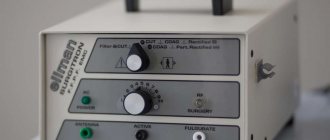
Removing fibrous formations using an electric knife minimizes the risk of bleeding (small vessels are sealed under the influence of current). As an alternative method for removing overgrown collagen fibers, the surgeon may choose a radio wave knife. The use of this equipment reduces trauma to the patient’s healthy tissue.
Fibroma: origin and essence of the disease
The causes of dermatofibroma and other forms of this type of tumor are not known.
Some researchers believe that fibroids can form as a localized tissue reaction after minor trauma. Sometimes fibroids can have a genetic component, especially in people of Northern European descent. Some medications, including beta blockers, have been reported to cause changes in fibrous tissue. In addition, some fibroids may be influenced by hormonal imbalances or pathologies of endocrine organs, including problems with the thyroid and pancreas. Hyperhidrosis (increased sweating), inflammatory processes on the skin, especially chronic ones, as well as the influence of prolonged UV radiation, poor nutrition and bad habits can have a certain impact.
Prognosis and prevention
When patients seek medical help in a timely manner, doctors can formulate a favorable prognosis in 75–80% of cases. The likelihood of a complete recovery of a child or adult is reduced when fibromatosis is advanced or secondary pathologies are attached to the disease.
Preventive measures are simple - patients are advised to avoid repetitive trauma to the skin, soft tissue and bones. Persons working in hazardous industries must undergo regular preventive examinations. Children and adults should eat a healthy diet and include adequate amounts of vitamins and minerals.
Removal of uterine and ovarian fibroids
Removal of uterine and ovarian fibroids is carried out after their diagnosis. A gynecologist or surgeon conducts an examination, studies the medical history and, if necessary, prescribes additional tests (ultrasound of the pelvic organs, laboratory tests).
After diagnosis, the gynecologist prescribes an individual treatment regimen for fibroids of the uterus and ovary, which includes drug therapy (anti-inflammatory and hormonal drugs) and surgical removal of fibroids. Methods for surgical removal of uterine and ovarian fibroids include:
- hysteroscopic surgery for fibroids of the uterus and ovary - performed through the vagina using a long thin probe with a built-in backlit video camera, the image from which is transmitted to the monitor;
- laparoscopy of fibroids of the uterus and ovary - a minimally invasive intervention during which the fibroid is removed through several small punctures in the lower abdomen;
- open surgery - access to the uterus is provided through an incision in the lower abdomen along a natural fold of skin; it is performed in the presence of large fibroids.
The duration of the operation to remove fibroids of the uterus and ovary is 1-2 hours. The evening before the operation and on the day of the intervention, you should not eat, and two hours before the start of the operation you should not drink.
Clinical picture of the disease
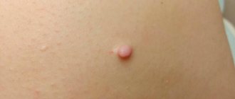
Fibroids are painless, except when they are injured by awkward movements. The color of the tumor can vary from flesh-colored to dark blue. It depends on how blood circulation occurs inside the tumor. Sometimes, when the fibroid stalk is twisted, the nutrition of the tumor is disrupted and it dies on its own.
The surface of the growth is uneven. There are additional formations and dents. Hard varieties have clear edges, soft ones are looser and shapeless.
Possible complications
Fibroma turns into a malignant form, therefore low-risk. The exception is location in the friction zone of clothing, or traumatically dangerous places, since the likelihood of violating the integrity of the cover and twisting of the growth can lead to swelling, inflammation, bleeding and the addition of a secondary infection.
After surgery, in 97% of cases, the problem is completely eliminated without the risk of relapse. But, if the wound is not properly cared for, infection and blood poisoning can develop. Therefore, you should strictly adhere to all the recommendations of our specialists.
Danger of illness
Fibroids on the inner thigh are dangerous if the size of the tumor increases rapidly. This indicates the absence of a capsule that inhibits tumor growth. If not removed in time, the neoplasm grows into the surrounding tissue and reaches a volume of several cubic centimeters.
Fibroids located in the groin or folds of skin are subject to friction, become damaged and may bleed, causing pain.
The main problem with having fibroids in people with hormonal disorders is the tendency to grow and increase in size.
Doctors surgeons
Dzhadzhiev Andrey Borisovich Surgeon
Cost of admission 1500
₽
Make an appointment
Marynich Alexander Alexandrovich Vascular surgeon, phlebologist, angiologist
Cost of admission 1500
₽
Make an appointment
Myshlyaev Anton Vsevolodovich Surgeon coloproctologist
Cost of admission 2000
₽
Make an appointment