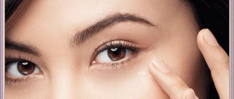
White dots (pimples, spots, bumps) that appear on the face and in the eye area are called millet due to their external resemblance to millet grains. The scientific name for this skin problem is milia.
Milia appear as small white balls under the surface of the skin. They are painless, have a dense consistency and do not change their size for quite a long time.
Such formations do not pose a danger to the body, are not a sign of any disease, etc. This is just one of the types of acne that can be easily treated by a cosmetologist.
Causes of milia
The appearance of whiteheads is caused by increased activity of the sebaceous glands and blockage of the hair follicles. Therefore, the risk of milia formation in those with oily skin type is much higher (both women and men). As a rule, such acne occurs during puberty, especially if there is poor hygiene.
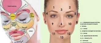
Such “troubles” can form not only in the eye area, but also on the cheekbones, under the lower lip, on the forehead and near the nose - in especially “fat” places on our face.
Some cosmetologists also tend to attribute excessive exposure to UV rays and damage to the upper layers of the skin as the causes of whiteheads.
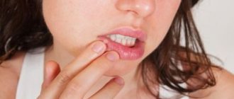
Ulcers caused by oral diseases
White sores in the mouth can appear due to diseases of the oral cavity. The most common cause is stomatitis. It comes in several types:
- aphthous;
- herpetic;
- bacterial;
- fungal.
| White ulcers in adults and children occur with aphthous and herpetic stomatitis. This disease can appear due to stress, untreated caries, lack of vitamin C, or overexertion. It is better not to self-medicate, but to immediately consult a doctor. Recommendations, in addition to medications, may include treatment of caries, a balanced diet, stress reduction, etc. |
| There are two separate forms of stomatitis - Bednar's aphthae and Setton's aphthae. The former develop only in a child due to injury to the mucous membrane or poor oral hygiene. Such rashes are also called erosions. Setton's aphthae is a more complex and painful phenomenon. It begins with the appearance of compactions, which then develop into painful ulcers. They most often form on the inside of the cheeks, in the corners of the lips and on the sides of the tongue. |
| Another cause of white sores is gingivitis. In smokers, such rashes are much more common due to the effects of nicotine on the mucous membranes. Those who have poor oral hygiene, reduced immunity and hormonal imbalances are also at risk of encountering gingivitis. If left untreated, it can progress to periodontitis: when the tissue that supports the tooth is affected. And this is a direct path to tooth loss. |
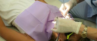
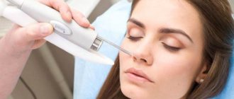
Whitehead Removal
To remove such a skin problem, a specialist may offer several cleaning procedures to choose from:
- Laser coagulation (painless treatment of problem areas using a laser).
- Mechanical removal (violation of the integrity of the eel using a sterile needle, squeezing out the contents).
- Electrocoagulation (exposure to problem areas with alternating current using an electrocoagulator needle).
Treatment of such “problems” by a cosmetologist guarantees their rapid disappearance and the absence of relapses if hygiene rules are followed.
Rules for choosing an effective lightening product for the face
- Buy only products from professional cosmetic brands . Cheap drugs and widely available products will not contain the necessary substances/concentrations for effective depigmentation.
(the advice is banal, but it will allow you to avoid unnecessary waste of money and time)
- Use preparations with a complex composition of lightening ingredients .
There are many mechanisms that bring pigment cells into a state of hyperproduction of the melanin pigment, and it is also necessary to block these processes in a comprehensive manner. Look for products with a composition of brightening ingredients that work at different points of application and enhance each other's effects.
- Remember that your daily care should at least include the use of a lightening cream + sunscreen !
Free consultations are available from leading cosmetologists of the Russian representative office of DermaQuest and Circadia.
Is it possible to get rid of milia on your own?
Trying to get rid of such acne at home is definitely not worth it. By squeezing or piercing them, it is difficult to achieve significant results. But getting an infection is very easy. In addition, unskilled punctures often leave scars and cicatrices on the skin. Therefore, after accurately comparing the costs of a cosmetologist and the potential costs of medications for inflammation and scars, it is better to give preference to a visit to a specialist.
All you can do at home is keep your skin clean and learn how to choose the right cosmetics.
Types of skin pigmentation
PHOTOAGING
The most common type of stain. They gain color after summer, lighten in winter, but do not completely disappear from the skin. You could say they often stay for life.
Ultraviolet radiation activates the synthesis of melanocortin, a pituitary hormone that stimulates melanogenesis in melanocytes and keratinocytes.
During photoaging, melanin is present in the form of granules in melanocytes, keratinocytes, dermal macrophages and in the intercellular space.
POST-INFLAMMATORY PIGMENTATION
Occurs in areas with long-term chronic inflammation: post-acne pigmentation, pigment spots after traumatic procedures, laser therapy and peelings.
Typically, this type of spots disappears once the inflammatory process resolves.
PROTECTING SKIN FROM LIGHT
Trigger factors - ultraviolet, blue light emitted by electronic gadgets (TVs, computers, smartphones...)
Since melanin serves as a shield from solar radiation and other types of rays, it is important to remember - in the fight against age spots of any origin, the first rule is to start using a protective cream with mineral SPF (zinc oxide and titanium dioxide) containing BlueLight filters for blue light and to infrared rays.
Prevention of milia
To prevent the occurrence of whiteheads on the face, you need to learn how to monitor the health of your skin, clean it properly, avoiding contamination, and buy suitable cosmetics. For example, if you have oily skin, it is better to refrain from using greasy, poorly absorbed creams. In this case, it is worth using gels.
To prevent the appearance of milia, it is also necessary to exfoliate the skin of the face and eye area. Apply cleansing masks, such as paraffin masks, which are good for eyelid skin.
And you definitely need to adjust your diet, minimizing fatty, sweet, flour, and fried foods.
In the medical department, everyone can undergo examination using the most modern diagnostic equipment, and based on the results, receive advice from a highly qualified specialist. We are open seven days a week and work daily from 9 a.m. to 9 p.m. Our specialists will help identify the cause of vision loss and provide competent treatment for identified pathologies.
You can find out the cost of a particular procedure or make an appointment at the Moscow Eye Clinic by calling 8 and 8 (499) 322-36-36 (daily from 9:00 to 21:00) or using the online registration form .
Symptoms
Seborrheic dermatitis is manifested by redness, peeling with “oily” scales with a certain localization of foci.
In children - isolated on the scalp (in the frontal area it is called the “baby cap” or “milk crust”), behind the ears and in the external auditory canal, on the face (in the area of the eyebrows, wings of the nose), on the back of the neck (along the edge of hair growth), in areas of skin friction (in folds), under diapers.
Sometimes seborrheic dermatitis in children is widespread. These are rashes in the form of spots with greasy crusts, merging into larger areas. In some cases it is accompanied by itching.
In adults, seborrheic dermatitis is presented in the scalp area, on the face: in the area of the eyebrows, behind the ears, inside the ears, on the wings of the nose and in the areas of the cheeks adjacent to the nose.
It can manifest itself as separate spots with peeling, total damage to the scalp, damage to the eyelids in the form of blepharitis, damage to the chest wall in the form of individual nodules and spots with crusts on the surface, inflammation of the hair follicles on the skin of the upper half of the body (ostiofolliculitis).
There are various causes of hypopigmented spots on a child's face, ranging from conditions that resolve on their own within a few weeks to conditions that can last a lifetime.
Causes of white spots on a child's face
White spots can be caused by the following skin conditions:
Milia
Milia are also called milk spots. These tiny white bumps usually appear on the face and rarely on the upper torso or limbs. Although milia can be seen at any age, it is common in newborns.
Causes and diagnosis of milia
Milia occurs due to entrapment of keratin (skin scales) beneath the surface of the skin. If there is no improvement after a couple of weeks, you should consult a doctor. Diagnosis is made by visual examination and no tests are required.
Prevention and treatment of milia
There is no cure for milia and it often goes away within a few weeks or months. There is no need to treat milia in children.
How can you reduce milia at home?
- Wash your child's face daily with mild soap and water.
- Pat your face dry after washing.
- Be careful not to scratch the milia to prevent skin damage and infection.
- Do not apply lotions or oils to your baby's face.
Note: Some may confuse baby pimples or Epstein pearls with milia. However, acne can cause red bumps and pustules on the face. Epstein's pearls are small, white-yellow cysts that often resemble milia, but they appear on the roof of the mouth and gums.
Pityriasis alba (lichen alba)
Pityriasis alba is a self-limiting skin condition that causes dry, thin, scaly and pale patches on the face. It is common in children and adolescents and is considered a type of eczema or dermatitis.
The name of this condition comes from the characteristic appearance of the skin.
Causes of white ptyriasis
The causes of white ptyriasis have not yet been clarified. However, atopic dermatitis and dry skin often coexist with it. Exposure to the sun can make it more visible by tanning the surrounding skin.
Suspected causes such as under- or over-bathing, low serum copper levels, ultraviolet radiation, or Malassezia yeast have not yet been proven to cause hypopigmentation.
Symptoms and signs of ptyriasis alba
One or more white spots, varying from 0.5 to 5 cm in diameter, are a characteristic sign of pityriasis alba. It may also cause mild itching in some children.
Diagnosis of pityriasis versicolor
The condition can be diagnosed through a physical examination by a doctor. A skin biopsy may show mild spongiotic (fluid accumulation between cells) dermatitis with decreased melanin pigment. A skin scraping may also be taken to rule out a yeast infection.
Treatment of white lichen
Treatment is not recommended in asymptomatic cases. Moisturizers are helpful for dry skin, and mild hydrocortisone (topical steroid) creams can help reduce itching and redness in the area.
Tacrolimus ointment, pimecrolimus cream, and calcineurin inhibitors have been shown to be effective in the treatment of pityriasis alba.
The appearance of the skin may gradually return to normal over several months or two to three years.
Prevention of white deprivation
Limited sun exposure may help reduce your risk of developing this disease.
Vitiligo
Vitiligo is a depigmentation of the skin due to the loss of melanocytes, which are the cells that produce the skin pigment called melanin. Vitiligo can affect both sun-exposed and unprotected areas of the body. Also, with the disease, depigmentation of the lips and gray hair are often observed.
Causes of vitiligo
The exact causes of vitiligo are unknown. The disease may be associated with dysfunction or loss of melanocytes (the cells that produce the pigment melanin), which give the skin its color. This may be due to genetic factors or an autoimmune condition in which the immune system attacks the melanocytes.
Vitiligo can occur at any age, but the onset of the disease is typical in children and adolescents.
Diagnosis of vitiligo
Vitiligo can be diagnosed through a physical examination. Rarely, a skin biopsy is performed to help confirm the diagnosis by the absence of melanocytes (pigment cells) on the skin. Thyroid disease and diabetes are checked as these may increase the risk of vitiligo in many people.
Treatment of vitiligo
Mild vitiligo may not require treatment: the spots will disappear over time. However, there are several treatments available to make your skin tone even.
Corticosteroid creams, photochemotherapy (PUVA), narrow band ultraviolet B (UVB) therapy, and depigmentation are a few treatment options. Using sunscreen can reduce tanning of the skin around vitiligo patches, and concealers can hide white spots on the skin.
Although vitiligo can be distressing to many people due to its appearance, it is not a medically dangerous condition. Vitiligo is not an infection, skin cancer, or a contagious disease.
Shingles
Shingles is a fungal skin infection that causes lighter or darker patches on the skin. The disease occurs at any age, but is most often seen in adolescents and young adults.
Causes of herpes zoster
Shingles is caused by a yeast that is usually present on the skin. Their excessive growth under the influence of environmental factors can lead to the appearance of spots on the skin. Malnutrition and excessive sweating (hyperhidrosis) can also lead to yeast growth.
Note: Shingles cannot be passed from one person to another (not contagious) since most people have Malassezia yeast on their skin. This condition is not caused by poor hygiene.
A weak immune system, oily and moist skin, or hot and humid climates may increase the risk of contracting shingles. Children taking corticosteroid medications may also be susceptible to this condition.
Signs and symptoms of shingles
Although white patches can appear on the face, they are often found on the chest, back, and forearms. The spots may be pink or light brown and have scales. Skin changes are usually limited to the outer layer of the skin and often do not cause any pain or itching.
Diagnosis of herpes zoster
A physical examination is sufficient to diagnose shingles. In rare cases, doctors may collect a skin scraping to confirm the diagnosis.
Treatment of herpes zoster
Shampoo containing selenium sulfide is the main treatment method. If the condition does not go away, then you can use antifungal or anti-dandruff shampoos. The skin may improve over a short period of time in many children. However, it may take several months to achieve an even skin tone. To prevent relapse of the disease, monthly use of shampoo is recommended.
Idiopathic guttate hypomelanosis
Idiopathic guttate hypomelanosis (IGH) is a skin condition with small, white, oval patches.
IGH is more common in older, fair-skinned people than in children and often goes undetected.
Causes of idiopathic guttate hypomelanosis
The exact cause of hypomelanosis is unknown. Although this has not yet been proven, it is believed that it may be related to sun exposure.
Diagnosis of idiopathic guttate hypomelanosis
A physical examination is sufficient to determine the condition. Rarely are biopsy samples taken. There is usually a decrease in the number of melanocytes in the affected areas, although they are not completely absent as in vitiligo.
Treatment of idiopathic guttate hypomelanosis
IGH spots are benign and do not require medical treatment. Most procedures are aimed at improving the cosmetic appearance of the skin.
Home remedies may include regular use of sunscreen and physical barriers to prevent sun exposure, as it can promote or accelerate hypomelanosis.
References:
- Milia; Healthychildren; The American Academy of Pediatrics
- Milia; MedlinePlus; The United States National Library of Medicine
- Pityriasis alba; DermNet NZ; The New Zealand Dermatological Society
- Donald N. Givler, et al.; Pityriasis Alba; The United States National Library of Medicine
- Pityriasis alba; C. S. Mott Children's Hospital; The University of Michigan 6. Pityriasis alba; The Australasian College of Dermatologists (ACD)
- Vitiligo; The mission of the National Institute of Arthritis and Musculoskeletal and Skin Diseases
- Vitiligo; National Health Service
- Diagnosing Vitiligo; NYU Langone Medical Center
- Vitiligo: Diagnosis and Treatment; The American Academy of Pediatrics
- Tinea Versicolor; Harvard Medical School
- Pityriasis Versicolor; National Health Service
- Falon Brown and Jonathan S. Crane; Idiopathic guttate hypomelanosis; The United States National Library of Medicine
- Idiopathic guttate hypomelanosis; DermNet NZ; The New Zealand Dermatological Society
Red spots
This type is the most common. They have the following features:
- The appearance of spots accompanied by itchy skin may be the result of a dermatological or fungal disease.
- Red spots, which are combined with the development of skin itching and flaking, may be the result of dermatitis, lichen or eczema.
- Small spots with a dark red or crimson tint appear against the background of chickenpox, measles and meningitis. Itching during illness can be unbearable.
- Red spots on healthy skin are the result of allergic reactions.
- Stress exposure is characterized by the appearance of diffuse dark red and burgundy spots in the neck or chest area. Their disappearance is observed after calming down.
Causes of hyperemia
Redness on the face is a clear and common sign of hyperemia.
Under the influence of external and internal factors, blood vessels dilate, which increases blood flow to the skin and leads to a change in their color. This is especially noticeable in people with fair and sensitive skin. Red spots on the face can appear for various reasons:
- Temporary stimuli . Among them are changes in external temperatures and wind, physical and emotional stress, sunburn or mechanical factors (friction, pressure). Redness of the skin can be caused by side effects of medications, incorrectly selected cosmetics and some types of caring procedures: peeling, resurfacing, microcurrent therapy.
- Physiological conditions.
Hormonal changes are often accompanied by systemic reactions, including skin reactions. Situations when red spots appear on the face cause psychological discomfort during puberty, pregnancy, menopause, etc. - Allergic and toxic agents.
They can enter the body through the gastrointestinal tract, respiratory tract, injection and contact (by direct contact with the skin). - Pathological conditions.
This is a large group of acute and chronic diseases, both of a purely dermatological and general nature, namely: disruption of the cardiovascular system and the hematopoietic mechanism; - acute infectious diseases and inflammatory processes in the body;
- damage to the gastrointestinal tract;
- pathology of ENT organs;
- endocrine and autoimmune diseases;
- disorders of the central and peripheral nervous system;
- dermatoses of various origins.
In such situations, it is possible to remove redness from the face only by simultaneously addressing the root cause and the symptom. Even true dermatological disorders are difficult to correct without improving the health of the whole organism.
How to remove pigment spots on the face from a cosmetologist
Aesthetic medicine provides a variety of techniques for removing defects. But before determining the method of influencing the pigment spot, the cosmetologist must conduct an examination. Consultations with specialized doctors may be required: endocrinologist, gynecologist, dermatologist, venereologist. Techniques for removing age spots in specialized institutions:
- chemical exfoliation (peeling) with preparations based on fruit acids - activates metabolic processes, cleanses the skin, evenly discolors spots, restores velvety;
- photorejuvenation – reduction of pigmentation brightness, cell regeneration, brightening effect;
- microdermabrasion - soft grinding of the surface layer of the skin, evens out and brightens the tone, improves microcirculation;
- cryotherapy - exposure to cold using liquid nitrogen, is more applicable for eliminating nevi and severe pigmentation;
- mesotherapy is an injection method of lightening with preparations containing peptides;
- Laser therapy is a painless procedure for eliminating pigmentation, suitable for treating large areas and intensely colored spots.
Any cosmetological technique requires preparation and compliance with the rules of care after manipulation. In some cases, one procedure is enough, in others, 3 to 5 sessions are required.