What is atheroma?
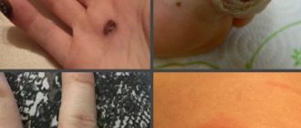
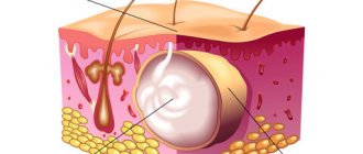
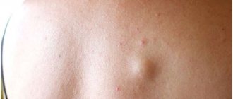
Atheroma is a cystic formation under the epidermis (upper layer of skin) filled with fat and keratin. It is formed in the ducts of the sebaceous glands and looks like a lipoma - both of these ailments are colloquially called wen.
Atheroma is quite rare - only 5% of the population, mainly in women. “Favorite” localization of formation is the place where the sebaceous glands are concentrated:
- face - lips, chin, cheeks, forehead;
- back;
- area near the ears;
- hair growth area;
- genitals;
- armpits.
Atheroma can be recognized by the shape of an oval or a regular circle with a diameter of 0.25 to 7 cm. It occurs individually or in large quantities. Atheroma appears for a number of reasons:
- hormonal imbalance during puberty, pregnancy or menopause;
- disorders of the liver and kidneys, central nervous system diseases, sedentary lifestyle;
- dysfunction of the thyroid gland, Moon's disease, in general - metabolic failure;
- hypothermia, a course of hormonal drugs - the functioning of the adrenal glands is disrupted;
- atopic or seborrheic dermatitis;
- poor skin care with high oil content and excess ultraviolet radiation;
- overdose of medications, resulting in malfunction of the sebaceous glands;
- lack of elastin in the skin and excess calcium;
- congenital pathology.
Atheroma has two stages: cold and progressive.
Cold atheroma is safe and can remain in this state for a lifetime. Unchangeable in density and size.
The progressive stage is dangerous due to complications that can lead to death. It quickly thickens and increases in diameter. If you do not treat it, the following consequences are possible:
- the formation can break into the blood, thereby causing sepsis with a 75% probability of death;
- when compacted, the tumor can develop into a malignant one;
- if atheroma appears near the nerve endings - partial numbness of the facial muscles;
- the occurrence of necrosis around the atheroma, if it grows quickly, then there is a violation of the blood supply and, as a result, blood poisoning.
The cold stage can turn into a progressive stage at any moment, so you should not hope for a stable state. It is better to start treatment as soon as atheroma is diagnosed. This will allow you to get rid of it faster than in the case of an advanced formation, and avoid many dangerous complications.
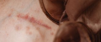
Characteristic symptoms of cyst suppuration
Inflamed atheroma may not cause any discomfort at first. If you do not seek help in time, local symptoms appear, and then the general condition of the body worsens.
Local symptoms:
- Education is painful;
- Rapid increase in size;
- Redness of the skin over the cyst;
- Twitching sensations at the site of the cyst, due to suppuration of its contents;
To make a diagnosis, the surgeon will rely on external data obtained from examination and palpation of the formation. It is important not only to seek treatment early, but also to choose a qualified specialist. At our clinic you can get all the help you need.
Useful information about visiting a surgeon at the clinic:
- How to prepare for a surgeon's appointment
- What diseases does the surgeon treat?
- Calling a surgeon to your home
- Surgical care in the clinic
- Surgical care at home
- What symptoms should you contact a surgeon for?
- Treatment of surgical diseases
- Treatment of intestinal pathologies
- Treatment of skin surgical pathologies
- Treatment of bedsores and necrosis
- Treatment of parasitic diseases
- Treatment of inflammatory processes of soft tissues
- Treatment of diseases of the musculoskeletal system
- Diagnosis of surgical diseases
Methods for removing atheroma
You can get rid of atheroma using laser, radio waves and surgical excision.
Laser techniques make it possible to eliminate growths in the cold and progressive stages of development. The procedure is performed under local anesthesia in several options:
- photocoagulation;
It is used if the atheroma is no more than 5 mm in diameter. It is burned with a laser beam, damaging the shell. After the procedure, a crust forms and no stitches are required. After a few weeks, the scab falls off on its own, revealing a healthy layer of skin.
- laser evaporation;
- laser excision.
Prescribed for the removal of cysts up to 20 mm in diameter. A small incision is made, damaging the shell, and the contents are squeezed out with sterile napkins. The remains of the atheroma are evaporated with a laser. After completion of the procedure, drainage is installed and stitches are applied. They are removed after 8–12 days.
Prescribed for formations ranging in size from 5 to 20 mm. An incision is made next to the atheroma, and a scalpel is used to move it upward to see where the capsule ends and healthy tissue begins. The capsule is cut out with a laser and removed. A drainage tube is placed at the operated site and stitches are applied. They are removed after 7–12 days.
Laser techniques are very effective, but have a number of contraindications:
- purulent nature of atheroma on the face;
- nerve receptors are affected;
- dry or sensitive skin.
Radio waves remove small cysts without inflammation. For the operation, as with laser methods, local anesthesia and special equipment are used. Suitable for dehydrated, dry and fair skin, but this method is not suitable for purulent inflammation.
The radio wave is directed to the top of the atheroma and burns it out, leaving a small depression at the site of formation. It is treated with an antiseptic and bandaged; stitches are not required in this case. Rehabilitation occurs in 3–5 days, and the reappearance of the growth is excluded.
Surgical excision is a traditional method that involves several options:
- with excision of the atheroma capsule;
- with preservation of the atheroma capsule.
Used for large formations. The shell is opened, the liquid is squeezed out or scooped out with a special tool, the capsule shell is removed, and the wound is sutured. If the incision was small, use medical glue.
The skin is incised near the formation, after which the entire capsule is removed through the incision. Next, the wound is sutured.
Surgical excision is prescribed if:
- atheroma more than 3 cm in diameter;
- there is a high probability of an abscess breaking through;
- degeneration into a malignant formation has begun (the nearest skin within a radius of 2 cm is also removed);
- Necrosis of the surrounding tissue develops.
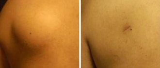
Rehabilitation after surgery takes 3 weeks, and a scar always remains.
Treatment of atheromas
Under no circumstances should you remove atheromas yourself at home and injure them. It is these actions that lead to suppuration processes and further complications.
The best treatment for atheroma is surgery. Surgical removal or excision is a radical method of treatment. As a rule, this is a minor operation that involves making an incision in the skin, removing tumors and suturing the wound. When special wound closure techniques are used and the suture material is non-traumatic, a thin (like a thread) scar is left behind. The method involves removing a tumor-like formation along with those areas of the skin to which they are fused. In case of suppuration, the treatment process involves opening and draining the cavity of the formation and resembles the treatment of abscesses.
Among the contraindications to surgical intervention, it is worth noting diseases of the cardiovascular system, congenital anomalies of the nervous system, diabetes mellitus, blood diseases, viral and infectious diseases of the skin.
Tumors up to five centimeters in size are operated on using local anesthesia and a cosmetic suture. The operation lasts approximately 10-15 minutes, and after a few days the patient can fully restore his ability to work.
Best materials of the month
- Coronaviruses: SARS-CoV-2 (COVID-19)
- Antibiotics for the prevention and treatment of COVID-19: how effective are they?
- The most common "office" diseases
- Does vodka kill coronavirus?
- How to stay alive on our roads?
Removing large tumors requires a stay in a surgical hospital, because when they are removed, the incision and suture will be large in size.
Possible complications
Possible complications after removal of atheroma:
- bleeding - after surgery, laser methods and radio waves weld blood vessels together after the procedure;
- infection - due to improper care, most often - behind an unhealed suture;
- burn - occurs when a doctor makes a mistake in laser and radio wave methods;
- loose seams;
- relapse - reappearance of atheroma if the membrane was not completely removed or the conditions under which the sebaceous glands were clogged persisted;
- the formation of a rough suture is associated either with insufficient experience of the surgeon, or with the characteristics of the patient’s body.
How atheroma is removed at the Platinental clinic
Platinental doctors have all the latest methods for removing atheroma in their arsenal. Depending on the indications, we use the following methods:
- surgical excision with a scalpel. In our work, we use tools made in Europe and the USA: their graphite nanospraying prevents the formation of scars;
- removal of atheroma using the radio wave method;
- laser removal of atheromas.
Expert opinion
“It is worth noting that not a single scheme of these three
cannot be considered worse or better than another.
The main task of the surgeon is to use one method or another to ensure a neat incision and completely remove the atheroma from under the skin without damaging it. The technique is chosen individually each time.
Specialized plastic surgery centers such as Platinental offer not only painless removal with a variety of methods to choose from.
Modern techniques allow me to do without stitches at all in 90% of cases.
«.
Maxim Vasiliev, plastic surgeon.
Scar formation
A scar almost always forms after removal of atheroma and means that rehabilitation is successful. It looks like a thin light stripe and disappears on its own over time. However, if the scar is convex or, conversely, below the skin texture, this is a reason to consult a doctor: the development of atrophic, hypertrophic or keloid scars is possible.
An atrophic scar is a pit or pockmark; it looks especially unsightly if the scar is after removal of atheroma on the face. The damaged area formed deformed collagen fibers in the dermis. If they are not destroyed and space is not cleared, healthy collagen will have nowhere to form.
Hypertrophic scar , on the contrary, rises above the skin level due to increased collagen production. It is usually red in color and occurs because abnormal scar tissue is compressing the blood vessels.
A keloid scar is similar in appearance to a hypertrophic scar, but over time it grows, affecting healthy areas of the skin and exceeding the size of the original wound. Brings discomfort, itching, burning, and a feeling of tightness. Eliminating a keloid scar is the most difficult.
Wound care at home
- Do not remove the wound dressing for __________ hour(s) after the procedure. Keep it clean and dry.
- If your doctor or nurse tells you to ice the wound, you can apply an ice pack every hour while you're awake for _____minutes, or as directed by your healthcare provider. You can do this for the first 24 to 48 hours after the procedure. This will help reduce bleeding, pain and swelling.
- You may shower __________ hours after the procedure. Let a weak stream of soul flow over the wound.
- Pat the wound dry with a clean gauze pad or a clean, dry terry cloth.
- Do not bathe, swim, or use a hot tub until the wound has healed.
Cleaning the wound
Clean the wound every day. Do this for _____ days/weeks after the procedure or until your next doctor's appointment. Follow the instructions below when cleaning the wound.
Prepare everything you need
To clean the wound you will need the following:
- one package of cotton swabs (Q-tips®);
- one package of non-stick gauze pads;
- solution ______________________________;
- ointment ______________________________;
- adhesive tape (Band-aid®), cut to fit the wound;
- paper patch.
- a clean gauze pad or a clean, dry terry cloth.
Instructions for cleaning a wound
- Prepare everything you need.
- Wash your hands with warm water and soap for at least 20 seconds or use an alcohol-based hand rub.
- Clean the wound with ______________________________ solution.
- Gently pat the wound dry with a clean gauze pad or a clean, dry terry cloth. Do not rub this area.
- Using cotton swabs, apply ointment ______________________________ to the wound.
- Cover the wound with non-stick gauze cut to fit the wound or Band-aid. If you are using non-stick gauze, secure it with paper tape.
- When finished, wash your hands with warm water and soap or use an alcohol-based hand sanitizer.
Follow these instructions for __________ days/weeks, or until the wound has healed.
Additional instructions:
to come back to the beginning
Possible methods of dealing with scars after removal of atheroma
It is possible to remove a scar after atheroma with the help of cosmetology, physiotherapy, and home applications with anti-scar agents. In extreme cases - with surgical intervention.
Cosmetology involves traumatizing the stratum corneum of the skin in order to renew it. This could be chemical peeling, dermabrasion, etc.
Applications are made with special anti-scar creams, gels and ointments 3-5 times a day. They are effective for small, fresh scars. Their combination with physiotherapy is effective.
Physiotherapy offers a course of electro- and phonophoresis procedures - exposure of the scar to electric current or ultrasound. These manipulations stimulate metabolic processes, improve blood circulation, and when combined with anti-scar agents help to significantly speed up the process of tissue regeneration. In this case, the main task of physiotherapy is to transfer particles of the medicinal substance into the deep layers of the skin.
Surgery is prescribed in extremely severe cases, for example, when the keloid scar has reached enormous sizes. After excision of the scar, the wound should heal, and therefore a new scar will appear at the site of the skin injury. With proper rehabilitation, it is normotrophic (light and flat). However, the risk of keloid recurrence is very high.