The appearance of a suspicious lump on the leg rarely goes unnoticed, because any attempt to walk without taking off shoes causes discomfort for the patient. But in most cases, seeing a doctor is postponed for months, or even years. It’s a pity, because going to the doctor in the early stages of arthrosis of the big toe allows you to “freeze” the disease and delay the appearance of irreversible foot deformities.
First, baths and applying onions in a sock are used, then, when the disease does not respond, ointments are used, and only then, exhausted by constant pain, the patient turns to an orthopedist or rheumatologist. What kind of diagnosis is this, how to treat arthrosis of the toe, how to recognize its first signs and what could be the consequences of postponing a visit to the doctor?
Pathogenesis
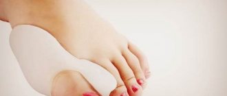
With valgus deformity of the first toe, the angle between the first and second metatarsal bones becomes larger, and displacement of the first metatarsal bone is observed. The big toe moves outward and the head of the bone protrudes outward, forming a bunion. This bone prevents the thumb from conforming to the norm, and it gradually deviates outward.
The tubercle (bone) interferes with wearing shoes, creates inflammation, friction and leads to bursitis (inflammation of the bursa of the first metatarsophalangeal joint). Gradually the bone becomes swollen, inflamed, and painful. Incorrect position of the joint provokes its premature wear, damage to the cartilage and an increase in the size of the bone growth. This in turn provokes foot trauma and further development of pathology.
As the disease progresses, the big toe displaces the other toes, causing them to develop a hammertoe deformity. As the disease develops, it leads to deforming arthrosis of the metatarsophalangeal joints, inflammation of the joint capsules, joints of the toes, chronic bursitis, internal displacement of the first metatarsal bone, flat feet (chronic and transverse), bone and cartilaginous growths of the heads of the metatarsal bones.
Forms
In normal condition, the metatarsal bones are parallel to each other. Under the influence of certain reasons, the first metatarsal bone deviates outward and because of this, a protruding small bump appears on the foot, ligaments and tendons lose their elasticity, and their dysfunction develops.
The disease goes through several stages:
- Early.
At this stage, the deviation of the big toe is less than 15 degrees.
- Average.
The deviation of the first finger to the side is from 15 to 20 degrees. At the same time, deformation of the second finger is observed. It rises above the thumb and becomes shaped like a hammer.
- Heavy.
The thumb deflection is 30 degrees. All the toes are already deformed, and a large bone growth is observed at the base of the first phalanx. In places where there is a lot of stress on the foot, rough calluses appear.
Causes
The main provoking factor in the appearance of hallux valgus is the wrong shoes. If a person wears tight shoes with a narrow toe or high heels, then the fingers are constantly in the wrong (compressed) position, which contributes to the development of hallux valgus. But this is not the only reason for the development of the disease.
The following reasons influence the appearance of pathology:
- traumatic injuries of the leg and foot;
- rickets;
- cerebral palsy;
- polyneuropathy;
- low arch of the foot;
- flat feet;
- congenital weakness of the muscular-ligamentous apparatus;
- chronic inflammation of the joints caused by psoriasis;
- arthritis;
- multiple sclerosis (accompanied by damage to the sheath of nerve fibers);
- diabetes;
- gout (deposition of urate in body tissues);
- joint hypermobility observed in Marfan and Down syndrome;
- juvenile foot (rapid increase in foot size during adolescence);
- Charcot-Marie-Tooth disease (hereditary neuropathy accompanied by muscle atrophy of the distal limbs);
- osteoporosis (bone loss);
- overstrain of the feet due to professional activities (waiters, athletes, ballerinas).
In the presence of provoking factors, the disease progresses rapidly.
Prevention
After treatment of corns, a relapse can easily occur, because it is not enough to relieve inflammation and accelerate tissue regeneration - you need to 100% eliminate the factors that provoke the development of the disease. To avoid relapse, follow 7 rules:
- Wear only comfortable shoes that do not cause pain or discomfort. Avoid high heels and very hard soles.
- Never skimp on shoes and prefer models made from natural materials,
- Use soft orthotics.
- Alternate heavy physical work and sports with periods of rest. Be sure to undergo a professional pedicure and foot massage, and take medicinal baths.
- Get regular medical examinations to identify problems with the endocrine and cardiovascular systems at an early stage.
- Avoid sudden weight gain (adjust your diet and exercise).
- If you experience itching, redness or severe pain, immediately make an appointment with a dermatologist. This will eliminate the disease at an early stage and avoid complications.
The multidisciplinary CELT clinic employs specialists with extensive experience. Here they will be able to determine the cause of the appearance of pathological formations on the soles and prescribe effective treatment. If the problem has already arisen, then you need to contact a specialist as soon as possible.
Make an appointment through the application or by calling +7 +7 We work every day:
- Monday—Friday: 8.00—20.00
- Saturday: 8.00–18.00
- Sunday is a day off
The nearest metro and MCC stations to the clinic:
- Highway of Enthusiasts or Perovo
- Partisan
- Enthusiast Highway
Driving directions
Symptoms
The symptoms of the pathology depend on the degree of damage to the foot. At the first stage, there is redness, swelling of the tissue in the area where the bone appears, pain in the phalanges of the fingers, which intensifies while walking. At the middle stage of development of the disease, pain, swelling, bone growths in the area of the metatarsal head, and dry callus appear under the middle phalanx of the finger. In the severe stage, severe, debilitating pain occurs in the sole of the foot and in the big toe. Dry calluses form, and keratinization of the skin appears under the second and third phalanges of the fingers.
Diagnostics
Diagnostic measures begin with collecting the patient's medical history. The doctor asks him about the disturbing symptoms, asks what provokes their appearance (stress on the legs, walking, tight shoes), whether there are metabolic diseases, systemic diseases, injuries of the lower extremities, hereditary bone diseases in the medical history.
Next, an external examination is carried out, during which the person is asked to walk. The doctor observes the patient's gait while simultaneously determining the intensity of pain. The position of the big toe, its location relative to other toes, and the range of its flexion and extension are studied.
The presence of other external symptoms is checked (swelling, redness, thickening of the stratum corneum under the bones of the fingers). An X-ray of the foot is taken in three projections to identify the degree of foot deformity, joint subluxation and associated pathologies. To exclude circulatory disorders in the legs, an ultrasound scan of the blood vessels is performed.
Our doctors
Tretyakov Anton Alexandrovich
Traumatologist-orthopedist
9 years experience
Make an appointment
Marina Vitaly Semenovich
Traumatologist-orthopedist, head of the minimally invasive traumatology and orthopedics service
Experience 37 years
Make an appointment
Zubikov Vladimir Sergeevich
Traumatologist-orthopedist, Doctor of Medical Sciences, doctor of the highest category, professor
45 years of experience
Make an appointment
Poltavsky Dmitry Ilyich
Traumatologist-orthopedist
Experience 29 years
Make an appointment
Treatment
Therapy for hallux valgus can be traditional or surgical. In the early stages, when you can still do without surgery, doctors advise choosing comfortable and proper shoes. It should not cause stress or friction on the foot. Comfortable shoes slow down the progression of the disease.
At the same time, the doctor will advise you to purchase special orthopedic devices:
- spacers for the joint capsule of the big toe (they eliminate the pressure of shoes);
- intertoe rollers, spacers, which contribute to the correct distribution of the load on the foot.
All orthopedic devices reduce pain, but it is impossible to completely get rid of discomfort with their help.
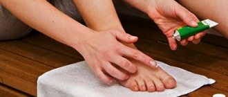
To eliminate pain and inflammation, non-steroidal anti-inflammatory drugs and corticosteroid injections are used. To relieve spasms and restore joint mobility, massage and special exercises to relax the feet are prescribed.
The effectiveness of physiotherapy
Physiotherapy plays an important role in the treatment of hallux valgus. To eliminate the disease, doctors prescribe electrophoresis with calcium, phonophoresis with hydrocortisone, paraffin and ozokerite applications.
One of the most effective methods for eliminating hallux valgus is shock wave therapy (SWT). This procedure involves short-term exposure of the painful area to acoustic low-frequency pulses. With the help of shock wave therapy, pain symptoms are eliminated and the factors contributing to their occurrence are affected.
The procedure is carried out using a special device that generates shock waves. They affect only pathological areas without affecting healthy tissue. With the help of ultrasound, metabolic processes are improved.
SWT is not performed in the presence of neurological, infectious, oncological, cardiac, somatic diseases, diabetes mellitus, or bleeding disorders. Shock wave therapy is not prescribed to pregnant, breastfeeding women and children under 18 years of age.
The effect of the procedure is observed after several procedures. Pain disappears, walking becomes easier. But it is impossible to completely remove the protruding bone using one UVT procedure. The doctor may prescribe at least 5-7 sessions. Depending on the stage of the disease, the order of procedures is prescribed. Some people are prescribed daily sessions, while for others one treatment per week is enough. The results of UVT treatment last for a long period.
Symptoms of osteoarthritis of the toes
An alarming signal for the patient, as a rule, is pain and fatigue in the legs. At first, they are mistaken for ordinary fatigue at the end of the day, the consequences of uncomfortable shoes, and a sedentary lifestyle. Discomfort is observed while walking; in the mornings, patients are bothered by stiffness of the toes and the entire foot, a feeling of numbness, and crunching. The pain increases after physical activity and subsides at night, but as arthrosis of the toes progresses, it becomes around the clock.
Also among the first symptoms to appear:
- swelling, sometimes up to the ankle;
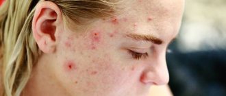
- hyperemia and hyperthermia (redness and increased skin temperature) as symptoms of the inflammatory process;
- frequent appearance of corns and calluses in the same places where they were not observed before;
- growing limitation of mobility in the joint (the joint space seems to be “overgrown” with osteophytes and the heads of the subchondral bones can no longer slide in the joint);
- audible rough crunching sound when walking;
- sometimes the patient can independently palpate osteophytes - hard tubercles under the skin.
Previously comfortable shoes become tight for the patient, it becomes more difficult to stand, and changes in gait appear. With arthrosis of the joint of the big toe, as people say, “the bone begins to bulge” on the inside of the foot, at the base of the first toe. Similar thickenings in the joint area can be observed on other fingers, incl. between the phalanges.
Weather-sensitive patients complain that before the weather changes, the joint begins to ache, twist, and ache.
Symptoms may vary depending on the stage of arthrosis of the toes. Stage 1 is characterized by aching pain and an increase in the size of the affected joints, however, due to smoothed symptoms, it may remain unnoticeable. For the 2nd - increased pain, crunching, inflammation. For the 3rd - deformation and inflexibility of the joints, displacement of the fingers.