The skin is an indicator of human health and is often the first to signal that unfavorable changes are occurring in the body. Moreover, any deviations from the norm in her condition, especially if they are observed on the face, cause significant psychological discomfort. One of the most common problems of this kind is hyperkeratosis, which can occur as a result of the adverse influence of external factors or be a symptom of a number of dermatological diseases or disorders in the functioning of internal organs. Therefore, if it develops, you should definitely consult a dermatologist and find out the cause of such unfavorable changes in the condition of the skin, as well as undergo comprehensive treatment to eliminate hyperkeratosis itself, as well as the reasons for its development.
Leather and its features
Human skin is a multifunctional organ, the condition of which is in close relationship with other organs and systems of the body. It has 3 layers, the thickness of which differs in different parts of the body. This:
- epidermis;
- dermis;
- subcutaneous fat tissue.
The epidermis is the top and thinnest layer of the skin. Moreover, it itself has a multilayer structure and is formed:
- stratum corneum;
- glassy or shiny layer (present only in the skin of the palms and soles);
- granular layer;
- spinous layer;
- basal layer.
The epidermis is 85% composed of keratinocytes - cells that contain keratin and successively undergo stages of differentiation, shifting from the basal layer to the spinous, granular and gradually losing water, the nucleus and turning into prismatic scales of the stratum corneum. The latter are called corneocytes. At the same time, the epidermis is constantly renewed due to the desquamation of corneocytes of the uppermost stratum corneum and its replacement by other keratinocytes, which also transform over time into corneocytes and are ultimately desquamated.
Thus, the skin is constantly renewed. If this process is disrupted due to the action of certain factors and desquamation slows down with thickening of the granular and stratum corneum of the skin or excessive formation of creatine is observed, hyperkeratosis develops.
Classification
There are several principles for classifying squamous cell skin cancer. According to the histological structure, there are 4 types of this neoplasm, and according to TNM staging - 4 stages, each of which reflects the prevalence of the process in the body.
According to histological classification, the following options are distinguished:
- The spindle cell type has a poor prognosis due to its rapid invasive growth as well as its tendency to metastasize and recur.
- Acantholytic type. Appears on skin affected by actinic keratosis.
- Verrucous squamous cell skin cancer is accompanied by symptoms of severe hyperkeratosis, which is clinically manifested by the formation of a horny growth (skin horn).
- The lymphoepithelial type consists of poorly differentiated cells. It is believed that this neoplasm is a tumor of the skin appendages, and not true squamous cell carcinoma.
The stage of development of this type of cancer is determined by the size of the primary tumor, the degree of invasion into the underlying tissue and the presence of distant metastases. The first stage corresponds to a formation less than 2 cm in size, the second and third - larger tumors with spread to nearby tissues, and the fourth - a lesion of any size with the presence of metastatic lesions.
What is hyperkeratosis
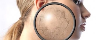
Hyperkeratosis is a pronounced thickening of the stratum corneum of the epidermis, which can reach from several millimeters to several centimeters. Depending on the changes occurring in the skin, thickening of the granular and spinous layers of the epidermis, which is called proliferative hyperkeratosis, or a slowdown in the rate of exfoliation of horn cells may be observed. In the latter case, the thickness of the granular and spinous layers, on the contrary, decreases. This is called retention hyperkeratosis.
Hyperkeratosis is not an independent disorder, but is a symptom of a whole group of skin diseases. It is characterized by excessive keratinization of individual areas or the entire skin. This can be due to a number of reasons, and only sometimes hyperkeratosis can be congenital or hereditary and act as a manifestation of ichthyosis, Mibelli porokeratosis and some other diseases. But if in this case the child has signs of skin disorders practically from birth, then with acquired hyperkeratosis they arise throughout life. They cause not only serious physical discomfort, but also psychological discomfort due to changes in appearance, especially if signs of the disease appear on the skin of visible areas of the body and especially the face.
Hyperkeratosis is also present in healthy people on the elbows, feet, and in rare cases on the knees, i.e., in places of greatest skin friction.
The reasons for the development of acquired hyperkeratosis can be external influences and disturbances in the functioning of the body, and their combinations are often observed. This:
- prolonged pressure on the skin of clothing or shoes;
- prolonged friction with items of clothing, accessories;
- constant contact with lubricating oils, petroleum products, coal and other similar substances;
- disruption of the endocrine system;
- deficiency of vitamins A and C or only one of them due to gastrointestinal pathologies or poor diet.
Hyperkeratosis can also be a manifestation of diseases such as:
- mycosis, i.e. fungal infection of the skin;
- atopic dermatitis;
- psoriasis;
- disseminated and discoid lupus erythematosus;
- seborrheic dermatitis;
- leprosy;
- lichen planus;
- neurodermatitis;
- erythroderma;
- celiac disease (celiac disease);
- eczema;
- xeroderma pigmentosum;
- diabetes mellitus, etc.
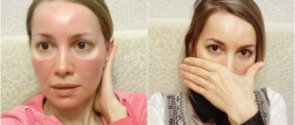
With facial hyperkeratosis, the presence of seborrheic dermatitis or discoid lupus erythematosus is often suspected. With the latter, round or oval pink spots with a bluish tint and sharp boundaries appear on the cheekbones, nose, cheeks, sometimes ears, neck and lips, which tend to increase to 5 cm and merge, and then become covered with dry dense scales.
Hyperkeratosis often occurs in older people. In such situations, it is usually caused by age-related changes in the skin and affects the back and extensor surfaces of the limbs, although it is possible that the face can also be affected.
Reasons for appearance
The exact causes of the disease have not yet been established. The main one is regular exposure to ultraviolet radiation. It affects the dermis, epidermal layers, blood vessels, sebaceous glands, and melonocytes.
Gradually, under the influence of sunlight, the disturbances increase, reaching the peak of the disease.
The following factors contribute to the development of pathology:
- genetic predisposition;
- weakened immune system;
- influence on the skin of chemicals (resinous substances, oil, sand, etc.);
- past infections;
- age-related changes (the disease most often affects people over 50 years of age).
Due to weak immunity, AIDS carriers, people with problems with the nervous and endocrine systems, as well as patients who have undergone chemotherapy or complex operations are more prone to the appearance of keratosis.
Some types of keratoses often affect young people. This usually applies to red-haired or fair-haired people with gray, blue or green eyes. Research shows that by the age of 40, 60% of the population has at least one element of keratosis.
Over the age of 80, everyone has some type of this pathology.
Types and symptoms
Hyperkeratosis is characterized by changes in skin texture and increased dryness, which may be accompanied by:
- peeling;
- the appearance of numerous dense dry scales, sometimes painful;
- decreased sensitivity in the affected area;
- change in skin color.
But in general, the manifestations of hyperkeratosis largely depend on the form in which it occurs. Depending on the clinical picture, the following types of this disease are distinguished:
- follicular;
- lenticular;
- disseminated;
- seborrheic;
- warty;
- multiform;
- diffuse.
Keratoderma, in which the skin of the palms and feet is predominantly affected, will be identified as a separate type.
However, the main and most common forms are follicular, lenticular and disseminated hyperkeratosis.
Follicular
Follicular hyperkeratosis is characterized by a disruption of the processes of keratinization of epidermal cells in the area of the mouths of hair follicles, including vellus hair, located not only on the body, but also on the face. As a result, they become clogged with scales, which leads to the formation of small nodules and plaques around the hair.
The consequence of this is that the skin in the affected area becomes dry and rough to the touch, since the ducts of the sebaceous glands, through which their secretions and hair come to the surface of the skin, are clogged with their contents. Due to mechanical friction, a red rim may form around the nodules. As a result, small white or reddish papules become clearly visible on the skin, and patients say that it has become like a goose or toad.
In adults, follicular hyperkeratosis often occurs in a diffuse form and often affects the skin of the face.
Follicular hyperkeratosis can be a symptom of a number of diseases or observed in isolation even in healthy people with unchanged skin functions against the background of:
- deficiency of vitamins A, C;
- violations of personal hygiene;
- negative effects on the skin of cold, hard water and other external factors.
The resulting nodules tend to become infected on their own or due to extrusion. This can cause the development of secondary pyoderma, i.e. purulent-inflammatory skin diseases.
Follicular hyperkeratosis can also be observed on the face. And although it does not pose a serious threat to human life and health, small but numerous papules can significantly spoil the appearance, which will become a reason for the development of complexes and psychological problems.
Lenticular and disseminated
These forms of hyperkeratosis are most typical for older men, although they can first appear at a fairly young age. It is not yet known for certain what exactly causes their development, but the hereditary nature of the disorder in the formation of the stratum corneum of the epidermis is assumed.
Lenticular hyperkeratosis is characterized by the formation of keratinized, rough papules 1-5 mm in size at the locations of hair follicles. Moreover, they have a reddish-brown or yellow-orange color, are located in a random, scattered order and are not prone to merging. However, they can sometimes cause itching, but do not cause pain or discomfort.
As a rule, such changes can be seen on the skin:
- dorsum of feet;
- shins;
- hips;
- hands;
- torso;
- ears;
- sometimes cheekbones and oral mucosa.
If you remove a papule, a moist depression remains in its place, in the center of which you can see a spot of blood. But it is not recommended to practice this, since mechanical trauma to the elements sharply increases the risk of secondary infections.
Lenticular hyperkeratosis occurs with periods of exacerbations and remissions.
For disseminated hyperkeratosis, the formation on the skin of single or groups of elements in the form of short and thick hair is typical. They also do not tend to merge, but can form in the form of a brush when 3-6 adjacent follicles are simultaneously affected.
Disseminated hyperkeratosis occurs chronically, without periods of improvement in the absence of treatment and proper skin care.
Seborrheic
Seborrheic hyperkeratosis is typical for middle-aged and elderly people, especially those whose relatives have experienced seborrhea. With this form of hyperkeratosis, the formation of benign neoplasms ranging in size from 0.2 to 3 cm in the area of the hair follicles is observed:
- head;
- face;
- neck;
- back surfaces of hands;
- in the elbow bends.
Initially, they appear as small spots or papules with clear boundaries of a yellowish or pink color. There are greasy crusts on their surface that can be easily removed. Over time, they become dense and thicken.
Seborrheic hyperkeratosis can also occur in a mushroom-shaped and dome-shaped form. In the first case, the formations are soft to the touch and have unclear boundaries, and their color ranges from dark brown to black. At the same time, inclusions like open comedones are often observed on their surface. They are retained horn cells that have acquired a black tint.
With the dome-shaped form of seborrheic hyperkeratosis, the formation of elements with a smooth surface is observed, in which, when viewed through a magnifying glass, small white or black inclusions can be seen, which are keratin deposits.
Warty
Warty hyperkeratosis is accompanied by the formation of horny layers, very similar to warts, yellowish-gray in color, sometimes with cracks on the surface, which are then covered with crusts. This is observed with seborrheic keratosis, cutaneous horn, warty nevi and other pathologies of this kind, as well as with aggressive effects on the skin of chemical, mechanical factors, and radiation exposure.
Multiform and diffuse
Hyperkeratosis multiforme is characterized not only by thickening of the stratum corneum of the skin, but also by disruption of the functioning of the nervous system, as well as deviations from the norm in the condition of the bones and skin appendages. Follicular hyperkeratosis in such situations can be combined with thickening of the nails and the development of leukoplakia on the mucous membranes.
Leukoplakia is keratinization of areas of the mucous membrane, which is dangerous from the point of view of the formation of malignant cells.
Diffuse hyperkeratosis is diagnosed when large areas of the body or entire skin are affected. In this case, severe dryness and flaking are observed.
Diagnostic methods
Since the direct dependence of the effectiveness of the treatment on the stage of the malignant neoplasm at which it was discovered has long been proven, there are two levels of diagnosis of squamous cell skin cancer: early and late.
An early diagnosis is considered to be made at stages I-II. In this case, complete recovery of the patient is possible, provided that the correct treatment tactics are chosen. Late detection means diagnosis at stages III and IV. The prognosis is usually unfavorable due to the complexity or impossibility of surgical treatment.
The “gold standard” for diagnosing squamous cell skin cancer is a biopsy followed by histological examination. The immunohistochemical method is considered especially informative. Since the tumor is external and it is not difficult to obtain biomaterial for histology, the tumor is verified in 99% of cases.
The dermatoscopy method is also widely used. In this case, the presence of central keratin plugs, dilated and branched vessels on the surface speaks in favor of skin cancer.
For any form of squamous cell carcinoma, along with a thorough history and physical examination, a specialist must evaluate the condition of the lymph nodes. If the presence of metastatic foci is suspected, the main diagnostic method is fine-needle aspiration. It is also possible to prescribe additional imaging methods (ultrasound, radiography, CT, angiography) to identify regional and distant metastases.
Treatment of hyperkeratosis
If symptoms of hyperkeratosis occur, you should consult a dermatologist. For an experienced, qualified specialist, it is not difficult to diagnose the presence of hyperkeratosis. Only in isolated cases, to clarify the nature of the formations, a biopsy of suspicious areas of the skin is required.
To effectively eliminate the problem of hyperkeratosis, especially affecting the skin of the face, complex therapy is required. It is also necessary to prescribe treatment for the disease that provoked its development, if one has been detected. To do this, the patient may need to consult not only a dermatologist, but also an endocrinologist or other specialist.
As part of the treatment of hyperkeratosis itself, drug therapy and cosmetic procedures are prescribed. The first is the use of retinoids, vitamin C, and sometimes topical corticosteroids. For faster skin restoration and elimination of cosmetic imperfections, the following is recommended:
- paraffin therapy;
- mild chemical peels;
- UV therapy;
- mesotherapy.
The use of scrubs and other mechanical exfoliation products is not indicated for hyperkeratosis. This will only worsen the situation and can lead to skin infection. But it is always recommended to apply emollients to the skin, which moisturize and soften the skin. They contain natural lipids or natural oils that nourish dry skin and saturate it with the substances it needs. As a result, it is possible to restore the skin’s natural protection and restore its natural smoothness.
In case of seborrheic hyperkeratosis, accompanied by the formation of growths, they can be removed using electrocoagulation, laser or cryodestruction.
Unfortunately, hyperkeratoses often accompany chronic diseases, which cannot be completely eliminated. Recovery can only be achieved in situations where thickening and peeling of the skin is caused by mechanical factors or treatable diseases, such as mycosis. In other cases, when dryness and thickening of the skin are caused by chronic dermatological or other pathologies, it is only possible to reduce the frequency of their exacerbations and the severity of the course, that is, to achieve a stable and long-term remission. But this requires the correct selection of treatment tactics, constant skin care, and often diet.
Drug therapy
First of all, vitamin A, also called retinol, and vitamin C are prescribed internally and externally. Normally, a person receives the necessary vitamins from food, but today rarely anyone can boast of a balanced diet.
Vitamin A, in addition to regulating cell growth, is necessary to maintain visual acuity, hair and nail growth, and also performs a number of other functions. For hyperkeratosis, vitamin A in the form of synthetic derivatives called retinoids is used for general and local therapy.
Some of the brightest representatives of this kind of drugs are Roaccutane, in which isotretinoin is the active ingredient, and Neotigazon, containing acitretin. These compounds are derivatives of retinoic acid. In the case of the development of follicular hyperkeratosis, the use of topical retinoids, in particular adapalene, is indicated.
If hyperkeratosis, especially follicular hyperkeratosis, is accompanied by an inflammatory process, topical corticosteroids are prescribed, for example, drugs based on hydrocortisone, prednisolone, fluacinolone, clobetasol. Most modern drugs in this group are available in several dosage forms: emulsions, creams, ointments. Moreover, the effectiveness of treatment largely depends on the correctness of its choice. Additionally, corticosteroids help accelerate the exfoliation of dead cells.
Paraffin therapy
Paraffin therapy is a cosmetic procedure in which special cosmetic molten paraffin is applied with a brush to a previously cleaned face or other areas of the body affected by hyperkeratosis. The procedure takes on average 20-60 minutes and promotes:
- gentle removal of dead cells;
- cleansing pores of impurities;
- activation of microcirculation;
- moisturizing the skin;
- reducing inflammation.
Peelings
For hyperkeratosis, chemical peelings are indicated, in particular using acids such as:
- salicylic;
- glycolic;
- dairy;
- lemon;
- apple;
- tartaric.
All of them have a good exfoliating effect, and also additionally open pores and moisturize the skin. As a result, it is possible to effectively solve the problem of excessive dryness and flaking of the skin, as well as improve its tone and texture, restoring velvety.
UV therapy
Ultraviolet irradiation helps eliminate the manifestations of hyperkeratosis by suppressing pathogenic microorganisms, including fungi, activating blood circulation and metabolism, suppressing the inflammatory process, and providing a regenerative and desensitizing effect.
Mesotherapy
Mesotherapy is a well-known cosmetic procedure, which consists of introducing into the skin the substances necessary for its proper functioning using numerous injections or using hardware. As a result, it is possible not only to saturate the tissues with vitamins, but also to achieve effective hydration, which helps reduce the manifestations of hyperkeratosis and improve the condition of the skin in general.
Thus, hyperkeratosis is a very common disorder, although it does not affect the face as often as, for example, the feet, elbows, skin of the legs and back. However, with excessive dryness and thickening of the facial skin, even in the form of small papules, a serious aesthetic disadvantage occurs. But with a timely visit to a dermatologist and the start of complex therapy, including the use of modern cosmetic procedures, it is possible to significantly improve the condition of the skin of the face and other parts of the body or even completely eliminate the signs of hyperkeratosis.
Are there any new developments in this direction on the world market?
Well, for example, from 1998 to the present, the Australian biopharmaceutical company Peplin has been studying the topical treatment of actinic keratosis with the drug Ingenol Mebutate, which is the first in a new class of formulations and is derived from milkweed juice. This ingredient has a long history of traditional use for a variety of skin conditions, including the topical treatment of skin cancer and precancerous skin lesions. The company plans to enter the third phase of the trial soon.
Stages of the disease
During the course of the disease, 4 successive stages can be distinguished:
| Stage | Description |
| I | Minimum tumor size. The process does not extend into the deeper layers of the skin. |
| II | The tumor is growing, but the lymph nodes and local tissues are not involved in the process, and there are no metastases. |
| III | Deep structures (bones, cartilage, muscles) located next to the tumor are affected; cancer cells can be found in at least one lymph node. |
| IV | Distant metastases are present. |
Diagnosis of PPH
At the initial appointment, the oncologist listens to complaints, asks in detail about when and under what circumstances the first symptoms appeared, whether there is a hereditary predisposition, how often he sunbathes, what medications he uses. After this, the specialist examines the skin and the neoplasm, palpating the lymph nodes closest to the formation.
Electronic mapping is available at MelanomaUnit, which allows you to create a “map” of all moles and tumors on the body. The results of the study are saved in the patient's electronic record and can be used to monitor their changes in the future.
To determine the nature of the tumor, the doctor performs a biopsy, that is, taking a small piece of tissue for histological examination in the laboratory.
Forecast
When squamous cell skin cancer measuring less than 2 cm is detected and adequate treatment is provided, the 5-year survival rate reaches 90%. If the tumor is larger and there is growth into the tissue, then this figure is reduced to 50%.
Tumors located on the skin of the periorbital region, external auditory canal, and nasolabial fold have a particularly unfavorable prognosis. With this localization, the tumor can grow into muscles and bones and can cause bleeding due to vascular damage, complicated by infections.
Prevention
The main measure for preventing squamous cell skin cancer is the timely detection and treatment of precancerous conditions. In this regard, young people are recommended to be examined by a dermatologist every 3 years, and people over 40 years old - every year. If you detect any changes in the skin, you should also contact a specialist.
Foci of chronic inflammation on the skin must undergo adequate sanitation. You should also avoid repeated damage to the same skin area (rubbing, compression by tight clothing and shoes, injury during monotonous manual work).
It is important to maintain a sun regime and use a cream with a sunscreen component, and avoid visiting a solarium. When working with chemical reagents that are sources of occupational hazard, it is necessary to use personal skin protection.
Book a consultation 24 hours a day
+7+7+78
When to see a doctor
You should see a doctor if you notice the following symptoms:
- the appearance of suspicious growths on the skin;
- non-healing ulcers;
- itching, bleeding, swelling in areas of the skin.
At the Sofia Oncology Center, patients can make an appointment with an oncologist. The Sofia Oncology Center is located at 2nd Tverskoy-Yamskaya Lane 10. All diagnostic measures are carried out using modern equipment, which allows us to obtain the most accurate research results.


