Hives are an allergic disease that appears as spots or red, intensely itchy plaques on the skin. Prevalence depends on age and environmental conditions, as well as comorbidities. The most common allergic dermatosis is recurrent urticaria.
The swelling and itching caused by hives can be severe. The feature that distinguishes this disease from others is red spots on the surface of the skin. They have irregular edges and small bubbles. Typically, hives are caused by allergies. When an allergen enters the body, histamine is released into the bloodstream and causes: swelling, redness and itching.
Types of urticaria
There are two forms of the disease: acute and chronic. Acute urticaria is often caused by food allergies, insect bites, medications, or exposure to an allergen. It lasts, as a rule, up to 1.5 months.
Chronic urticaria lasts more than 6 weeks. It can begin when the immune system attacks the body's tissues, thereby causing an autoimmune reaction. Antibodies (proteins that normally fight bacteria and viruses) cause the release of histamine, which leads to lesions on the skin.
The rash can be localized or appear anywhere on the body, from the neck to the knees. Rarely does the allergy affect the arms, legs and face, unlike angioedema. Characterized by the appearance of rashes and spots on the skin. There is also swelling in the eyelids, lips and larynx.
Angioedema urticaria is a medical emergency because it can cause breathing problems and suffocation.
It is believed that 30 to 50% of cases with chronic urticaria have an autoimmune cause. The reason why autoimmune urticaria develops is unknown, even though it often occurs along with other autoimmune diseases such as:
- rheumatoid arthritis – the immune system attacks the joints;
- lupus – the immune system attacks the joints and skin.
In rare cases, chronic hives can also be caused by other infections (eg Helicobacter), hyperthyroidism (overactive thyroid), hepatitis B or C (liver infections), intestinal parasites.
Table - Types of urticaria in children
| Name | Peculiarities |
| Immune (IgE-dependent - food, drug, helminth; complement components - anaphylotoxins C3a and C5a; complement-induced - hereditary or acquired deficiency of the C-1q inactivator) | Characterized by skin damage and itching. Quincke's edema, urticarial type of vasculitis and anaphylactoid urticarial reaction are possible. |
| Non-immune (based on a pseudo-allergic reaction) | There is no true reaction of interaction between antibody and antigen with the formation of an immune complex. Ig-E does not increase. Occurs due to consumption of foods that contain vasoactive amines. |
| Pigmented | Accompanied by an increase in the number of mast cells. Often found on the upper and lower extremities or trunk. May damage internal organs and be accompanied by systemic mastocytosis. |
| Vascular urticaria or vasculitis | It is characterized by several lesions that have an unusual shape and disappear after a few days. Other symptoms of urticaria vasculitis include fever, joint and muscle discomfort, abdominal pain and headache. |
Contact dermatitis
- Symptoms
This hypersensitivity reaction is accompanied by redness and swelling of the skin immediately after contact with the allergen or several hours later. Small blisters, papules, and weeping erosions may appear on the affected area. A characteristic symptom of skin allergies is itching. As the inflammation subsides, scales and crusts form at the site of the blisters and inflammation. With chronic contact dermatitis, peeling of the skin is common.
- Treatment
To stop the development of an allergic reaction in an infant, it is necessary to avoid contact with the irritant substance. These can be food products, certain components of detergents and cosmetics, the juice of certain plants, etc. To treat red spots due to skin allergies, local anti-inflammatory drugs in the form of creams and ointments can be used. If swelling is severe, wet-dry dressings may be indicated. In severe cases, systemic hormonal therapy is used to treat the symptoms of contact dermatitis.
Causes of urticaria
As soon as the hypothalamus registers an increase in body temperature in the brain, it sends an impulse to neurons that activate the sweat glands. Neurons release acetylcholine, which stimulates the release of histamine and heparin from mast cells in the skin (mast cell degranulation). This degranulation results in a diffuse release of histamine near the sweat glands. Histamine activates an inflammatory response in surrounding tissues and causes itching and redness. When you sweat (which is difficult for people with cholinergic urticaria), the skin reaction stops.
Chronic stress and nervousness can trigger an attack of psychosomatic urticaria. When constantly anxious, the body releases higher amounts of cortisol and adrenaline (stress hormones). But when the body is constantly in a state of tension and hormones continue to be released into the bloodstream, hormonal imbalance occurs. Adrenaline and cortisol combine with mast cells in the skin to produce histamine. Elevated levels of cortisol in the body cause allergic skin reactions such as hives and may even slow down healing.
Urticaria is usually a consequence of an allergic reaction in children when the body is exposed to the following allergens:
- pollen of flowering plants;
- house dust (dust mites);
- pet hair;
- some preservatives;
- Food.
Hives can also occur when a child wears clothes that are too tight and made from irritating materials such as polyester. It can also be caused by infections caused by bacteria, viruses or other microorganisms.
Some medications, such as ibuprofen, can cause hives in children as a side effect. Also, such a reaction can occur if the child has an individual intolerance to one or another substance included in the medicine. Most often these are antibacterial and non-steroidal drugs.
If children are suddenly exposed to large temperature fluctuations, hives may result. Likewise, prolonged exposure to the sun or cold can cause a similar skin reaction. In rare cases, urticaria can develop due to diseases such as leukemia and lymphoma. This happens because the immune system is weakened.
Hives in children can also be caused by insect bites, which can lead to significant swelling. If it's a mosquito or spider bite, sensitive skin may respond by releasing histamine, which causes hives. Contact with various viruses or bacteria can cause a diffuse rash. Reddened plaques can be observed throughout the body.
Symptoms of urticaria
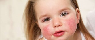
Hives appear as red spots, swelling and blisters. The main symptoms are itching and burning sensation on the skin. Symptoms may progress and spread to other areas of the body.
Additional symptoms of urticaria in children include:
- nausea and vomiting;
- increased body temperature;
- discomfort in the joints.
The spots can be as small as a dot or the size of a coin. Typically, spots appear and disappear within a few hours. However, chronic hives can last for more than 6 weeks.
Diagnosis of urticaria in children
A preliminary diagnosis is made by the doctor after an initial examination of the skin.
The doctor knows exactly what urticaria looks like in a child, so he will not confuse it with manifestations of other diseases. For allergies, the following tests are prescribed:
- general blood test to exclude immune and inflammatory processes;
- allergy tests to identify the allergen.
In some situations, a biochemical blood test and examination for helminthic and parasitic diseases are performed. If necessary, a thyroid examination is prescribed to identify hyper- and hypothyroidism.
Methods of treating diathesis
When dealing with diathesis in a young child, the main thing is to choose the right diet, excluding foods that may cause an allergic reaction.
For a child in his first year of life, it is very important to receive breast milk.
The proteins of human milk are easily broken down by the baby's enzymes and are completely devoid of allergic properties. However, a nursing mother must also follow a diet excluding fish, poultry, tomatoes, chocolate, smoked meats, spices and other allergenic foods.
When mixed feeding, the child should not be given some juices: orange, carrot, tomato. Introduce any new product carefully - from a small amount. It is important to prevent an allergic exacerbation, and at the same time, to give the baby’s body everything it needs for its development.
Specialist consultation
The Family Doctor pediatric allergist-immunologist will help you deal with your child’s skin problems, determine the cause of allergies, and also build a rational diet that is suitable specifically for your child.
Make an appointment Do not self-medicate. Contact our specialists who will correctly diagnose and prescribe treatment.
Rate how useful the material was
thank you for rating
Treatment of urticaria
Pharmaceutical drugs used to treat urticaria should be taken as recommended by your doctor. The best treatment for this condition is to know and avoid trigger factors. The hives will probably go away on their own. But at the same time, medical supervision is a prerequisite to exclude the development of complications.
Treatment for urticaria in children includes:
- taking antihistamines to reduce the rash;
- eliminating foods that cause hives;
- prescribing diuretics to eliminate severe edema;
- using antipruritic ointments to relieve the condition.
- in some situations, hormones are used (topically and by injection).
You need to keep a diary about what the child does and eats. This approach allows us to identify predisposing factors and eliminate them.
Gradually, one can narrow down the real causes and respond accordingly.
With such a complication as Quincke's edema, coughing attacks appear and breathing becomes difficult. In this case, urticaria in children requires emergency care.
Young children often experience swelling of the gastrointestinal mucosa, which is accompanied by vomiting. In severe cases, damage to the nervous system and brain occurs. Such conditions are dangerous to health and life, so delaying a visit to the doctor is unacceptable.
Most children switched to a hypoallergenic diet recover quickly and recover even without medication.
Hives
- Symptoms
Your baby may develop a rash in the form of blisters ranging in size from a few millimeters to several centimeters. Against the background of an acute allergic reaction, swelling may develop. Sometimes individual blisters merge with each other and affect large areas of the skin.
- Treatment
The doctor chooses a course of treatment depending on the cause of the condition. Thus, urticaria often appears as a reaction to insect bites, medications, pollen, foods, etc. This condition can be closely associated with systemic diseases, and sometimes occurs spontaneously. The basis of therapy is the exclusion of the factor that provokes this specific reaction. Your doctor may recommend a hypoallergenic diet even if your hives are not caused by food. If a reaction to cold, heat, or ultraviolet radiation occurs, the baby’s skin must be reliably protected from this factor. The doctor may prescribe antihistamines to treat allergies on the child's cheeks.
Forecast and prevention of urticaria
Hives usually go away over time. However, it is impossible to predict how long this problem will persist. In many cases, the disease can be controlled by avoiding trigger substances and taking antihistamines.
Prevention methods include diet control and avoidance of contact with volatile and food allergens. You should wear loose clothing made from natural fabrics and not cause stress in your child. It is unacceptable to neglect chronic diseases.
Before treating hives in a child, you should consult your doctor. Contact a pediatrician or allergist at the RebenOK clinic in Moscow. We employ specialists with extensive practical experience. Thanks to modern equipment, it is possible to quickly diagnose and, based on the results obtained, prescribe effective therapy.
Hemangioma in children - symptoms and treatment
Currently, there are various ways to treat infantile hemangiomas. Many of them have already lost their significance, others are successfully used at the present time. The main vector of treatment is active observation or conducting one or another type of therapy from the moment of diagnosis.
When choosing treatment tactics, you should be guided by several factors:
- age of the child (the younger the patient, the higher the risk of further growth of the hemangioma);
- type of hemangioma and its location;
- presence of complications;
- parents’ desire to conduct this or that therapy;
- doctor's experience [24].
To help doctors decide on treatment tactics for infantile hemangioma, Russian surgeons D. A. Safin and D. V. Romanov proposed a special rating scale. With its help, you can assess the need for drug treatment with beta-blockers. This scale takes into account both the age of the child and the location of the hemangioma, as well as its size, quantity and thickness (presence of pathological volume) [28].
Rating scale for determining indications for systemic drug treatment of infantile hemangiomas with beta-blockers
| Criteria | Description | Points |
| Age | 0-4 months | 4 |
| 5-8 months | 3 | |
| 9-12 months | 2 | |
| 1 year and older | 1 | |
| Localization | Orbit, nose, lips, ears | 4 |
| Perineum, buttocks, genitals | 4 | |
| Hemangiomatosis | 4 | |
| Parenchymal organs and glands | 4 | |
| Scalp and other parts of the face | 3 | |
| Neck | 3 | |
| Joint area | 2 | |
| Hands and feet | 2 | |
| Torso | 1 | |
| Limbs | 1 | |
| Size (diameter) | More than 5 cm | 2 |
| 1-5 cm | 1 | |
| Up to 1 cm | 0 | |
| Pathological volume | Eat | 1 |
| No | 0 | |
| Quantity | 5 or more elements | 1 |
| 1-4 elements | 0 | |
| Complications | Eat | 1 |
| No | 0 |
The doctor examines the patient, evaluates the specified criteria and sums up the points. When the score is from 9 to 14, systemic drug treatment is indicated, and when the score is from 4 to 8, drug therapy is not required.
When assessing the location of a hemangioma, it is necessary to take into account not only the cosmetic defect, but also the risk of various complications. These “critical zones” include the ears, nose, lips and eyelids (orbits of the eye). For example, if the cartilage of the ear is destroyed, a permanent deformation of the auricle will occur, the correction of which will require plastic surgery. When infant hemangiomas are located in the perineum and buttocks, ulcerations often appear that are difficult to heal, so this localization is also critical.
The large size of the tumor indicates an active process of proliferation, which is accompanied by the risk of further growth of the hemangioma and a high probability of various complications.
In the treatment tactics of infantile hemangioma, it is also necessary to take into account the presence of a deep (subcutaneous) component of the tumor. It is not amenable to local treatment and laser treatment, which leads to tumor progression in the deep layers of the subcutaneous fat layer. Therefore, in this case, it is recommended to resort to drug therapy.
The greater the number of hemangiomas on the skin, the more difficult it is to carry out local therapy. The presence of more than five vascular tumors significantly increases the risk of hemangiomas in the internal organs (diffuse neonatal hemangiomatosis).
All methods of treating infantile hemangiomas can be divided into two categories:
- Conservative (non-invasive) treatment : oral medication, lotions, watchful waiting (time treatment) and laser treatment.
- Surgical (invasive) treatment : injections (sclerotherapy) or surgery (removal of hemangioma).
Conservative (non-invasive) treatment
Waiting tactics
This treatment was widely practiced before the advent of beta blockers. Now this method can also be used, but it is necessary to take into account: the younger the child, the higher the risk of tumor growth. Watchful waiting requires frequent and systematic follow-up examinations to determine the possible growth of hemangioma.
Drug treatment
Beta blockers. In 2008, French dermatologist C. Leaute-Labreze discovered the effect of propranolol on infant hemangiomas. From this period began the “golden age” of beta-blockers in the treatment of infantile hemangiomas. Given the low risk of side effects and the high effectiveness of treatment, they have become the “first line” of treatment for this disease.
The exact mechanism of action of beta-blockers is still unknown. Presumably, it includes vasoconstriction, suppression of VEGF-A (vascular endothelial growth factor), stimulation of apoptosis (the natural “disassembly” of pathological tissues). Studies have revealed the presence of β2-adrenergic receptors and VEGF-A in the capillaries of expanding infantile hemangioma, which decrease when β2-adrenergic receptors are suppressed.
Beta blockers can be used topically as a lotion or systemically by administering the medication orally. For local treatment, timolol-based eye drops or eye gel are used. For systemic therapy, the drugs Propranolol or Atenolol are used. Each of them has its own characteristics in dosage, frequency of administration and the risk of side effects.
Comparison of the pharmacological activity of the drugs Propranolol and Atenolol.
| Indicators of pharmacological activity | Propranolol | Atenolol |
| Bioavailability | 20-30 % | 40-60 % |
| Half-life of the drug | 3-5 hours | 6-9 hours |
| Lipophilicity (permeability through the cell membrane) | +++ | – |
| Removing the drug from the body | Liver 100% | Kidneys 90%, liver 10% |
Steroid hormones are one of the oldest methods of drug treatment for infantile hemangiomas. This method has been used from 1960 to the present and is a “second line” of therapy. Until 2008, it was the main method of treating hemangiomas, especially with active tumor growth.
The mechanism of the effect of glucocorticosteroids on infantile hemangioma is still not completely clear. However, it is known that steroid hormones influence adipogenesis, inhibit the formation of new vessels and reduce the production of proangiogenic proteins (VEGF-A, etc.).
Steroid hormones in tablets can be used as systemic drug treatment. Given the high risk of side effects of glucocorticosteroids and the emergence of beta-blockers, this method has become much less common. The main indications for its use are complicated hemangiomas and unresponsiveness of the formation to treatment with beta-blockers. The average course of treatment is from 4 to 12 weeks (maximum dose). Sometimes steroid hormone therapy can last up to 9-12 months of the child’s life.
It is also possible to inject glucocorticoids into the tumor. Typically, this treatment uses Triamcinolone injections or a mixture of Triamcinolone and Betamethasone. As a rule, such treatment is prescribed for small volumetric formations.
Cytostatics (Cytoxan, Vinblastine and Avastin), according to foreign authors, help stop the division of tumor cells in metaphase. However, this method has not found application in Russia, since Cytoxan acts on the growth of sensitive, rapidly proliferating cells, thereby inhibiting the erythrocyte lineage of the blood and the development of the egg. There is also an opinion that after using Cytoxan there is a risk of developing a secondary malignant tumor - angiosarcoma.
Vincristine is a cytostatic drug, an alkaloid from the pink periwinkle plant (Vincarosea). It causes apoptosis (the regulated process of cell death) of endothelial cells and reduces the production of their growth factors. As a rule, treatment with Vincristine is carried out for vascular pathology, which is not a true infantile hemangioma, but is associated with kaposiform hemendothelioma or tufted angioma with Kasabach-Merritt syndrome. The medication is given weekly through a central catheter.
This treatment method may be useful if steroid hormone therapy does not respond. Its appointment is carried out with the direct participation of oncologists. Side effects of the drug include skin irritation and rash, neurotoxicity, constipation, cranial nerve palsies, bone pain, alopecia, and muscle weakness. Typically, side effects are short-lived.
Interferons (Interferon-alpha-2a, Interferon-alpha-2b and Imiquimod - 5% Aldara ointment) stimulate the secretion of interferons, which suppress endothelial and fibroblast growth factors, and also promote tumor necrosis. They are used when steroid therapy is ineffective. The effect of these drugs is noticeable only after four months of use.
Since interferons cause a lot of complications (anemia, neutropenia, hypothyroidism, fever, neuroplegia), their use in the treatment of young children is not justified.
Laser correction
To treat vascular pathology, lasers with wavelengths of 532 and 585 nm, less often 1064 nm, are used. These lasers do not damage the skin, therefore they are considered conservative treatment.
The operation of lasers is based on the theory of selective photothermolysis, which was described in 1980. Laser radiation has a constant wavelength, but is absorbed differently by tissues. This is explained by the presence of chromophores (water, melanin and hemoglobin) in the skin. The result of this energy absorption is heating of the tissue. For example, wavelengths of 532 and 585 nm are best absorbed by oxyhemoglobin, due to which isolated heating of the vessel occurs, leading to damage to its endothelium without harm to surrounding tissues. As a result, the vessel closes.
After laser treatment, there are no scars or scars.
Surgical (invasive) treatment
Sclerosis of hemangioma
This method can be used for small surface formations. To glue tumor vessels together, a special drug is injected into them, which damages the endothelium of pathological capillaries and stimulates the formation of a blood clot. As a result, a fibrous subcutaneous scar is formed [4][5][7][16].
This method requires stages - carrying out a course of treatment at certain time intervals, for example, performing sclerotherapy once every 1-1.5 months. This is due to the rapid blood flow in the hemangioma, which reduces the time of exposure of the sclerosant to the endothelium, thereby reducing its effect.
The proposed sclerosants, for example, Aethoxysclerol and Fibro-vein, are intended for sclerosis of the veins of the lower extremities. A prerequisite for their use is the introduction of the drug into the bloodstream. They can cause disruption of microcirculation around the hemangioma up to tumor necrosis, which will lead to gross cicatricial changes. Alcohol should also not be used for sclerosis of vascular formations. Although it has a pronounced sclerosing effect, its use is often accompanied by side effects.
Cryodestruction (removal with liquid nitrogen)
The tumor focus can be destroyed using liquid nitrogen at a temperature of −195.6 °C. It is a colorless and odorless liquid that is sterile, non-toxic, inert to biological tissues and non-flammable. During cauterization of the hemangioma with liquid nitrogen, the tumor focus is clearly demarcated, which is replaced by an organotypic regenerate by 21-30 days after cryotherapy.
It is impossible to stop the growth and completely eliminate extensive and deep hemangiomas that have abundant blood circulation using cryodestruction. Usually it does not bring the desired result, as it leaves cosmetic defects, and in many cases leads to increased growth of hemangioma. Therefore, this method is best used in the treatment of local, non-extensive hemangiomas [5][8][21].
Surgical method of treatment
The radicality of surgical treatment of hemangiomas lies in the complete removal of all tissue affected by the tumor. Previously, it was believed that surgical removal of a tumor was dangerous due to complications: heavy bleeding during surgery and damage to the nerves, lymph nodes, and large arterial and venous trunks involved in the tumor. Now, with the advent of modern research methods, it has become possible to simultaneously remove a vascular tumor within healthy tissues, without affecting important anatomical structures [5][7][8].
Not all hemangiomas are subject to surgical removal: it should be used only in the presence of life-threatening conditions, for example, bleeding or when the airways are blocked, but most often it is resorted to at the final stage of involution, when there is almost no blood flow in the hemangioma - most often in 4- 5 years.
The scope of the operation is determined by the ability to perform it without irreversible cosmetic defects, the formation of a rough scar and the risk of dysfunction of nearby organs. In this regard, surgical treatment has its contraindications [5][7][8][21].
Outdated treatment methods
Radiation therapy (RBRT)
The essence of this method is to irradiate an area of skin with X-rays from a short distance. Such radiation is absorbed predominantly in superficial tissues and is effective only for infantile hemangiomas located exclusively on the surface of the skin.
As a result of X-ray therapy, “scars” form in the hemangioma, and telangiectasias (spider veins) form on the surface of the skin. As a result, to obtain a good cosmetic result in the future, either plastic surgery or laser treatment of the area of close-focus therapy is required [7][10].


