A COURSE OF TREATMENT:
one-time manipulation
+7
+7
Make an appointment
Removal of earlobe atheroma is a procedure for removing a benign neoplasm (cyst) resulting from a blockage of the sebaceous gland.
Removal of earlobe atheroma is a procedure for removing a benign neoplasm (cyst) resulting from a blockage of the sebaceous gland.
Contraindications:
- No
Doctors:
- Zaitsev Vladimir Mikhailovich (20 years of experience)
- Magomedov Murad Magomedovich (6 years of experience)
Equipment used:
- Laser
- Radio wave surgical device FOTEK E81M
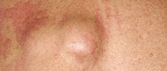
Atheroma of the earlobe is a benign neoplasm (cyst) resulting from blockage of the sebaceous gland. It is a spherical tumor of the lobe, which is filled with a light-colored cheesy substance. Fluid and dead cells accumulate inside it. The size of atheromas reaches half a centimeter in diameter. The problem can affect absolutely every person, regardless of gender and age. Women who wear earrings are at particular risk: in this case, atheroma significantly increases the risk of developing infection.
An earlobe with such a tumor not only does not look aesthetically pleasing, but can also be harmful to health. The cyst can fester and provoke inflammation of nearby tissues, which will sharply worsen the person’s condition. Therefore, it is necessary to remove the atheroma in time.
The formation of atheroma is associated with a malfunction of the sebaceous glands, as a result of which sebum is not removed outside, but accumulates inside. This is facilitated by factors such as:
- metabolic disease;
- hormonal imbalance in the body;
- endocrine disorders;
- improper ear cleaning;
- incorrectly performed ear piercing;
- skin damage;
- acne.
At the initial stage, the formation on the lobe is invisible. Then it begins to increase in size. Characteristic symptoms gradually appear. With atheroma in the ear, pain appears in this area, redness of the skin, swelling, itching, burning, and a feeling of accumulation of water inside the “ball”.
If you find such a seal on the lobe, you need to take treatment responsibly: to open the festering tumor and remove it, you need to consult a doctor - an otolaryngologist.
| Medical service | price, rub. |
| Opening of suppurating atheroma of the earlobe (on one side) | 5000 |
| Initial appointment with an ENT doctor | 2000 |
| Repeated appointment with an ENT doctor | 1500 |
| Initial consultation with the head of the clinic | 4000 |
| Repeated consultation with the head of the clinic | 2000 |
| Additional consultation during procedures | 500 |
| Adaptation of a child to the ENT office | 2000 |
| Adaptation of a child to the ENT office by the head of the clinic | 4000 |
A ball has appeared in my earlobe and it hurts. What could it be?
There may be several reasons why a painful ball has formed inside the earlobe. To establish what exactly causes its appearance, you need to take into account the location of formation, the type of compaction, how mobile it is and how it behaves when pressed (pain, change in color and temperature of the skin).
- The most common reason for the appearance of a ball in the earlobe, which hurts when pressed, is atheroma (wen). Don't immediately panic at this name. Atheroma is, of course, a tumor, but a benign one. It is formed from fat cells.
Doctors do not know the exact reasons for the appearance of wen, but they may be associated with blockage of the excretory ducts of the sebaceous glands, poor lifestyle, frequent overeating or disruption of the endocrine system.
Atheroma is a dense, often painless compaction in color and temperature no different from the skin. When pressed, it moves easily inside.
As a rule, when such balls appear, there is no inconvenience, except for cosmetic ones, unless, of course, we are talking about inflammation and suppuration. The inflammatory process is indicated by an increase in size and reddening of the surrounding skin. When you press the ball, you feel pain and you can notice that its temperature is higher than the temperature of the skin.
- An epidermoid cyst, which is also the cause of the appearance of lumps in the earlobe, is practically indistinguishable from atheroma in appearance. Its formation is caused by excessive proliferation of epidermal cells, as a result of which a capsule of epithelial cells of a fairly dense structure appears. In the case of suppuration of the epidermoid cyst, pain appears when pressing the ball and its increase in size is observed.
- Traumatic tumor. A ball in the ear that hurts can be caused by injury, damage, or an insect bite.
The most common reason why a ball-shaped lump forms in the earlobe, which hurts when pressed, is ear piercing. Unpleasant sensations are caused by increased production of histamine. If you notice that the ball in your earlobe hurts and becomes hot, there is redness of the skin around it and purulent discharge appears, this indicates the beginning of an inflammatory process caused by the addition of an infection.
- Inflammatory infiltrate. The appearance of a small red lump on the earlobe is often associated with blockage and suppuration of the skin glands or hair roots. The distinctive features of the inflammatory infiltrate are that it rises above the surface of the skin, while redness of the skin around is observed, and an abscess can be seen inside.
If the lump in your earlobe hurts and begins to increase in size, you will not be able to fix the problem on your own. In such a situation, medical attention is needed. The appearance of pain indicates that the inflammatory process and suppuration have begun. If they are not eliminated, a benign tumor can become malignant, or the infection can spread throughout the body.
Dermatological cyst
When a ball forms on the ear lobe, we can talk about a cyst. It looks very similar to atheroma. A cyst occurs due to the influence of pathogenic proliferation of skin cells, as a result of the accumulation of which a dense capsule appears. If the process of inflammation begins in it, it increases significantly in size and causes severe pain.
Symptoms and etiology of atheroma
In most cases, a ball in the earlobe is nothing more than atheroma, which is a benign neoplasm. In this case, a small lump initially appears, but if left untreated, it can increase in size to 5-6 centimeters and cause discomfort. Redness of the skin around the lump indicates the beginning of the inflammatory process and it is possible that pus has accumulated inside.
The boundaries of the atheroma (wen) are clearly defined, its surface is smooth, and its shape is often round. The edematous excretory duct present in the center of the compaction is increased in size, and the capsule itself is filled with a porridge-like mass, which consists of epithelial cells and glandular secretions.
The appearance of a lump on the earlobe that hurts when pressed indicates an infection of the wen. The onset of the inflammatory process is indicated by the following signs:
- The area where the lump appears is too painful, especially if you press on it;
- Increased body temperature;
- Increased level of blood flow (hyperemia);
- When you press on the lump, an ichor with blood and purulent impurities appears;
- Release of the mushy mass with which the capsule is filled upon spontaneous opening.
If a painful lump occurs in your earlobe, it is recommended to seek medical help at the very beginning. At the beginning of the development of pathology, the disease is treated easier and faster. Under no circumstances should you try to open the wen yourself. This may lead to re-infection. See also: Geranium for ear pain
Acne
Fixed and soft balls in the earlobes can be ordinary pimples. They appear due to blockage of hair follicles and clogged pores. This phenomenon can be easily distinguished from other formations. In this case, the formation is located on the surface of the skin. As a rule, the color of pimples is red, and in the center you can see white dots, which represent an accumulation of pus. Pimples can also hurt when pressed, but after they break out and their contents leak out, the discomfort goes away.
In rare cases, boils form in the ear area. Such pimples cannot be squeezed at home. Their removal should only be entrusted to a specialist.
Signs of atheroma
Initially, special attention is not paid to the seal due to the fact that it is perceived as an ordinary pimple on the earlobe. As the lump grows, distinctive features will appear. These include:
- Deterioration of blood supply to the ear. It is caused by pathological growth of adipose tissue.
- Changes in the appearance of the skin on the earlobe and in the auricle itself. There is the appearance of shine, tension and pronounced swelling.
- Inflammation of the wen is accompanied by redness of the skin and an increase in temperature at the site of its formation.
- When a ball appears on the earlobe, the development of otitis media cannot be ruled out.
- Untimely and incorrect treatment can lead to the appearance of cystic formations and other lipomas.
- Complaints of feeling unwell and fever.
If the ball that hurts. appeared behind the earlobe, then you should not delay a visit to a specialist. If it breaks under the skin, purulent exudate can enter the brain, which will lead to the development of meningitis.
It is very important to consult a doctor immediately after a lump appears on your earlobe and undergo the appropriate course of treatment. At the initial stage of development, atheroma can be cured with medications. In the advanced stage, surgical intervention is indispensable.
Traumatic lesion
When a ball appears on the earlobe, we can also talk about a traumatic cause of the tumor. That is why it is necessary to protect your ears from blows and other injuries.
Bites of various insects can also cause this phenomenon. Often a ball appears in a person’s lobe after a puncture. That is why it is recommended that a girl get earrings from a qualified specialist using sterile instruments. This process cannot be trusted with friends and acquaintances. The ball that appears after an ear piercing usually does not cause any discomfort. However, in some cases, an inflammatory process may begin. Pus accumulates in the punctured area, the skin hurts greatly, the temperature of its surface rises, and the color turns red.
Treatment methods
Even if a lump that appears in the ear area does not cause you any discomfort or pain, it is not recommended to delay going to the doctor. Untimely removal of atheroma leads to re-inflammation, as a result of which suppuration begins, swelling appears and the temperature rises. In such situations, long-term treatment is required and always with the use of antibiotics.
If the inflammatory process has already begun, surgical intervention is necessary, during which the cyst is opened and its contents are removed.
After the end of the inflammatory process, it is necessary to perform a second operation to remove the capsule. If this is not done and treatment is stopped after removing the contents of the cyst, the atheroma will periodically grow and become inflamed.
The tumor is removed under local anesthesia. During the operation, the surgeon makes a thin incision through which the capsule and its contents are removed. If you have to make a large incision, stitches are applied, which can be removed after 4-5 days. The sooner the atheroma is removed, the less noticeable the trace of the operation. After timely removal of the ball on the earlobe, virtually no traces remain.
In addition to surgical intervention, atheroma can be removed using laser or radio wave methods.
- Laser removal
If the ball that has formed in the earlobe does not exceed 5 mm in diameter, it is not necessary to remove it through surgery; it is quite possible to use a carbon dioxide laser. If the diameter of the lump is more than 5 mm, removal can be done using a combined method, which includes the use of a laser beam and a scalpel.
With this method of treatment, tissue injury is minimal and relapses occur less frequently. The main advantage of laser removal of atheroma is the short recovery period.
- Radio wave method
The use of radio waves to remove wen on the earlobe began quite recently. Since this method is the most gentle, it is better to focus on it. The atheroma is removed by the so-called “evaporation” of the cyst along with the capsule. The radio wave method means the use of high-frequency waves, the energy of which is concentrated on the corresponding electrode. The procedure does not involve heating the tissues or causing thermal damage. It is performed under local anesthesia.
The equipment used to remove a cyst allows you to kill cells only in the desired area. Thus, healthy cells are not exposed to radio waves. After removing the formation, a crust forms in its place, under which the wound heals.
Under no circumstances should you try to squeeze out the wen yourself. The blocked duct is so narrow in diameter that all attempts will not only fail, but will also cause the onset of the inflammatory process.
Distinctive features
As the ENT doctor notes, all options should not be compared with a herpes infection that manifests itself on the lips. For example, herpes in the ear leads to otitis media.
“We know that there are three types of otitis in humans - external, that is, the one that forms near the ear canal of the ears, otitis media and internal otitis, when a person suffers from serious health problems and dizziness. If we are talking about a herpetic infection, then most often we are talking about damage to the outer ear and skin here. It happens less often that herpes affects the area deeper or behind the membrane,” says Vladimir Zaitsev.
A herpetic infection is much stronger than a viral or even bacterial one. This is due to the fact that such infections take longer and are more difficult to treat. One of the reasons is the difficulty of diagnosis, since not all doctors immediately suspect that the causative agent is herpes.
“There are often situations when a person has already had otitis media before and even without the development of purulent processes. Accordingly, everything went easily and quickly for him, he remembered that there were no problems. And so the situation repeats itself, but let’s say the pain is worse, even after a few years, he again starts treatment according to a similar scheme, but it doesn’t work. The infection here is different, and it requires a special approach,” explains Zaitsev.
My ear is blocked. How to distinguish otitis media from wax plug Read more
Prevention
Thanks to the modern level of surgery and new methods of treating wen, it is not necessary to shave the hair during manipulation. This point is important for women who have delayed a visit to the doctor, fearing precisely this.
Despite the fact that the appearance of a wen is not dangerous, aesthetically it is a very unpleasant disease and I would like to know how to prevent it. Firstly, you need to get rid of chronic diseases, and secondly, pay due attention to the topic of proper nutrition, especially for people who are prone to obesity. And of course, don’t forget about the rules of hygiene.
Possible reasons
Lumps in the neck area are often a cause of concern, as this area contains large arteries that supply the brain as well as the spinal cord. But in fact, malignant tumors in this part of the body are quite rare. Most often, patients are diagnosed with so-called safe seals.
| Name | Description |
| Lipoma | In common people it is called wen. Lipomas are accumulations of fat in a variety of places on the human body. Tumors of this type are completely safe and are removed only if the defect is very noticeable. Then a cosmetic procedure is carried out to remove it. |
| Furuncle | This is a purulent inflammation in the area of the hair follicle. Boils form against the background of reduced immunity, if a person is often sick or suffers from vitamin deficiency. Boils can also be accompanied by infections and diabetes. The main feature of a boil is that it forms very quickly and is very painful. If severe swelling occurs, it is better to go to the hospital, but most often the boils go away on their own. |
| Fibroma | This is a benign tumor that can appear at the site of an injury or scratch that may have become infected. Such a compaction extremely rarely degenerates into a malignant formation. It also does not cause pain and is considered safe. |
| Lymphadenitis | With the development of infectious diseases, enlargement of the lymph nodes very often occurs. As a rule, the swelling goes away on its own. But if, when pressing on such a seal, severe pain appears or hard balls are felt inside, then it is better to consult a specialist. You should also seek help if a person has a very high temperature. |
But there are also problems that require urgent attention from specialists, and sometimes surgical intervention.
Atheroma
A lump under the earlobe on the neck may indicate a blocked sebaceous gland. As a rule, atheroma forms in the area where the hair follicle is located, that is, on the head, in the groin area, and in the axillary area. During the development of atheroma, fat cells, as well as epithelium, accumulate inside it, which can provoke suppuration.
Externally, atheroma is very similar to lipoma, but differs from it in that in this case severe pain appears. As a rule, those who are diagnosed with seborrhea, hyperhidrosis or acne suffer from such blockage.
Neck cyst
This is a hollow formation with liquid inside. The danger of a cyst is that a large amount of pus can very quickly form inside it and a malignant tumor can develop.
More often, the cyst appears at an early stage of embryonic development and sometimes develops simultaneously with a neck fistula. Usually the problem is noticed immediately after the birth of the child, but sometimes the lump appears in adolescence or in adults. In 50% of cases, the cyst suppurates, and a fistula develops due to the fact that the abscess is emptied through the skin.
Neurogenic tumor
As a rule, such tumors form at the ends of nerve trunks. Formations of this type are considered safe, but in rare situations a malignant formation is diagnosed. In this case, the patient will have to undergo chemotherapy or radiotherapy. But the risk of developing a malignant tumor is only 20%.
Tumors of this type appear as dense formations that are easily palpated. In the initial stages, patients do not complain of any additional symptoms. But sometimes tingling or “pins and needles” sensations may appear in the area of nerve damage. Gradually, this feeling intensifies, and partial numbness of this area may periodically occur.
Lump under the earlobe on the neck
A benign tumor of the neurogenic type is distinguished by the fact that it is mobile, but does not move along the so-called length of the affected trunk. Also, the seal remains motionless when neighboring muscles contract. Muscle weakness and severe numbness of the affected and adjacent areas are observed in the late stages of development of a neurogenic tumor. This is explained by the fact that the conduction of impulses is blocked.
If the tumor is malignant, the patient complains of severe pain, which noticeably increases with palpation. People also experience cutting (decreased muscle strength), numbness, paleness, and thinning of the skin. The tumor itself is very dense and connects to neighboring tissues, so it does not move.
Lymphogranulomatosis
LGM is an enlargement of the lymph nodes of a malignant type. In this case, the person may not feel any discomfort or additional symptoms. Lymphogranulomatosis affects mainly young patients aged 20 to 30 years, as well as those who are over 60 years old. The exact reasons for the development of forest products have not yet been established. It is believed that LGM develops against the background of viral infections, but it can also be a hereditary pathology.
Symptoms of lymphogranulomatosis include intoxication and the appearance of extranodal foci (lymphoma). As a rule, the pathology develops with the onset of fever, when the patient’s body temperature reaches 39°C. People also complain of excessive sweating, which worsens at night, a feeling of weakness, lack of appetite and itching in the affected area. However, such symptoms are characteristic of many pathologies, so identifying the disease can be difficult.
The first sign of lymphogranulomatosis is enlarged lymph nodes. In this case, the increase will not necessarily be visible, but will only be felt upon independent palpation.
Oncology of neighboring organs
A lump under the earlobe on the neck does not necessarily indicate problems directly in this area. The cause may be damage to the thyroid gland, trachea, throat or larynx. Against the background of the disease, metastases develop, which are difficult to diagnose at an early stage, since the compaction does not cause discomfort or pain. As a rule, tumors of this type are detected only during a routine complete examination of the patient.
Lump behind the ear in children
If a lump appears in a child, this may also indicate liphadenitis, boils and other non-dangerous diseases that also affect adults. Children often develop atheromas and lipomas.
But lumpiness in this area can also be a sign of mumps (better known as mumps). We are talking about an inflammatory process that occurs in the parotid salivary glands.
During mumps and children, other symptoms are observed:
- increased body temperature;
- general malaise, drowsiness;
- pain in the neck and ears, which gets worse when chewing.
Mumps is a dangerous infection that can cause serious complications. Therefore, when the first symptoms appear, you should consult a pediatrician.
In young children, an ear fistula is more often diagnosed due to abnormalities during the intrauterine development of the embryo. Usually doctors promptly detect the presence of such a fistula and prescribe anti-inflammatory drugs. In more advanced cases, surgery may be required.
Manifestations in children
Most often, a ball on the earlobe appears in people aged 25-50 years, but the possibility of its formation in children cannot be ruled out. Children encounter this problem before the onset of puberty; from a medical point of view, we are talking about atheroma of the auricle. If you notice such a formation in a child, you should immediately consult a doctor, since in a child’s body such lumps can reach large sizes.
If a ball has formed near the girl’s earlobe and it hurts, the reason may be related to ear piercing. In this case, it is recommended to wear silver earrings rather than gold ones for some time.
From a medical point of view, the appearance of atheroma does not pose any danger, however, this phenomenon may indicate infection with AIDS. In some cases, a wen appears after unprotected sexual intercourse and is accompanied by deterioration of health, the appearance of acne and herpes. In such cases, atheroma is localized not only in the ear area, but spreads throughout the body, in the locations of the sebaceous glands. In addition to consulting with an infectious disease doctor, it is necessary to undergo appropriate tests.
See also: Ear cannot hear but does not hurt
According to doctors, the appearance of atheroma may be associated with:
- Hypothermia;
- Prolonged exposure to the sun;
- Improper hygiene;
- Injuries to the earlobes;
- Unsuccessful ear piercing;
- Acne;
- Hereditary predisposition;
- Hormonal imbalance caused by disruption of the endocrine system;
- Seborrhea;
- Wearing jewelry that is made from low-quality materials;
- Long stay in a hot, dusty room.
Folk recipes
Any folk remedy can be used only after consultation with a doctor. Some of the most effective drugs include:
Aloe. A product made from such a plant perfectly absorbs the entire contents of the ball. To do this, the flower is crushed in a blender, and the finished pulp is placed several times a day on the affected area. For complete recovery, a course lasting three weeks will be required. The ball should open up and pus will come out of it. For pain on the surface of the ear, you can treat the skin with peroxide.
Balm “Star”. They have been using it for several generations. The drug is applied twice a day to the seal. If it hurts slightly, turns red and burns, then the healing process has already begun.
Essential oils. Their benefits for the human body are limitless. You need to purchase tea and living tree oils at the pharmacy and alternately lubricate the tumor with them until it completely disappears. However, it must be remembered that with such treatment the integrity of the ball must not be violated. Otherwise, an infectious process may begin. Therefore, you cannot squeeze out the pus yourself, much less cut the lump near the earlobe, as the consequences can be disastrous.
Possible consequences
According to doctors’ recommendations, it is better to remove the atheroma in order to avoid its degeneration into fatty phlegnoma in case of infection. Since phlegnoma is located close to the blood vessels that provide nutrition to the brain, it is very dangerous. Delayed treatment can lead to adverse consequences, including death.
Treatment of atheroma should be carried out only by a specialist. To prevent atheroma, it is recommended to exclude all fatty, sweet and starchy foods from the diet. You need to clean your ears in a timely manner; for this it is better to use soap and water or a special scrub. By following these simple preventive measures and avoiding mechanical damage to the ear, the likelihood of a ball appearing on the earlobe is minimal.
Do not forget that self-squeezing the ball can result in infection and the beginning of the inflammatory process. If it is not possible to see a doctor, it is better to wait until the wen disappears on its own.
Precautionary measures
To clarify the diagnosis, tests must be taken. But at the same time, a person must take care not to re-infect others. So, it is advisable to observe bed rest. Naturally, it is worth suppressing the desire to warm up, which often arises in everyone who is faced with ear pathologies.
“All spa treatments are prohibited - bathhouse, hammam, sauna, solarium, swimming pool. It is also worth giving up traditional warming methods, for example, hot salt or cereal in a bag or egg. The explanation is simple - all this can lead to generalization of the infection. That is, it will spread throughout the body with the bloodstream and will only get worse,” says Vladimir Zaitsev.
Article on the topic
It has grown so much. What formations appear in the nose and why are they dangerous?
Diagnostics
A lump under the earlobe in the neck area may not appear in any way in the first stages, even if it is a malignant tumor. The person does not complain of discomfort, which is why he puts off visiting the doctor.
Let specialists choose the best ones based on the type of seal. If the patient has no idea what this could be, then it is worth visiting a therapist who can identify the suspected pathology or refer the patient to a more specialized specialist. If we are talking about skin diseases, then the treatment is carried out by a dermatologist, you can also consult with a specialist in cosmetic surgery. Sometimes, against the background of a neck cyst, infections can form in adjacent areas.
This means that a person is additionally diagnosed with caries, sore throat or tonsillitis. In such cases, the treatment is carried out by a dentist and an otolaryngologist. In case of pathologies of the thyroid gland, it is necessary to contact an endocrinologist, and the oncologist will rule out the possibility of developing a malignant tumor.
At the appointment, the doctor first conducts a visual examination and palpation. Additionally, an MRI of the cervical spine will be required. Studying the soft tissues will determine how large the formation actually is and whether it is connected to neighboring tissues, as well as blood vessels. Additionally, to study the compaction, it is necessary to undergo an ultrasound procedure and take an x-ray.
In more complex situations, when the doctor finds it difficult to make a diagnosis, he prescribes a biopsy. This procedure involves taking a tissue sample and analyzing it. During histological examination, it will be possible to say with absolute certainty whether the formation is malignant or benign.


