Reasons for appearance
There are many reasons for the appearance of a lump, most of them are not life-threatening, but still you should not put off visiting a doctor.
- Cyst. This benign formation practically does not hurt, is small in size, about the size of a pea, and does not differ in rigidity when pressed from other parts of the body. While palpating, the ball rolls under the skin and even disappears, but then appears again. However, the cyst is dangerous because the lump often contains lymph, pus, or other fluid. The reason for the appearance of a cyst is an infection under the skin with further blockage of the sebaceous glands.
- A lipoma is a soft lump under the skin on the neck. Its development occurs painlessly, and during palpation does not cause discomfort. Remove if the hard ball under the skin hurts when pressed or is very itchy. If there are no symptoms, the lump may not be cut out.
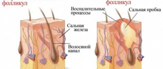
- Folliculitis is another reason why a slight thickening appears on the skin. The disease appears when infection or dirt gets into the hair follicles, where the inflammatory process develops. In most cases, this disease affects people who are overweight, have diabetes, or have weak immune systems. In this case, there is no need to remove the formation. A course of antibiotics and anti-inflammatory drugs is sufficient for treatment.
- A furuncle is a purulent inflammation of the hair follicle. The problem can be identified by severe itching and pain when pressing. This compaction appears when personal hygiene rules are violated, the immune system is weak, or hypothermia occurs. The development of the boil occurs within 2-4 days. These days, the lump gradually increases in size, acquires a red tint, is very painful and has a white core - pus.
- Inflammation of the lymph nodes on the side of the neck on the left or right. It is characterized by mild pain when pressed in the area of enlargement, as well as near the ear or under the lower jaw. Several lymph nodes can become inflamed at once, for example, in the armpits or groin. In addition, the person experiences a severe headache, weakness or chills, and there is also an increase in temperature to 37-38 degrees. This happens when a virus enters or during an inflammatory process of any organ. Enlarged lymph nodes on the neck on the right or left without pain are a dangerous signal. At this time, the body may develop lymphogranuloma or lymphocytic leukemia.
- A malignant tumor is characterized by a hard structure that resembles a stone when pressed. During palpation, severe pain appears, the formation practically does not move to the side. With the development of a malignant process, the tumor does not go away on its own, but only increases in size. A painful lump often occurs when cancer cells appear in the lymph nodes or thyroid gland.
Making a diagnosis on your own and starting treatment is not the best idea. Otherwise, the signs may go away and the disease will be asymptomatic. This will make it difficult to make a correct diagnosis and prescribe effective therapy.
The mechanism of formation of a blood bubble on the oral mucosa
Bloody blisters in the mouth in most cases are not life-threatening. They are formed as a result of mechanical damage to the mucous membrane. When microtrauma occurs, harmful microorganisms attack the damaged area.
After this, a number of responses are activated in the human body:
- The immune system is activated. Monocytes and leukocytes, as well as macrophages, instantly arrive at the damaged area, attacking the harmful pathogen and quickly destroying it.
- Immune cells die. This is a signal for other cells and substances are released in the affected area that are mediators of inflammation of the mucous membrane - serotonin, histamine and bradykinin.
- These substances cause a strong spasm of the circulatory system and the outflow of blood is hampered. After the spasm is relieved, all accumulated blood immediately flows to the site of inflammation. It moves at high speed and under pressure. A detachment of the mucous membrane occurs in the mouth, and a bloody blister appears.
How to recognize the disease
When a ball appears under the skin on the neck, a person may not feel any worsening of the condition at all. You should carefully monitor the well-being and behavior of the seal.
During the development of malignant tumors, a person sometimes does not experience pain in the area of the lump. The lump often reaches large sizes and is visible to the naked eye even under clothing. Most often, cancer is accompanied by weakness, chills, loss of strength, itching, loss of appetite, sudden weight loss and a temperature of up to 38 degrees.
If the lump is a wen or atheroma, then it does not hurt and moves under the skin. The boil develops quickly and causes severe pain, as well as itching and redness of the tissue.
Types of neoplasms
Damage to the skin as a result of cell proliferation forms compactions of various types and sizes: from a pea to a walnut. Depending on the nature of the pathology of the formation, there are:
- benign;
- malignant;
- borderline (precancerous).
The formations cause physical and aesthetic discomfort to the child, but the main danger is their degeneration from a benign tumor to a malignant one. Only a doctor can determine the nature of the formation under the skin.
In this regard, we note that in addition to the otolaryngologist, solving problems of this kind may involve visiting other highly specialized specialists. Depending on the etiology and nature of the seals, treatment can be carried out by a dermatologist, surgeon, endocrinologist and oncologist. If necessary, the ENT doctor will refer the patient to the appropriate specialist.
Diagnosis and treatment of the disease
To accurately find out the diagnosis and begin treatment, you should first visit a surgeon. The doctor sends the patient for an ultrasound examination and, possibly, to an oncologist. However, this does not mean that a malignant tumor has been found in the body. A thorough examination by several doctors can help rule out certain diagnoses and prescribe effective therapy. Other necessary research procedures may also be prescribed: general blood test, x-ray, biochemistry and biopsy. In order not to blur the picture of the disease, it is not recommended to use medications before going to a specialist, and especially not to try to squeeze out or open the subcutaneous ball.
After undergoing the necessary diagnostic procedures, the patient is prescribed treatment. The following treatment regimen is possible:
- taking antibiotics, anti-inflammatory or antiviral drugs when an infectious disease is confirmed;
- a small incision or puncture to remove fluid from cysts and wen;
- surgical removal of a cancerous tumor, wen or cyst.
You can relieve inflammation using folk methods, for example, compresses with medicinal herbs, honey, lard or Vishnevsky ointment. It is strictly prohibited to use iodine or alcohol! They have a warming effect and increase the inflammatory process.
Sakania Luiza Ruslanovna
Dermatovenerologist, cosmetologist, trichologist
Ask a Question
If after treatment there is no relief within 2 weeks, you should definitely visit your doctor again. Also, while taking medications, you must give up alcoholic beverages and smoking.
Causes of development of lateral neck cyst
A lateral neck cyst or branchiogenic tumor (the name of the disease comes from the Greek word “kystis” - bubble) refers to congenital benign tumor formations, which is a consequence of pathological embryonic development of the fetus. Long-term studies indicate a direct connection with abnormal intrauterine development of the fetus in the early stages of pregnancy.
Already from the fourth week of pregnancy, the formation of the gill apparatus in the embryo occurs, consisting of 5 pairs of cavities or “gill pockets”, gill slits and arches connecting them, involved in the development and formation of the structures of the child’s head and neck. With normal anatomical development of the fetus, the gill pouches gradually become overgrown, but in some cases, they persist and form various types of cysts (lateral, median).
A ball under the skin on a child's neck
The appearance of a lump on the left or right side of a child’s neck will tell not only about the diseases described above, but also the so-called childhood diseases - mumps, mononucleosis and others. Children may also develop lipomas, atheromas, tumors and boils. To determine the exact cause, you need to visit a pediatrician or pediatric surgeon.
Most often, young patients are diagnosed with a wen, which is not dangerous to health. The cause of compaction is also the lymph nodes. Most often, such a ball does not reach more than 1 cm. Lymph nodes become inflamed during infectious diseases in the body, diseases and teething, otitis, measles, acute respiratory infections or inflammation of the tonsils. It is necessary to urgently visit a doctor if:
- the compaction became very hard and increased sharply;
- the lump hurts a lot and cannot be moved with your fingers when pressed;
- the area around the growth has changed color to bright red or blue, and is also covered in a rash;
- a core of pus appears on the ball, which begins to ooze over time;
- Several more lumps appeared both near the first bump and in other places.
If the child was previously diagnosed and prescribed treatment that does not bring results within 7 days, this is a reason to undergo the examination again. Perhaps there was an error in the analysis or the wrong medications were selected.
There is no need to be afraid of a lump, even if it has appeared and is rolling under the child’s skin. It is better to identify the pathology at an early stage and begin treatment. Therefore, if even a small nodule is detected, it is recommended to seek help from a surgeon. Reduced immunity, severe stress against the background of a suspicious ball and refusal of therapy will only worsen the condition.
You can ask your question to our author:
Characteristics of a blood bubble on the oral mucosa
The mucous membrane protects the entire body from the negative influence of the environment, from harmful microorganisms, various types of pollution, and also has a fairly high level of regeneration. If blood blisters regularly appear on the oral mucosa, then you should take this signal seriously and take action.
A bloody ball in the mouth is a hematoma (bruise), which is characterized by the accumulation of blood in a certain place in the oral cavity. The appearance of bloody blisters is a kind of hemorrhage that occurs due to trauma to the capillaries and thin vessels of the mucous membrane.
A blister on the mucous membrane may contain clear serous fluid without the presence of blood. This means that the vessels were not damaged and the resulting wound is superficial. Such blisters on the mucous membrane heal much faster. The presence of blood in the bladder indicates a deep injury and a longer period of healing and blood resorption.
Prognosis for treatment of lateral neck cyst
The prognosis of a lateral neck cyst depends on the following factors:
- general health of the patient; - age; - the size of the cyst and its localization in relation to important organs and large vessels; — duration of cyst development; - forms of cysts - inflammation, suppuration; - contents of the cavity - pus or exudate; - type of fistula; - presence or absence of chronic diseases.
In general, the prognosis for treatment of a lateral neck cyst can be considered favorable. The risk of a lateral cyst degenerating into a malignant form is extremely low.
Diagnosis of lateral neck cyst
To confirm the diagnosis of “ lateral neck cyst ,” a differential approach to medical examination is required using modern diagnostic equipment - computed tomography, ultrasound, and, if necessary, puncture.
As a rule, the diagnosis of a lateral neck cyst is performed if the patient has a complication in which a noticeable increase in the size of the cyst, phlegmon or a characteristic abscess is already observed. When diagnosing a neck cyst, it is very important to exclude other diseases in the neck area that have similar manifestations and similar symptoms. A branchiogenic cyst can be differentiated from the following neck diseases: neck lipoma, lymphosarcoma, lymphangioma, neck teratoma, lymphadenitis, including the nonspecific tuberculous form, glomus tumor (vagus nerve), branchiogenic carcinoma, metastases from thyroid cancer.
Diagnosis of a lateral neck cyst consists of the following steps:
- complete collection of medical history, including hereditary factors; — external examination with palpation of the neck and lymph nodes; — computed tomography of the neck with contrast to clarify the size of the tumor, its location, type of fistula, contents of the cavity; — Ultrasound of the neck; — cyst puncture (according to indications); - fistulogram or staining of the fistula tract.
Is there a way to prevent branchiogenic cervical cysts?
Unfortunately, there are no preventive measures to prevent the development of this disease due to its nature of abnormal intrauterine conception. Most likely, this is a question for geneticists dealing with the problems of the etiology and pathogenesis of congenital anomalies of embryonic development. But, if a lateral cyst is nevertheless identified, then dynamic observation is recommended for children up to three years of age with periodic examinations every 3 months. Children should undergo regular medical examinations with a mandatory examination by an ENT doctor who can monitor the development of the tumor and eliminate various risks and complications.