29.11.2019
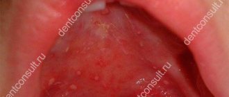
A growth on the mucous membrane of the mouth, located on the upper palate, causes discomfort to the wearer, sometimes accompanied by pain. A lump on the roof of the mouth occurs due to several completely different diseases. Each disease has its own characteristics and causes, but their clinical characteristics are similar. Without the qualified help of a doctor, it is impossible to determine an accurate diagnosis and effective method of therapy.
Factors contributing to the formation of bubbles:
- Oral diseases: caries, pulpitis, periodontitis, stomatitis;
- Unbalanced diet;
- Allergy to toothpaste or mouthwash;
- Injuries;
- Poor hygiene;
- Hereditary predisposition;
- Hormonal imbalance;
- Immunity impairment;
- Alcohol and nicotine addiction.
Watery blisters on the mucous membrane in some cases can indicate the occurrence of serious diseases. Such manifestations occur with tuberculosis, syphilis or diabetes. Therefore, if such a pathology occurs, it is recommended to immediately make an appointment with a doctor and undergo the necessary examination. The first sign is the sensation of a strange object in the mouth that is in the way.
Preliminary determination of lump symptoms
A lump on the palate is the cause of a dental disease or inflammatory process. Each type of growth has its own causes and subtleties of symptoms, and therefore requires separate consideration.
Angioma
Angioma is a tumor in the palate caused by disruption of processes in the tissues of the blood vessels of the soft palate. The shape of the cone resembles a rolled corkscrew. The tubercle has a blue or purplish-black characteristic color. The ball is a product of a disorder in the development of blood vessels, so the color is caused by the excess amount of blood in the formation.
Pressure causes bleeding, which should not be experimented with. There is a pulsating response to pressure.
Such a lump on the upper palate is life-threatening for the patient. Bleeding is difficult to stop, and large blood loss is fatal.
At the first signs of such compaction, you must seek qualified help.
Cyst
A lump in the mouth on the palate is classified as a cyst when a hard ball appears on the palate and its size does not exceed 12 cm. The cyst appears due to a disorder of the sebaceous glands. It won’t hurt, but it will make it significantly more difficult to eat, and then completely disrupt the correct functioning of these secretions.
A cyst on the palate requires surgery and can only be treated with it.
Pemphigus
Children are most susceptible to developing pemphigus. It pops up in the shape of a white ball at the top of the mouth. The bumps are a consequence of erosion and later develop into ulcers. When pressed it bleeds and hurts. Diagnosed by visual examination and using the Nikolsky method.
If the tumor is not removed, erosion will develop into massive exfoliation of the oral epithelium, disruption of digestive processes, and general weakness. If there is no treatment, the bumps become widespread. Pressure causes rupture of tumors, the contents of which cause intoxication of the body.
Myxoma
A myxioma is a hard, white growth at the top of the mouth. The disease affects the hard part of the palate, and upon visual examination it is almost invisible; pressure on the tumor is not felt. This significantly complicates diagnosis and delays the patient’s visit to the doctor.
The diagnosis can be confirmed or refuted by a biopsy of the palate.
Oncological disease
Cancerous lump is classified into two diseases:
- Hard palate cancer - starting from the bone tissue between the nasopharynx and the palate, the disease spreads to all mucosal tissues.
- Cancer of the soft palate - a lump appears due to an oncological process in the muscle and mucous tissues of the mouth.
Additionally, the oncological tubercle on the roof of the mouth is divided according to the tissue from which the spread of the disease began:
- Cylinder - the maternal tissue is the tissue of the glands, the cancer spreads quickly and invades the oral cavity;
- Adenocarcinoma - begins expansion of the mouth from the soft tissues of the cavity;
- Squamous cell carcinoma is a malignant formation that begins to develop from the tissues of the mucous membrane.
Symptoms
- Painful sensations when eating occur if a bubble has formed in the gum area or on the tongue;
- Pain when talking or smiling appears when a formation occurs on the inner surface of the lip;
- Pain in the throat area occurs if the bubble is located at the root of the tongue.
Transparent and bloody blisters can be not only inflammatory in nature. If the formation does not cause pain and is not accompanied by swelling, then you should consult a dermatologist. It is not recommended to treat yourself in such a situation, as it can be dangerous to health.
If the formation on the gum causes discomfort
If a bubble near a molar causes pain, this indicates an infectious inflammatory process. Discomfort is also typical with gum injuries and hematomas. Painful formations can be caused by a cyst, cancer, fibroma, periostitis, periodontitis.
Gingivitis - the disease affects only one gum, and the periodontium near the molar remains uninjured. The main symptoms of the pathology are swelling, bleeding, and peeling of the epithelium. Gingivitis often occurs against the background of halitosis. Sometimes pathology develops as a result of endocrine and metabolic disorders.
When treating, it is necessary to eliminate the cause of gingivitis. Professional oral hygiene, diagnosis and treatment of metabolic disorders are performed. To eliminate inflammation and prevent further spread of bacteria, antibiotic therapy is administered. For pain and severe discomfort, analgesics are prescribed.
Periodontitis - purulent bumps on the gums appear as a result of degeneration of the alveolar process, through which the tooth root is held in the alveolus. Tissues can be destroyed due to poor oral hygiene and internal pathologies.
In the initial stages, periodontitis is treated by teeth cleaning at the dental clinic and further proper care. An advanced form of the disease, in which the teeth become loose, pus accumulates, requires surgical intervention - restoration of the alveolar processes or removal of molars.
Periostitis or flux is a dense formation near a problematic molar with carious lesions. Patients complain of pain radiating to the temple, chest, neck, ear. The condition gradually worsens and the temperature can rise to 38 degrees. In the oral cavity, lesions due to periostitis are hyperemic. A purulent fistula forms. When the discharge is removed, the discomfort decreases.
Causes of periostitis: trauma, periodontitis, osteomyelitis, weak immune system, infectious pathologies occurring in the body and vitamin deficiency.
Treatment methods depending on the cause:
- If the formation occurs due to a viral infection, then treatment will be based on the use of antiviral drugs;
- If the cause is injury, then it is necessary to eliminate the consequences and rinse the mouth with antiseptics;
- If an allergic reaction provokes an illness, then you should find out what the irritant is and not use it anymore. The doctor will also prescribe antihistamines;
- If a large bloody bubble has formed, then surgery is performed. After this, the doctor prescribes medications for rapid tissue regeneration and restoration.
Ball in mouth - what is it?
When the inflammation begins to gradually progress, a blister appears on the gum. The main reason for the formation of a lump is poor oral hygiene and prevention. But the ball can also form under the influence of other factors: inflammation occurring in the roots of the teeth, gums, mucous membranes, and periosteum.
The problem is that the disease can be diagnosed in the later stages, with the development of a purulent process. This happens because the patient does not visit the dentist. For patients who experience fear while in a chair, the doctor can give sedative anesthesia, in which the receptors are turned off and the patient falls into a shallow sleep.
Attention! It is better to diagnose the disease early in order to prevent more dangerous consequences, including blood infection and inflammation of the entire jaw.
Treatment of cones
The treatment methodology depends on the specific case. The approach is determined by:
- the presence of pain;
- part of the cavity that has been infected (upper, lower);
- time elapsed since the onset of the disease;
- reaction of the cone to pressure.
Treatment of angioma
Removing a lump of this type occurs in three stages:
- Drug treatment and treatment;
- Surgery;
- Radiation therapy.
At the first stage, alcohol is used: it constricts blood vessels and helps eliminate high blood loss during surgery.
Then, in the case of the capillary form, radium therapy is used, it consolidates the effect of treatment, and sometimes can act as an independent drug and cure the disease completely.
It is prohibited to treat the cavernous form of angioma with radium. It can help transform the lump into a malignant one, which can greatly worsen the patient’s condition.
Treatment of pemphigus
A lump of this type that appears on the palate is treated mainly with antibiotics and a diet that includes a high content of proteins and vitamins, but without salt.
If such a lump appears, it is necessary to use disinfectant solutions. In severe forms, blood transfusions are used.
Treatment of myxoma and cysts
In these forms of the disease, the attending physician prescribes antiseptic drugs and prepares the cavity for surgery (if there are signs of accelerated growth of the lump into the tissue).
Additional methods of electrical treatment are also used, which make it possible to artificially kill tissue without the use of a scalpel, but all methods of this type are dangerous, thanks to them, a malignant formation may appear in place of a benign one.
Treatment of cancer
Cancer requires immediate intervention by a qualified specialist. The earlier the stage of the disease, the less harm the treatment and the disease itself will cause to the body. If a cancer lump appears, the following treatment approaches are used:
- Radiation therapy – the cancerous growth is irradiated with X-rays. In the early stages, it completely cures the disease.
- Surgical intervention - not only the harmful lump is cut out, but also the tissue around it, in order to exclude relapses. Such an intervention leaves defects on the face, which can later be corrected with plastic surgery.
- Chemotherapy - taking cytostatics.
For the cancer in question, chemotherapy is effective only in combination with radiation and surgery.
It is easier to defeat any disease at its inception stage. But in the case of bumps on the palate, it is better not to have the disease than to treat it later.
The root causes of the disease have not been fully established. Therefore, following the hypothetically formed rules will not completely protect you from the disease, but it will definitely reduce the chance of developing a similar problem. A preventative visit to the dentist will ensure timely treatment, even if the lump pops up unnoticed.
Category Miscellaneous Published by Mister dentist
Prerequisites for the occurrence of a lump
Modern medicine has not created a complete list of reasons why bumps may appear. Doctors' reasoning is based on hypothetical cause-and-effect relationships. The appearance of a lump on the palate is caused by:
- Bad habits (smoking, alcohol, oral drugs);
- The presence of micro and macro injuries to the oral cavity (surgeries, scratches of the upper parts of the oral cavity);
- Availability of dentures;
- Viral infections;
- Intrauterine disorders (hemangioma is a congenital disease and acquired from the mother);
- Violation of the activity and integrity of the mucous membrane (typical of angina);
- Congenital and acquired dysfunction of the glands (the main cause of cysts).
There are two more hypothetical reasons for the appearance of malignant tumors:
- Eating too hot and spicy foods, constantly disrupting the structure of the cells of the oral cavity;
- The presence of papillomatosis or leukoplakia - diseases that are precancerous lumps that can develop into an oncological disease.
Diagnostic methods
To determine the method by which a lump on the roof of the mouth will be treated, the causes of its occurrence are studied. The diagnostic procedure is carried out by a dentist.
Regular visits to the dentist are important to prevent the disease and determine whether a lump has appeared on the roof of your mouth. It needs to be treated at an early stage.
For diagnosis use:
- Radiography;
- Biopsy;
- Blood analysis;
- Radioisotope study;
- Ultrasound.
If you comprehensively examine the ball on the roof of your mouth, this will confirm or deny the presence of a malignant formation, and if it is absent, it will determine the exact diagnosis of a benign one.