Last update: 03/03/2021
In case of burns, regardless of their severity, the skin takes the first blow. The most serious harm is caused to it: even a 1st degree burn leaves redness on the surface of the skin, what can we say about deeper lesions (after which the removal of dead tissue is sometimes required).
How do you understand that a burn requires treatment? Our skin took care of this.
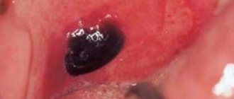
When you receive a burn of 2nd degree or higher (there are 4 in total), blisters filled with a yellowish liquid - exudate - form on the damaged surface of the body. These bubbles appear directly where the burn occurred, so even if the victim has lost consciousness or has a temporary loss of sensation, the burn site is easy to detect by these characteristic bubbles.
Causes of watery calluses
A water callus on the toe, heel, or foot occurs as a result of wearing tight shoes.
Most often, traumatic friction on the surface of the skin is created by decorative membranes, toes, heels or hard elements of new shoes. The occurrence of water callus is promoted by:
- tight shoes;
- sweaty feet;
- swelling of the limbs;
- shoes made of artificial materials;
- sand or other abrasives on the surface of the skin.
The mechanism of formation of water calluses
As a result of pressure or friction on the surface of the skin, redness, swelling and peeling of the epidermis occurs. The resulting cavity (bubble) is quickly filled with intercellular fluid. The occurrence of dropsy is a physiological process of protecting the body from the penetration of staphylococcal or streptococcal infections into injured skin.
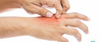
At the slightest touch or pressure, the water callus causes significant pain. If friction against the surface of the skin continues, the filled bubble bursts, leaving a red, weeping wound on the inflamed area.
Signs of water blisters infection:
- temperature increase;
- purulent discharge from the callus;
- expansion of the boundaries of redness;
- change in color of water callus;
- the appearance of yellow flaky areas around the callus;
- pain in the inflamed area even without mechanical impact.
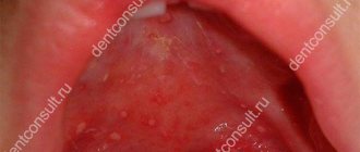
Blisters on the skin
Bubbles are the primary morphological elements of an exudative nature, cavitary, contain fluid and slightly rise above the skin level.
In the vesicles there is a cavity filled with serous, less often serous-hemorrhagic contents, a tire and a bottom. The blisters can be located under the stratum corneum, in the middle of the epidermis, and between the epidermis and dermis. They can be single-chamber and sometimes multi-chamber. In this case, it seems that the patient has a bladder, but it does not have partitions. The bubble size ranges from the size of a pinhead to the size of a lentil. The contents of the vesicle may be transparent, serous, or less often bloody. It often becomes cloudy and purulent, which occurs when the vesicle turns into an abscess. The liquid separated from the bubble dries into a crust or its tire bursts, an erosive surface is formed and weeping occurs.
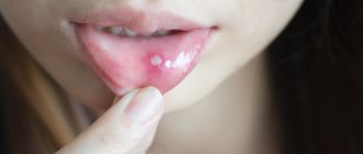
Blisters can be located on unchanged skin, but more often have an inflammatory erythematous base. On the oral mucosa and on the contacting surfaces of the skin, the bubbles quickly open, revealing erosive surfaces. In places with thicker tires, for example, on the palms with dyshidrosis, they last longer. The blisters pass without a trace or leave behind temporary pigmentation , as, for example, with Dühring's dermatitis herpetiformis.
When vesicles form, spongiosis (eczema, dermatitis), ballooning degeneration (simple vesicular and herpes zoster, chicken pox), intracellular vacuolization (dyshidrosis) are observed histologically.
Diagnostics
Water calluses on the feet are diagnosed as follows:
- an examination is carried out by a dermatologist
; - You may need additional consultation with a rheumatologist
, podologist or
orthopedist
.
Calluses should not be confused, for example, with inflammatory changes in joints, pinched nerves, inflammation of hangnails, genetically determined keratinization of the skin, and warts.
When diagnosing water callus, it is important to determine whether the patient has concomitant diseases. For example, varicose veins, diabetes mellitus, venous insufficiency, neuritis and so on.
What to do with a blister after a burn: treat it or get rid of it?
The burn bubble needs to burst, BUT a doctor must do this.
Before starting the procedure, the burn site and bladder are treated with an antiseptic solution.
The bladder is punctured with a sterile needle. The puncture should be shallow - you just need to burst the bubble itself from above; The procedure is practically painless and may sting slightly.
Using a sterile napkin, remove the liquid that has leaked from the bladder. The film is carefully removed from the surface of the skin, and the wound is treated with an antiseptic. A sterile bandage with an antimicrobial agent is applied to the burn site.
A healing ointment is also used to accelerate skin regeneration. The La-Cri restorative cream, which contains only natural ingredients, is perfect for these purposes. Its natural non-hormonal composition is designed specifically to eliminate all kinds of skin problems and cope with the consequences of burns:
- Panthenol and avocado oil accelerate the healing and regeneration process and soften the skin.
- Licorice has anti-inflammatory properties.
- Walnut extract protects against germs.
After applying the cream, the burn site should be covered with a sterile bandage to prevent dirt from getting into the wound.
If the procedure is carried out under sterile conditions, then you are not at risk of infection. And with regular use of the La-Cri healing ointment, the burn will go away very quickly and will not leave any traces.
How to cure water blister
What to do if a water callus has formed on your leg? Small dropsies quickly go away on their own. To protect against injury, it is recommended to cover the inflamed area with a bactericidal plaster for 1-2 days and not wear tight shoes.
Large water calluses on the feet cause aesthetic and physiological discomfort and therefore require treatment. The optimal therapeutic method is opening or puncture. It is recommended to pierce the callus on the first day the blister appears.
Is it possible to pierce an inflamed callus yourself? In order to avoid wound infection, all manipulations should be carried out with clean hands and a sterile needle. If it is not possible to ensure sterility and thorough surface treatment, it is better to open the dropsy in a doctor’s office.
Self-puncture of a watery callus on the leg is carried out as follows:
- Treat the callus and surrounding skin with an antiseptic solution (alcohol, brilliant green, hydrogen peroxide, iodine).
- Take a sterile needle from an injection syringe. If you don’t have such a needle, you can heat a regular pin over a fire or disinfect it in medical alcohol.
- The needle is inserted into the water callus parallel to the surface of the skin (on the side of the bladder). It is not recommended to pierce water blisters on the legs in the central part because... this can injure the bottom of the callus.
- For good outflow of fluid, it is necessary to make 2-3 punctures, while maintaining the integrity of the walls of the bladder.
- When the punctures are made, the water bubble is slowly pressed against the surface of the skin with a sterile bandage or gauze to squeeze out all the liquid present in the cavity.
- A bactericidal patch is applied to the dropsy. The walls of the released bladder cover the inflamed skin inside the callus, protecting the wound from infection.
- The patch or bandage on calluses must be changed 2 times a day and removed at night.
There are cases when, after a few days, the water callus is again filled with intercellular fluid. Then the puncture should be repeated.
Water bladder suppuration
If there was suppuration in the callus, it is necessary:
- open the water callus and remove the infected bladder walls;
- treat the wound with an antiseptic;
- apply antibiotic ointment to the weeping wound;
- cover the affected area of skin with a gauze pad and adhesive tape.
The inflammatory process and suppuration in the water callus, which are accompanied by an increase in body temperature and the growth of the area of redness, are localized on an outpatient basis, in the surgical office. The specialist will clean the wound area, apply a bandage with drainage if necessary, and prescribe antibacterial therapy.
Treatment of water callus with folk remedies
- A proven folk method for treating water blisters on the legs and arms is salt baths. A warm saline solution (1 teaspoon per 1 liter of water) or a strong solution of potassium permanganate disinfects well and softens the callus, accelerating healing.
- Aloe juice is considered another effective remedy. Aloe pulp is a natural antiseptic that relieves inflammation and accelerates healing. A plant leaf cut along the surface or a swab soaked in aloe juice is placed on the water bladder. This compress is fixed with a bandage and changed every 4-6 hours. To treat a small callus, you can use regular plantain. A leaf washed in cold water must be mashed until the juice appears and applied tightly to the callus. After 4-5 hours, the application is repeated.
- Lemon has a good antiseptic and softening effect. For water blisters, it is recommended to apply an application of grated pulp and lemon peel to the damaged area of skin and hold for up to 5 minutes. It will be enough to apply 2 compresses during the day so that the next day the pain goes away and the callus decreases.
If the water bubble bursts on its own
Due to excessive pressure or mechanical damage, the water callus may burst. Spontaneous opening is accompanied by painful discomfort and burning. The open wound should be disinfected and covered with a bactericidal bandage. If after a few days the area of redness around the callus increases and suppuration is noticeable, such symptoms indicate infection of the burst callus and require immediate contact with a specialist.
Water calluses on the soles require special attention. If the skin on your feet is rough and prone to calluses, dropsy can become a painful ingrown callus.
What is the danger of blistering after a burn?
First of all, you should know that the appearance of blisters in itself indicates that the skin damage is quite serious. Bubbles form directly at the burn site (in contact with hot objects and open flames, contact with chemicals, prolonged exposure to the sun, irradiation). Therefore, the nature of the resulting skin lesion can be judged by the condition of the skin covered with blisters. You should immediately consult a doctor in the following cases:
- If the burn is 1st degree, but more than 10% of the body area is burned (you can take your palm as a “measuring device”, the size of which is about 1% of the skin area), if the burn is 2nd degree, and the affected area is more than 1%, and if the burn is received 3rd and 4th degrees.
- If a large number of blisters appear on the skin, which quickly combine into large blisters against the background of swelling of the damaged part of the body.
- Severe pain, loss of sensation in the burn area.
- Burn of any degree in the face, groin area, joints.
- A child and an elderly person were injured.
Clinical researches
A clinical study conducted by the company together with the Union of Pediatricians of Russia proves the high efficiency, safety and tolerability of products for daily skin care of children with mild and moderate forms of atopic dermatitis and during remission, accompanied by a decrease in the quality of life of patients. As a result of therapy, a decrease in the activity of the inflammatory process, a decrease in dryness, itching and flaking was noted.
It has been proven that La Cree cream for sensitive skin reduces itching and irritation, relieves redness of the skin, moisturizes and gently cares for it. The product is recommended by the Union of Pediatricians of Russia.
Sources:
- Kovyazina N.A., Fedosimova N.A., Illek Ya. Yu. Diagnosis of atopic dermatitis in young children, Vyatka Medical Bulletin, 2007
- Smirnova G.I. Managing the course of the disease: atopic dermatitis in children, Russian pediatric journal, 2014
- Dermatology: an illustrated guide to clinical diagnostics according to Professor A.N. Rodionov. / Rodionov A.N., Zaslavsky D.V., Sydikov A.A. Edited by Professor A.N. Rodionova.M.: Border. 2022.- 944 p.