The appearance of a lump in the groin in men always indicates one or another pathology of varying severity. The compaction can be located either directly under the skin or in the deep layers of tissue, accompanied by painful sensations or not causing any discomfort at all. In any case, the cause of this condition must be clarified.
What is an abscess and what causes it?
An abscess is a painful pustule surrounded by inflamed tissue. Often it can be easily felt. Such a formation can appear on any part of the body, but most often it affects:
- armpits;
- area around the anus and vagina (Bartholin gland abscess);
- tissue around the tooth;
- skin in the groin area;
- hair follicles.
During their development, abscesses are filled with necrotic masses and can open on their own. However, it is best to consult a doctor in case of such a disease, who will open the purulent focus and clean (drain) it.
Causes of abscesses:
- as an independent disease, abscesses of the skin and soft tissues most often occur, caused by blockage of the openings of the sebaceous and sweat glands, the formation of cysts and the proliferation of pathogenic microorganisms there;
- purulent soft tissue abscess can be a complication of skin damage;
- a post-injection abscess develops when the drug is administered with a non-sterile syringe; in severe cases, a purulent process – phlegmon – may spread;
- such a process often complicates the course of any infectious diseases of a bacterial nature, for example, a peritonsillar abscess develops as a complication of tonsillitis;
- Abdominal abscesses can develop when microbes are transferred through blood vessels;
- in some cases, the cause of the disease is not bacteria, but protozoan microorganisms, for example, amoebic liver abscess;
- pathology may arise primarily due to the penetration of a large number of pathogens with high virulence (damaging ability) into the tissues, and a lung abscess may form.
Signs of an abscess occur more often in people with weakened immune systems. Risk factors for developing pathology:
- long-term treatment with glucocorticoids and chemotherapy drugs;
- diabetes mellitus, malignant tumors;
- diseases of the blood and hematopoiesis – sickle cell anemia, leukemia, HIV infection;
- Crohn's disease, ulcerative colitis;
- severe injuries or burns;
- alcoholism, drug addiction.
Other risk factors are exposure to a polluted environment, contact with dust, hydrocarbons, poor skin hygiene, atherosclerosis of peripheral arteries or severe varicose veins.
1.General information
The testicle and its epididymis, the spermatic cord, as well as orchitis, epididymitis, funiculitis - all these terms refer only and exclusively to men. Due to the evolutionary division into two sexes and the corresponding anatomical dimorphism, functionally similar formations in women are located inside, in the abdominal cavity; they are called differently, and only their inflammations are designated according to the same principle - by adding the “inflammatory” suffix “-itis”, generally accepted in medicine.
The term “abscess” is also universal. Its features (unlike inflammation in general) are the obligatory presence of a pyogenic infectious pathogen, localization in a limited area, disintegration (purulent melting in medical terminology) of functional tissue and the formation of a high-pressure capsule filled with exudate.
The testicles, as you know, are a paired organ of the male reproductive system, hidden in a leather pouch - the scrotum, which, in turn, is located in the groin at the base of the penis. Each testicle in its rear part, mainly at the upper pole, is equipped with the so-called. epididymis, a narrow labyrinth-incubator of sperm, which, when maturing, at the right moment enter through the duct in the spermatic cord (which also serves as a mechanical “suspension” of the testicle) into the seminal storage vesicle, are saturated with the energy carrier fructose, and from there they are released into the urethra, where mixed with the secretion of the prostate gland and form the final composition of the fertilizing ejaculate, or sperm.
Sign up for a consultation
Inflammatory processes of the testicle are generalized by the term “orchitis”, of the epididymis - “epididymitis”, of both formations at the same time - orchiepididymitis. Any of these types of inflammation is dangerous in itself, and any of them can be further complicated by the formation of an abscess. In approximately every fifth case, inflammation leads to infertility; with a probability of about 65%, an atrophic process may begin in the testicular structures. In many cases, abscessation of the testicle and/or epididymis leaves no choice other than orchiectomy (surgical removal of the testicle). In other words, any signs of inflammation in these organs require immediate specialized medical care.
A must read! Help with treatment and hospitalization!
Symptoms and diagnosis of abscess
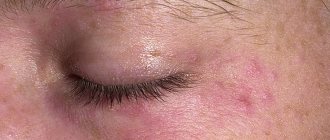
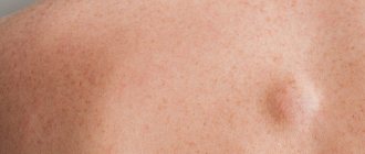
Symptoms of an abscess vary depending on its location. When skin and soft tissues are affected, the following is noted:
- skin redness;
- swelling and soreness;
- palpation of a soft space-occupying formation under the skin.
Abscesses of internal organs are accompanied by corresponding symptoms: cough, sore throat, sore throat, right hypochondrium, lower back, back, headache, dysfunction of the affected organs.
The infection causes signs of intoxication - high fever, headache, weakness, nausea. Sometimes the course of abscesses is not accompanied by pronounced symptoms; they become chronic, and in these cases, diagnosing the disease is difficult. Complications of an abscess are associated with the entry of microorganisms from its cavity into the blood vessels. At the same time, they spread throughout the body, causing sepsis (“blood poisoning”). This condition is life-threatening. Therefore, it is very important to identify and eliminate the purulent focus in time.
Diagnosis of an abscess is carried out by external examination. To detect ulcers in internal organs, ultrasound, CT, MRI, and radiography are used. Blood tests reveal signs of inflammation, but they do not provide information about the location of the pathology.
Inguinal purulent funiculitis. Review of clinical observations
Journal "Experimental and Clinical Urology" Issue No. 2 for 2015
Prokhorov A.V.
The term “funiculitis” means inflammation of the membranes, fiber and elements of the spermatic cord (vas deferens, blood and lymph vessels, nerves) caused by banal or specific microflora [1, 2]. Currently, this term has become broader and unites many acute and chronic inflammatory diseases of the spermatic cord of various (autoimmune, infectious, pseudotumor and parasitic) natures, differing in clinical manifestations, course and prognosis [3, 4]. Funiculitis is often combined with inflammation of the vas deferens (deferentitis) and can be its complication (or vice versa), which confuses the terminology of these diseases [2].
BRIEF REVIEW OF LITERATURE. RELEVANCE
Inguinal purulent funiculitis (PGF) is a rare, insufficiently studied and unfamiliar disease to a wide range of surgeons, urologists and radiologists. This pathology was first described by NR Smith in 1834 as inguinal phlegmon of unknown etiology [2]. According to K. Eddy et al., in the English-language literature for the period from 1933 to 2011. Only 4 cases of PHF are described [5, 6]. In total, the literature reports 12 cases of PHF in patients of different ages, mainly middle-aged and elderly [2-11].
The etiopathogenesis of PHF remains unknown. PGF is considered as a phlegmon or abscess of the spermatic cord [2, 7], as a segmental infected dilatation of the inguinal part of the vas deferens [8], as a purulent vasitis (resp., deferentitis) [3-6, 9-11]. It is assumed that PHF is caused by aerobic and anaerobic pyogenic flora, Mycobacterium tuberculosis, Haemophilus influenzae Pfeiffer, and fungi. In most cases, the causative microflora and the source of infection at the time of clinical manifestation of PHF cannot be identified, which is considered as one of the features of this disease [11]. In this regard, it has even been proposed to name the disease as idiopathic PHF [2]. It is believed that the route of infection spread to the membranes of the spermatic cord is hematogenous [2-6].
In PHF, the inguinal part of the spermatic cord is affected [2-11]. PHF occurs in two clinical and pathomorphological forms (in the form of phlegmon and abscess of the spermatic cord), occurs as an acute surgical disease and can be the cause of diagnostic errors, simulating a strangulated inguinal hernia, an abscess of the anterior abdominal wall or acute disease of the scrotal organs [3-6, 10 , eleven].
In the diagnosis of PHF, many authors have noted the high effectiveness of computed tomography (CT), which in the presented literature observations made it possible to identify phlegmon or abscess of the spermatic cord before surgery and exclude other acute surgical diseases, in particular, strangulated inguinal hernia [5, 6, 8-14] .
Treatment of PGF is only surgical (by percutaneous puncture and aspiration of the spermatic cord abscess under ultrasound navigation or by open surgery - revision and drainage of the spermatic cord membranes - in case of phlegmon) [8-10]. With timely surgical treatment, the prognosis of the disease is favorable. Death due to sepsis was observed in 3 (25%) of the described cases of PHF [2].
The prognosis for PHF can be different and depends on the clinical and pathomorphological form of the disease, the timeliness of diagnosis and treatment, as well as the immunoreactivity of the body. In immunocompromised patients, PHF is characterized by a septic course and leads to death [2, 5, 6]. In patients of reproductive age, the course of PGF can be complicated by the excretory form of infertility due to the development of stricture or obliteration of the vas deferens as a result of inflammation [1, 2].
Over a 20-year period (from 1994 to 2014), two cases of PHF were observed in the City Clinical Urological Hospital No. 47 and the City Clinical Hospital No. 57 of the Moscow Department of Health, working in emergency urological care, which caused serious diagnostic difficulties. We present these observations as an illustration.
CLINICAL CASE NO. 1
Patient K., 42 years old, was admitted to the clinic in September 2009 for urgent reasons with a referral diagnosis: acute left-sided epididymo-orchitis, hydrocele on the left. Complains of enlargement and pain in the left groin area and the left half of the scrotum, hyperthermia up to 38° C with chills for 1.5 weeks, weakness, sweating. Got acutely ill. The disease is associated with hypothermia. He had a history of repeated urogenital infections (gonorrhea, trichomoniasis), and in childhood he had a right orchiectomy for inguinal cryptorchidism, which led to testicular atrophy. Denies tuberculosis, HIV, hepatitis B and C. General condition of moderate severity due to inflammatory intoxication. Body temperature – 37.5°C. Correct physique, normal nutrition. The skin is pale with an earthy tint. No pathological abnormalities were identified in the internal organs and organ systems. Blood pressure – 130/70 mm. Hg Art., pulse – 92 beats/min. On examination, an increase in the left groin area and the left half of the scrotum was noted. The skin of the left groin area is hyperemic, edematous, and the local temperature is elevated. In the projection of the left inguinal canal, an ovoid-shaped, tight-elastic and sharply painful formation of 7x3 cm, limited by the anatomical boundaries of the inguinal canal, is palpated. The color of the skin of the scrotum and the local temperature are not changed, the folds of the skin of the left half of the scrotum are somewhat smoothed out. The left testicle, epididymis and scrotal part of the spermatic cord are not changed upon palpation. There is a small accumulation of fluid in the membranes of the left testicle (hydrocele). The right testicle is not visible in the scrotum (removed). No pathological discharge from the urethra was noted. During digital rectal examination, the rectum, prostate gland, seminal vesicles and ampullary sections of the vas deferens are not changed. Blood tests revealed moderate anemia (hemoglobin - 108 g/l, erythrocytes - 4.0x10⁶), leukocytosis to 16x103, a shift of the blood count to the left to juvenile forms, an increase in ESR to 43 mm/hour. No pathological changes were detected in urine tests. X-ray of the chest organs did not reveal focal infiltrative changes in the lungs; diffuse enhancement and deformation of the pulmonary pattern (chronic “smoker’s” bronchitis) were detected. Ultrasound examination (US) in the projection of the left inguinal canal revealed a liquid heterogeneous formation of a “cigar-shaped” shape 9.5x3.5x4.2 cm (280 cm3), completely occupying the inguinal canal and limited by its walls, pushing the elements of the spermatic cord posteriorly (Fig. 1 , 2, 3, 4). Magnetic resonance imaging (MRI) was performed, which confirmed the ultrasound data and additionally detected a high protein content in the effusion of the left inguinal canal, which indicated its purulent nature (Fig. 5, 6). Differential diagnosis was carried out between a suppurating cyst (as the most likely diagnosis) and a hematoma (as the least likely diagnosis) of the spermatic cord. During an exploratory inspection of the inguinal canal, the presence of a purulent cystic formation of the spermatic cord was confirmed, which was opened, excised and drained. About 250 ml of thick pus was evacuated; the walls of the inguinal canal and the membrane of the spermatic cord were saturated with pus, thickened and compacted. During the operation, it was suggested that the congenital funiculocele may be suppurating. A pathohistological examination revealed a picture of diffuse purulent inflammation of the membranes and tissue of the spermatic cord. When inoculating the purulent contents, no flora growth was detected. The outcome of the disease is recovery.
Rice. 1. Case No. 1. Patient K., 42 years old. Panoramic longitudinal echogram of the left inguinal-scrotal region. 1 funiculopyocele, occupying the entire inguinal canal, 2 – unchanged testicle and epididymis, 3 – small hydrocele
Rice. 2. Same patient. Targeted ultrasound of the left inguinoscrotal area. 1 – funiculopyocele, 2 – unchanged funicular part of the vas deferens
Rice. 3. Same patient. Transverse echogram of the inguinal canal on the left in the middle third. 1 – funiculopyocele, 2 elements of the altered spermatic cord
Rice. 4. Same patient. Doppler angiogram of the spermatic cord on the left in the middle third of the inguinal canal. 1 – funiculopiocele, 2 – vessels of the spermatic cord
Rice. 5. Same patient. MRI. Frontal cut. A – T1VI, B – T2VI. 1 – funiculopiocele on the left, 2 – hydrocele on the left, 3 – the right testicle is absent in the scrotum (removed)
Rice. 6. Same patient. MRI. T2VI. Axial section at the level of the middle third of the inguinal canal on the left. 1 – funiculopiocele, 2 – spermatic cord
CLINICAL CASE NO. 2
Patient S., 63 years old, underwent radical prostatectomy in August 2010 due to T1N0M0 prostate cancer. The early postoperative period was complicated by a suppurating pelvic lymphocele with a volume of 650 ml, which was drained openly. Wound healing by secondary intention. He was discharged on the 24th day after surgery in satisfactory condition. 1 month after discharge, he was admitted to the clinic again with complaints of enlargement and pain in the left groin area and the left half of the scrotum, hyperthermia up to 38°C with chills for 5 days. General condition of moderate severity, caused by purulent-inflammatory intoxication. In the left groin area, a fixed, tight-elastic and sharply painful formation 10x4x3 cm is palpated, the skin over it is swollen and hyperemic (Fig. 7). Acute epididymo-orchitis and strangulated inguinal hernia were excluded during clinical and echographic examination. The assumption of an inguinal lymphocele was also rejected, since dissection of the inguinal lymph nodes was not performed during the previous operation. Ultrasound of the inguinal canal on the left revealed an encysted accumulation of heterogeneous fluid in the membranes of the spermatic cord (Fig. 8). Preliminary diagnosis: acute inguinal funiculitis with the development of reactive (inflammatory) funiculopyocele. During the revision of the inguinal canal and spermatic cord, about 150 ml of interthecal purulent exudate was evacuated, thickening and compaction of the walls of the inguinal canal and the membranes of the spermatic cord were noted. Pathohistological examination of the resected membranes of the spermatic cord revealed signs of acute purulent diffuse inflammation (Fig. 9). When sowing the contents of the spermatic cord membranes, no flora growth was detected. The outcome of the disease is recovery. The patient was discharged on the 20th day after surgery in satisfactory condition with a healed wound. Ultrasound 2 weeks after surgery revealed restoration of the structure of the spermatic cord and the walls of the inguinal canal (Fig. 10).
Rice. 7. Case No. 2. Patient S., 61 years old. Asymmetrical enlargement of the right groin and scrotum (arrow). Hyperemia of the skin of the scrotum and groin area on the right. Right inguinal funiculitis
Rice. 8. Same patient. Panoramic longitudinal echogram of the right inguinoscrotal region. 1 funiculopyocele simulating an inguinal hernia, 2 – intact testicle and epididymis, 3 – hydrocele, 4 – abdominal cavity
Rice. 9. Same patient. Microscopic specimen of the spermatic cord membranes. Hematoxylin and eosin. Magnification: x 100. Foci of polymorphonuclear leukocyte and eosinophilic infiltration (1) with foci of hemorrhages (2). Acute purulent funiculitis
Rice. 10. The same patient. 14th day after surgery - opening and drainage of phlegmon of the spermatic cord on the right. Panoramic longitudinal echogram of the right inguinoscrotal region. Restoration of the structure of the spermatic cord and inguinal canal. 1 – spermatic cord, 2 – testicle, 3 – hydrocele, 4 abdominal cavity
DISCUSSION
The clinical manifestations and course of PHF in our observations did not differ from the cases of PHF described in the literature [2 – 11]. PGF clinically simulated various acute diseases of the inguinal-scrotal region: ranging from acute purulent epididymo-orchitis and ending with a suppurating hematoma of the spermatic cord. The final verification of the disease involved intraoperative and pathomorphological research methods.
The etiopathogenesis of PHF in the two cases presented remains not entirely clear. In the first case, taking into account the presence of a history of abnormalities of the genital organs (cryptorchidism), repeated urogenital infections and the results of exploratory revision of the inguinal canal, suppuration of the congenital funiculocele against the background of latent infectious deferentitis can be assumed as the pathomorphological substrate of PGF. As is known, congenital incomplete obliteration of the vaginal process of the peritoneum (processus vaginalis peritoneae Halleri) occurs in approximately 20% of children and can cause the development of funiculocele complicated by suppuration [12].
In the 2nd case, the occurrence of PHF was most likely preceded by pelvic purulent cellulitis and pelvic pyolymphocele, which arose as a complication of radical prostatectomy. It is known that complications of radical prostatectomy are common: every 4th – 5th patient [13]. Among them, purulent-inflammatory diseases of the kidneys, urinary tract, scrotum and pelvic tissue occupy first place, accounting for 40–60% of all postoperative complications [13, 14]. At the same time, the most likely route of spread of purulent infection from the bed of the removed prostate gland to the membranes of the spermatic cord is well studied: along the fascial sheath of the vas deferens to the membranes of the spermatic cord along the continuation - per continuitatem with the formation of funicolopyelocele [15].
Our observations noted the high diagnostic efficiency of high-resolution ultrasound (6 – 16 MHz) and MRI. Ultrasound played a major role; it made it possible to assess the nature of the space-occupying lesion of the spermatic cord and carry out a differential diagnosis with acute surgical diseases of the inguinal-scrotal region. MRI, in our opinion, is somewhat more accurate than ultrasound in assessing the extent of lesions of the spermatic cord, as well as in clarifying the nature of the effusion (pus, blood, lymph).
Treatment of PGF was surgical: revision of the inguinal canal, excision and drainage of the funiculopyocele. Surgical intervention was performed according to urgent indications for diagnostic and therapeutic purposes and in compliance with the principles of purulent surgery; As a result of treatment, both patients recovered.
It is proposed to differentiate PHF, first of all, from a strangulated inguinal hernia and acute diseases of the scrotal organs (acute epididymitis and testicular torsion), as well as hematoma, vein thrombosis and tumor of the spermatic cord, osteomyelitis of the pubic bones and symphysis [2-11]. In the differential diagnosis of PHF and acute diseases of the scrotal organs, the method of choice is gray scale ultrasound, supplemented by Doppler ultrasound; while when distinguishing between PHF and strangulated inguinal hernia, priority belongs to computed tomography (CT) [5, 6, 8]. To diagnose a tumor or hematoma of the spermatic cord, the use of MRI and high-resolution ultrasound is effective; for osteomyelitis of the pelvic bones, the use of pelvic radiography and CT is effective [2, 5, 6]. In our observations, the use of a comprehensive radiation examination, including high-resolution ultrasound (6-16 MHz) and MRI of the inguinal-scrotal region, allowed us to speak out about the nature of the disease of the spermatic cord and choose the necessary treatment tactics.
CONCLUSION
Inguinal purulent funiculitis is an extremely rare disease with an unknown etiopathogenesis. One of the likely mechanisms for the development of PGF is suppuration of congenital funiculocele or the development of acquired funiculopyocele against the background of urogenital infection or pelvic purulent cellulitis. A causative infection, presumably from the urethra, prostate gland, seminal vesicles and pelvic tissue, spreads to the membranes of the spermatic cord along the lumen of the vas deferens (canalicular way) or along its fascial sheath and further - from the vas deferens to the spermatic cord - per continuitatem . PHF occurs as an acute surgical or urological disease and causes serious diagnostic difficulties. In diagnosing PHF, the use of high-resolution ultrasound and MRI of the inguinal canal and scrotal organs is effective. The final verification of the diagnosis is carried out by intraoperative and pathomorphological methods. PHF must be differentiated using radiation methods with acute diseases of the inguinal scrotum area, and above all, with a strangulated inguinal hernia, acute epididymitis and testicular inversion. Treatment of PGF should be surgical (revision of the inguinal canal, opening and drainage of the membranes of the spermatic cord). With timely diagnosis and treatment, the prognosis of PHF is favorable.
LITERATURE
1. Tiktinsky O.L., Mikhailichenko V.V. Andrology (manual). St. Petersburg, 1999. pp. 62 – 80.
2. Wilensky AO, Samuels SS. Acute deferentitis and funiculitis. // Ann Surg. 1923. Vol. 78, N 6. P. 785 – 794.
3. Bissada NK, Redman JF. Unusual masses in the spermatic cord: report of six cases and review of the literature. // South Med J. 1976. Vol. 69. P. 1410 –1412.
4. Chan PTK, Schglegel PN. Inflammatory conditions of the male excurrent ductal system. Review. Parts II. // J Androl. 2002. Vol. 23, N 4. P. 461 – 469.
5. Eddy K, Pierce B, Eddy R. Vasitis: clinical and ultrasound confusion with inguinal hernia clarified by computed tomography. // Can Urol Assoc J. 2011. Vol. 5, N 4. P. E74 – E76.
6. Eddy K, Connell D, Gooodacre B, Eddy R. Imaging findings to prevent unnecessary surgery in vasitis: an under-reported condition mimicking inguinal hernia. // Clin Radiol. 2011. Vol. 66, N 5. P. 475 – 477. 7. Maitra AK. Odd inguinal swelling. // Lancet. 1970. Vol. 1. P. 45.
8. Gomez Herrera JJ., Zabia Galindez E, Carrera Terron R, Borruel Nacenta S. Dilatacion unilateral completa de conducto deferente como causa de massa inguinal. // Radiologia. 2013. Vol. 55, N 6. P. 533 – 536.
9. Bissada NK, Redman JF, Finkbeiner AE. Unusual inguinal mass secondary to vasitis. // Urology. 1976. Vol. 8. P. 488 – 499. 10. Ryan SP, Harte PJ. Suppurative inflammation of vas deferens: an unusual groin mass. // Urology. 1988. Vol. 31. P. 245 – 246.
11. Wolbarst AL. The vas deferens, generally unrecognized clinical entity in urogenital disease. // J Urol. 1933. Vol. 29. P. 405 – 412.
12. Garriga V, Serrano A, Marin A, Medrano S, Roson N, Pruna X. US of the tunica vaginalis testis: anatomic relationships and pathological conditions. // RadioGraphics. 2009. Vol. 29. P. 2022 – 2032.
13. . Froehner M, Novotny V, Koch R, Leike S, Twelker L, Wirth MP. Perioperative complications after radical prostatectomy: open versus robot-assisted laparoscopic approach. //Urol. Int. 2013. Vol. 90, N 3. P. 312 – 315.
14. Salomon L, Levrel O, Anastasiadis AG, Saint F, de La Taille A, Cicco A, Vordos D, Hoznek A, Chopin D, Abbou CC.Outcome and complications of radical prostatectomy in patients with PSA 10 ng/ml: comparison between the retropubic, perineal and laparoscopic approach. // Prostate Cancer Prostatic Dis. 2002. Vol. 5, N 4. P. 285 – 290.
15. Kovanov V.V., Anikina T.I. Surgical anatomy of human fascia and cellular spaces. M.: Medgiz, 1961. P. 169 – 185.
Magazine
Journal "Experimental and Clinical Urology" Issue No. 2 for 2015
Comments
To post comments you must log in or register
Abscess treatment
If you experience pain, swelling of the skin, the formation of a tubercle or a lump with a softened center, you should contact a surgeon.
- Treatment of an abscess is carried out under local anesthesia.
- The skin is disinfected, an anesthetic is injected, and the abscess is opened.
- After emptying the cavity, it is washed with an antiseptic solution and dried.
- A drain is inserted into the wound for 1–2 days and covered with a sterile bandage.
Immediately after such an operation, the patient’s well-being improves significantly. Additionally, the doctor may prescribe antibiotics and anti-inflammatory drugs. After a few days, the drainage is removed.
For abscesses of internal organs, the principle of treatment is the same - opening, cleansing, draining. Access to the desired organ may require extensive surgery or the use of endoscopic techniques.
Surgeons do not recommend treating an abscess at home, as this can lead to a delay in the process and the development of sepsis. However, people often use lotions with aloe juice, onion gruel, and compresses with rye bread. We advise you to immediately contact the clinic by making an appointment by phone.
Do not puncture or squeeze out the contents of the abscess. This can cause the infection to spread through the blood vessels.
Treatment at the Mama Papa Ya clinic
The network of family clinics “Mama Papa Ya” offers medical services for the treatment of abscesses by a qualified surgeon. The clinic's branches are located in Moscow and Moscow Region.
Our advantages:
- quick appointment of a patient without a long wait in line;
- immediate surgical care - opening and draining the abscess in a sterile office;
- comprehensive recommendations for caring for the postoperative wound to prevent complications;
- if necessary, consultation with other specialists (gynecologist, ENT doctor, neurologist and others);
- diagnosis of internal abscesses using multislice computed tomography.
To obtain a doctor’s consultation, you must contact our manager by phone or through the feedback form.
Reviews
Good clinic, good doctor!
Raisa Vasilievna can clearly and clearly explain what the problem is. If something is wrong, she speaks about everything directly, not in a veiled way, as other doctors sometimes do. I don’t regret that I ended up with her. Anna
I would like to express my gratitude to the staff of the clinic: Mom, Dad, and me. The clinic has a very friendly atmosphere, a very friendly and cheerful team and highly qualified specialists. Thank you very much! I wish your clinic prosperity.
Anonymous user
Today I had a mole removed on my face from dermatologist I.A. Kodareva. The doctor is very neat! Correct! Thanks a lot! Administrator Yulia Borshchevskaya is friendly and accurately fulfills her duties.
Belova E.M.
Today I was treated at the clinic, I was satisfied with the staff, as well as the gynecologist. Everyone treats patients with respect and attention. Many thanks to them and continued prosperity.
Anonymous user
The Mama Papa Ya clinic in Lyubertsy is very good. The team is friendly and responsive. I recommend this clinic to all my friends. Thanks to all doctors and administrators. I wish the clinic prosperity and many adequate clients.
Iratyev V.V.
We visited the “Mama Papa Ya” Clinic with our child. A consultation with a pediatric cardiologist was needed. I liked the clinic. Good service, doctors. There was no queue, everything was the same price.
Evgeniya
I liked the first visit. They examined me carefully, prescribed additional examinations, and gave me good recommendations. I will continue treatment further; I liked the conditions at the clinic.
Christina
The doctor carefully examined my husband, prescribed an ECG and made a preliminary diagnosis. She gave recommendations on our situation and ordered additional examination. No comments so far. Financial agreements have been met.
Marina Petrovna
I really liked the clinic. Helpful staff. I had an appointment with gynecologist E.A. Mikhailova. I was satisfied, there are more such doctors. Thank you!!!
Olga