The eyelid is a structurally complex organ that serves the eye as both protection and a “windshield wiper,” so to speak. To perform these functions in the structure of the eyelid, evolution provides, in particular, numerous glands (Zeissian, Meibomian), producing mucous-oily secretion and liquid fractions of the tear fluid. The excretory channels of these glands are found both at the base of the eyelashes and on the inner mucous membrane of the eyelid facing the eyeball - the conjunctiva.
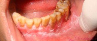
An experienced and enlightened reader, looking at the term in the title and paying attention to the ending “-it”, will immediately understand: we are talking about inflammation. Indeed, meibomitis is an acute inflammatory process of the meibomian gland inside the eyelid (in most cases, the upper eyelid, since there are up to one and a half times more glands there than in the lower eyelid, and, accordingly, the likelihood of certain problems occurring is higher). The more common colloquial name “internal barley” is, at the same time, less accurate and correct.
Table of contents:
Manifestation of xanthelasma
Causes of xanthelasma of the eyelids Diagnosis of the disease Methods for removing xanthelasma of the eyelids Stages of the fight and prevention of xanthelasma
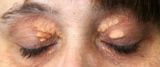
Skin formations are not only a cosmetic defect, but also a signal of an existing disease. Therefore, you should not ignore their appearance. Yellowish plaques, single or multiple, appear on the upper eyelid and are called xanthelasma. Xanthomas protrude on the lower eyelid or other parts of the body. These are benign formations, soft to the touch, with a characteristic color. As a rule, degeneration into malignant does not occur. They are usually diagnosed by a visual examination by a doctor. If necessary, he prescribes additional tests and examinations so as not to confuse them with tumors that pose a threat to health.
Treatment of meibomitis
In most cases, especially with timely treatment, meibomitis can be cured with conservative, non-surgical methods. First of all, disinfection of the ciliary edge of the eyelid is necessary, then local medications are prescribed.
Antibacterial drops and ointments
- Albucid
- Levomycetin
- Tetracycline eye ointment
- Floxal (ointment and drops), etc.
Anti-inflammatory drugs
- Dexamethasone eye drops
- Hydrocortisone eye ointment
Combination drugs
- Maxitrol
- Tobradex
- Sofradex and others.
The duration and frequency of use is determined by the attending ophthalmologist individually, depending on the specific situation.
Manifestation of xanthelasma
The following external manifestations are characteristic of xanthelasma:
- appearance: the shape of a plaque protruding above the skin;
- size: from pea to bean;
- location: often close to the bridge of the nose on one or both eyelids;
- consistency: soft, painless;
- color: yellowish;
- quantity: single or multiple, merging into an uneven tuberous strip;
- speed of spread: appear suddenly and grow slowly, becoming a serious cosmetic problem.
Prognosis for recovery from melanoma of the eyelid
In general, eyelid melanoma is an aggressive tumor and is diagnosed at late stages, when the prognosis for treatment is unfavorable. Five-year survival can be achieved in only half of the patients. But in the initial stages, with a melanoma thickness of less than 0.75 mm, the chances of a complete recovery are much higher.
| Read more about dermatological research at Euroonko | |
| Consultation with a dermatologist-oncologist | from 5,100 rub. |
| Skin examination using the German FotoFinder device | RUB 13,400 |
| Diagnosis of melanoma | from 5,100 rub. |
Book a consultation 24 hours a day
+7+7+78
Causes of xanthelasma of the eyelids
The cause of the development is considered to be metabolic disorders, most often lipid metabolism, the presence of problems with the heart, liver, pancreas in people suffering from obesity, diabetes, and atherosclerosis. Growth occurs slowly, mostly observed in older women. Can be inherited. They will not disappear on their own, so it is necessary to eliminate them. There are several options.
The plaque is affected by:
- liquid nitrogen for several seconds until the cells are completely destroyed;
- targeted laser, painless, bloodless, without scarring or unpleasant consequences;
- radio waves contactlessly evaporate unsuitable layers;
- promptly using scissors and tweezers using local anesthesia; the edges of the wound are treated with medications or cauterized with electrodes.
In order to prevent a secondary occurrence, you need to look for the abnormalities in the body responsible for this and cure them.
Reasons for appearance
In order to select the most effective treatment, it is necessary to identify the exact cause of the formation. These include:
- allergic reaction
- internal and external stimuli;
- demodicosis;
- infections;
- Moll cyst;
- papillomas;
- conjunctivitis;
- ophthalmoherpes;
- immune system disorders;
- gastrointestinal problems;
- low-quality cosmetics.
In most cases, diseases develop for several reasons, which negatively affect the functioning of the body.
Allergic reaction
A pimple on the upper or lower eyelid, in the corner of the eye, may appear under the influence of irritants that provoke allergies. These include:
- dust;
- cosmetics and soaps that are unsuitable for your skin type;
- pollen.
Eating exotic fruits, the body's reaction to the consumption of which has not been tested in advance, can lead to the development of allergies. Neglecting personal hygiene rules can accelerate the formation of acne. Allergies are often accompanied by additional symptoms: redness, swelling of the eyes, irritation, itching and increased tearing.
Internal and external stimuli
Bubbles around the eyes, on the eyelids and mucous membranes can form under the influence of internal and external irritants. Establishing the exact factor is possible only after a medical examination, on the basis of which the doctor prescribes treatment. Internal factors are considered:
- hormonal disbalance;
- long-term use of antibiotics;
- diseases of the digestive tract.
External factors that accelerate the appearance of acne include:
- failure to comply with personal hygiene rules;
- decorative cosmetics;
- long exposure to the sun.
Foundation, powder and other concealing cosmetics cause clogged pores and heat the skin: oxygen deficiency leads to the withering of the dermis.
Demodicosis
The causative agent of demodicosis is the eel, which lives in dust. The pathology has characteristic signs:
- itching;
- tearfulness;
- eyelash loss;
- feeling of “sand” in the eye.
The eye mite affects the eyebrows, skin in the forehead and chin area, and ear canals. Pathology manifests itself against the background of:
- chronic disorders of the functional activity of the digestive tract;
- endocrine system disorders;
- hepatitis A;
- metabolic disorders in the body;
- ophthalmological pathologies;
- increased oily skin;
- seborrhea.
The causative agent of demodicosis causes an acute allergic reaction.
Infection
Infectious lesions of the organs of vision provoke the appearance of purulent pimples and dense pimples. These include:
- trachoma;
- blepharitis;
- keratitis;
- ulcerative lesions of the cornea by staphylococci;
- dacryocystitis.
Ophthalmological disorders of infectious etiology require immediate contact with a medical institution. Infectious diseases have characteristic symptoms:
- thick discharge;
- redness;
- swelling of the eyes;
- pain accompanied by blurred vision and photophobia;
- dilation or constriction of the pupils;
- lacrimation;
- peeling of the skin.
The pathological process can be triggered by fungi, viruses and bacteria.
Moll cyst
A cyst forms on the eyelids during a change in the functional activity of the sweat glands of the eyes. 2 or more bubbles with transparent contents appear. After some time they disappear, the disease is often chronic. The pathology is accompanied by lacrimation and photophobia; hyperemia and swelling of the eyelids may appear. Pimply rashes take on a purulent character and are accompanied by the formation of crusts.
At the initial stage, spots may appear on the eyelids, which disappear on their own after a few days. During an exacerbation, the bubbles merge with each other. In the severe stage, Moll cysts are accompanied by the formation of scars and scabs, which increases the risk of entropion and eversion of the eyelids. In the absence of timely treatment, the patient's condition worsens: body temperature rises, general malaise and hyperesthesia are observed.
Papilloma
Papilloma is understood as a benign skin growth formed as a result of the rapid proliferation of epithelial cells. Pathological neoplasms are caused by the HPV virus. The disease affects men and women equally. Papilloma can occur on any part of the eyes, both on the upper and lower eyelids. The virus can spread in several ways:
- sexually;
- during childbirth;
- by everyday means.
The virus can be transmitted during hair removal or shaving through instruments not treated with an antiseptic.
Conjunctivitis
Pimples on the eyes of children and adults often appear due to viral conjunctivitis. The disease is accompanied by a pathological inflammatory process, which is localized on the outer mucosa of the eye. The disease can affect the inner surface of the eyelids. Conjunctivitis is caused by the penetration of an infection into the human body that affects the respiratory tract (a type of adenovirus or herpes). The contagiousness of the pathology is high, so it is epidemic in nature.
In addition to bubbles with transparent contents, clinical signs of the disease include:
- increased lacrimation;
- rupture of blood vessels inside the eye;
- itching;
- pain;
- photophobia;
- swelling of the eyelids.
With viral conjunctivitis, the lymph nodes are affected, so the person experiences pain in the mandibular and parotid region.
Ophthalmoherpes
Ophthalmoherpes affects the ocular appendages and the eyeball. The disease is caused by herpes simplex virus type 1 or 2. The characteristic symptoms of the disease are pain, photophobia, lacrimation and partial blurred vision. There are several main forms:
- primary;
- front;
- rear;
- recurrent.
A seal forms on the inner surface of the upper eyelid, and the eye swells. Purulent or mucous discharge is accompanied by itching and severe burning. Vision is blurred, the visible image is distorted.
Immune system disorder
One of the main reasons for the formation of acne on the eyes is considered to be a weakened immune system. Weakening can be temporary due to hypothermia, fatigue, and in women - before the onset of menstruation and menopause. Permanently weakened immunity is observed in people suffering from chronic diseases, immune deficiency or leading an unhealthy lifestyle.
Weakened immunity creates conditions favorable for the proliferation of pathogenic microorganisms. Bacteria rapidly multiply near the mouth of the sebaceous glands, causing their inflammation. The immune system is not able to independently stop the spread of inflammatory processes and ensure the normal functioning of the skin.
Gastrointestinal problems
The causes of acne around or inside the eyes include diseases of the gastrointestinal tract. This:
- low acidity;
- dysbacteriosis;
- gastroduodenitis;
- duodenitis;
- erosive and ulcerative lesions of the stomach and duodenum;
- pancreatitis;
- gastritis.
Improper nutrition leads to disruption of the functional activity of the digestive organs. The body stops quickly digesting food, the balance of fats, proteins and carbohydrates is disrupted. Useful components (vitamins, micro- and macroelements) are supplied with food in limited quantities, which leads to a weakened immune system.
Low-quality cosmetics
Low-quality decorative cosmetics often contain parabens, which can provoke the development of an allergic reaction. An allergy to powder, foundation or eye shadow is caused by the body’s individual reaction to some of the components included in the product. At the initial stage of the allergic reaction, pimples are small, often single, and have transparent contents. When a bacterial infection joins an existing allergy, pus forms inside the pimple.
Diagnosis of the disease
The first thing to do when xanthelasma manifests itself is to make an appointment with a specialist.
After a visual examination, if xanthelasma is suspected, the doctor performs diascopy. The formation is pressed down with glass to bleed it so that the yellowness appears more clearly. Examinations are needed to exclude suspicions of a malignant neoplasm, syringoma, or elastic pseudosanthoma.
Additionally, a blood test for lipid spectrum is prescribed.
It is necessary to identify the risk of developing atherosclerosis, heart attack, and hypertension.
Let's clarify
| Question | Answer |
| Is it possible for xanthelasma to degenerate into a malignant neoplasm? | The patient is not in danger of such degeneration. |
| What complications accompany plaque removal? | There may be bleeding or scarring of the skin at the site of removal. It depends on the skill of the doctor and the correct choice of method. |
| Is there a re-emergence? | Yes. To prevent this from happening, it is necessary to carry out a course of special treatment and try to eliminate the provoking factors. |
| Who is at risk? | Women, people with chronic diseases, age category over forty-five. |
| What can be a contraindication to surgery? | The procedure should be postponed for pregnant women, if there is fever or inflammation. It is not recommended to do this if you have oncology or exacerbation of chronic diseases. |
| Is it worth exposing yourself to operational risk? | It is enough to compare photos of patients before and after surgery. Moreover, the risks are minimized. |
| What should be done to prevent this manifestation? | It is important to eliminate possible causes of metabolic disorders in the body.
|
| Is it possible to use decoctions and infusions of plants in addition to medications to reduce cholesterol? | Yes. Some plants contribute to this. Have fewer side effects than chemical drugs. But it can only be used strictly as prescribed by a doctor. Along with other actions, they stimulate bile secretion, so any violations of these organs can lead to serious complications. |
| Which removal method is most popular? | The use of a laser is highly effective for any size of formation and has minimal consequences, since the integrity of the tissue is practically not compromised. The last word always belongs to the specialist. |
| Does laser pose a threat to the eyes? | The laser beam precisely targets the problem area. The mucous membranes of the organs of vision are not in danger. |
| What determines the cost of the operation? | The price for this service is dictated by the chosen removal method and the size of the growth. The larger it is, the more expensive it is. The category of specialist and the level of equipment used also have an impact. |
How is eyelid melanoma diagnosed?
Although eyelid melanoma is a superficial tumor that is easily accessible for visual inspection, it is not always possible to diagnose the disease at an early stage, since patients do not pay attention to it and do not go to see a doctor. Therefore, much attention is paid to self-examination. You can suspect a malignant process based on the following signs:
- The neoplasm has fuzzy, blurry edges.
- Asymmetrical shape.
- Uneven coloring of the tumor. In melanoma, areas of hyperpigmentation may be replaced by depigmented areas.
- The neoplasm increases in size.
- The skin pattern changes. Melanoma is characterized by a smooth surface; peeling or ulceration is less common.
- There is redness around the tumor.
- The melanoma itself may be itchy, painful, and cause a tingling or burning sensation.
As it grows, eyelid melanoma spreads to the underlying tissues, involving the conjunctiva, sclera and other structures of the eyeball in the process. In order to diagnose melanoma in time and begin treatment, it is necessary to regularly see an ophthalmologist, especially over the age of 50 years.
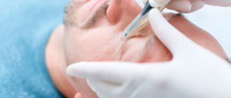
Methods for removing xanthelasma of the eyelids
Removal of eyelid xanthelasma is carried out using different methods on an outpatient basis:
- Traditional - using a scalpel. Surgical intervention is accompanied by suturing. Scar formation is possible. Bleeding inevitably occurs and there is a possibility of infection. This method is used for large growths or if there is sagging of the upper eyelid. At the same time, formation and excess skin are removed. There is a possibility of relapse. Therefore, plaque fusion should not be allowed to occur.
- Low temperature most often uses liquid nitrogen as a refrigerant. At a temperature of minus one hundred and ninety degrees, freezing and destruction of the structure of the fatty plaque occurs with excellent cosmetic consequences. It is difficult to protect the eye from cold temperatures, but there is minimal risk of infection.
- Radio waves act with waves of a given frequency. Under the influence of heat emanating from a thin electrode, the neoplasm cells die, providing a good aesthetic effect. Minimal risk of complications and recurrence after resection.
- A chemical is used to burn out the plaque, usually using trichloroacetic acid. Rarely used due to the risk of the reagent getting into the eyes.
- Electrocoagulation works using high frequency current. An electrode is placed on the tumor, current is applied to it, and the cells of the growth are destroyed. The cauterization site finally heals after ten days. Special ointments are applied to it. The method is suitable for small formations. There is no risk of retinal burns.
- Laser evaporates the formation with a laser beam. The method of laser treatment of tumors is considered today the most effective and safe, but quite expensive. The procedure is performed under local anesthesia. The patient's eyes are protected with special glasses. The wound heals under a crust in a few days. An experienced doctor is able to remove all formations in one visit.
Advantages of laser xanthelasma removal:
- No injury to adjacent tissues of healthy skin;
- bloodless, sterile method;
- high accuracy of the beam hitting the affected area to the required depth;
- absence of scars;
- fast recovery;
- harmless to the mucous membranes of the eye.
ethnoscience
Folk remedies must be used in combination with drug treatment. The complex effect helps to cope with the tumor in a few days. Warming is very popular; the procedure can be used even for barley. The heat should be dry; the barley itself and the skin around it should not be soaked. This increases the risk of pathogenic microorganisms spreading through healthy tissues. Dry heat is most effective at an early stage in the absence of an abscess.
A chicken egg must be boiled, wrapped in natural fabric and applied to the sore eye until it cools completely. Puree the boiled potatoes, wrap them in cloth and apply them to the swollen eyelid. You can heat sea salt, the application algorithm is the same.
Lotions based on medicinal plants are considered no less effective. The prepared product must be moistened with a tampon or a bandage folded several times and applied to the sore spot for 5-15 minutes. Cooking recipes:
- Pharmaceutical chamomile (15 g) is poured with a glass of boiling water and left for 30 minutes. The finished infusion is decanted, cooled and used for its intended purpose as a rinse or lotion.
- Black tea (1 tsp) is poured into 100 ml of boiling water. Insist for 10 hours. You can rinse your eyes with the liquid or make lotions 3-4 times a day.
- Plantain (fresh or dried leaves) is poured with water in a ratio of 3:1. The product is infused in a warm place for 40-50 minutes, then expressed. You can wash your face with the infusion 2-3 times a day.
- Soak a cotton swab in pharmaceutical tincture of propolis and cauterize the stye. The procedure can be performed up to 3 times a day.
- A leaf is cut from the bottom of the aloe, wrapped in cling film and placed in the refrigerator for 10-12 hours (overnight). In the morning, the thorns are cut off the leaf, the skin is removed, cut into small pieces and ground in a blender. The juice is squeezed out of the resulting pulp and used to burn the pimple 3-4 times a day. Children should not apply pure aloe juice to their skin: it should be diluted with water (in equal parts).
Honey is effective at an early stage. The beekeeping product has anti-inflammatory, disinfectant, decongestant and wound-healing effects. The compress should be used with caution by hypersensitive people prone to allergies.
Stages of fight and prevention of xanthelasma
The only reliable way to combat xanthelasma is to eliminate it. Only the doctor will decide which method is preferable based on the specific situation. The laser beam is one of the latest achievements of high-tech medicine. Our clinic is equipped with modern equipment, and the procedure for removing xanthelasma of the eyelids is carried out by experienced professionals. Laser removal is characterized by a minimal risk of relapse, high sterility, low likelihood of injury to adjacent skin and the absence of subsequent inflammation. Contraindications for this highly effective procedure include diabetes, pregnancy, and the presence of dangerous tumor processes.
Stages of the fight against xanthelasma:
- preparation - visiting an ophthalmologist, taking tests, choosing a removal method taking into account the indications;
- destruction of education;
- restoration with the implementation of appropriate appointments and follow-up examinations.
As preventive measures, you need to choose those that are aimed at the normal functioning of the body as a whole.
To prevent xanthelasma you should:
- monitor your weight and achieve its optimal value;
- eat wisely, including fruits, vegetables, foods with beneficial properties in your diet, reduce the consumption of flour, fatty foods, and sweets;
- stabilize your drinking regime, drink at least two liters a day;
- lead a healthy lifestyle, eliminate bad habits, stress, regularly give the body adequate physical activity;
- prevent damage to the skin;
- observe personal hygiene standards;
- strengthen immunity;
- systematically undergo preventive examinations.
Forecast
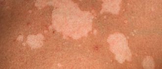
A pimple under the eye and on the eyeball is considered not only an aesthetic defect; the neoplasm is accompanied by inflammatory processes, profuse lacrimation, swelling of the eyelid, severe itching and visual disturbances. Education in the eye area is dangerous due to complications and therefore requires timely treatment. In most cases, the prognosis is favorable; it depends on the size of the pimple, the reasons for its appearance, the age of the patient, the individual characteristics of his body and skin type.
Squeezing pimples or taking medications on your own is strictly prohibited. In this case, the prognosis will worsen, and amateur activities often lead to the development of complications.