Do men have cellulite? We need to start with a definition. Cellulite is a clinical condition observed primarily in women (at all stages of the life cycle, starting from puberty). Caused by disorders of subcutaneous fatty tissue (structural, inflammatory, metabolic and biochemical), causing topographic changes in the skin.
In medicine, these changes are called “gynoid lipodystrophy” (GLD). It does not threaten human life. Nevertheless, it is classified as an undesirable phenomenon due to the fact that the external manifestation is expressed in the unsightly appearance of the skin, and therefore affects the quality of life.
And what about the male part of the population? Does cellulite occur in men? Yes. But rarely.
Why Most Men Don't Have Cellulite
DHF is more common in women. This is rather a female “cosmetic defect”. Here's why men usually don't have cellulite:
- The development of “female” cellulite occurs because women have weaker connective tissue and more fat cells, which is due to the action of female hormones.
- Men have stronger connective tissue and less subcutaneous fat, so cellulite usually does not appear in them.
In order to develop the topic further, we need to briefly consider the process of cellulite manifestation.
Female and male anatomical structure of the skin
The difference in the anatomical structure of the skin may already be the answer to the question of whether men have cellulite. This is not the only explanation, but we need to start with it.
The skin has a multi-layer structure. To make it easier to understand the process of cellulite manifestation, you need to pay attention to the layers of the skin:
- The top layer is the epidermis.
- Below it is the dermis (corium) - this is a thicker, deeper layer of skin consisting of connective tissue. The dermis is thicker than the epidermis because it contains extensive collagenous connective tissue. Collagen is the main insoluble fibrous protein in the extracellular matrix and in the cells of the body.
- The next layer is the first of two layers of subcutaneous fat (the areolar layer with chambers of fat cells).
In women, the anatomical structure of the areolar layer is such that the fat cell chambers are located vertically. This is the basis for the scientific basis for the concept of cellulite, that is, a small protrusion of fat (called “papillae of adipose tissue”) into the dermis. This also happens due to weakness (or weakening) of the connective tissue in the dermis.
This structural change in the subcutaneous layer of fat protruding into the dermis (similar to a hernia protrusion) gives the skin an orange peel appearance.
Additionally, during the female menstrual cycle, connective tissue is gradually broken down by enzymes called MMPs. Which speeds up the process of cellulite manifestation.
Men have a different organizational structure of connective tissue: there is a network of intersecting connective tissue structures, which can be figuratively represented as a fine-mesh network. It does not allow subcutaneous fat deposits to protrude into the dermis so clearly. The fat in this case can expand in the lateral (and internal) direction. In addition, men have a thicker layer of epidermis and dermis. Especially in the hips and buttocks area. But this does not mean that male cellulite is impossible.
Various locations of fat deposits
Subcutaneous fat is where most fat accumulates, causing concern among people watching their figure. Adipose tissue is a collection of fat cells (adipocytes) that act as a human energy reserve and also act as an insulating material that retains heat. In addition, adipose tissue performs a protective function, protecting the internal structures of the human body from injury.
As fat cells increase in size, they break down the connective tissue around them to expand. This leads to a decrease in the amount of collagen around the enlarged fat pads.
The deposition and accumulation of fat in the subcutaneous tissue depends on:
- hormones (testosterone, estrogen, insulin, glucagon, leptin and others);
- genetic factors;
- age-related changes;
- body mass index;
- nutrition.
Men accumulate fat in the neck, lower back, arms, and stomach. In women, fat accumulations are more common in the chest, thighs and buttocks.
Statistical data
Signs of lipodystrophy are detected in 75% of girls aged 20-35 years. After reaching the age of 40, cellulite lesions appear in 95% of women. The affected areas are the buttocks, thighs, abdomen, knees and arms.
The accumulation of fluid in the inner layers of the skin provokes the formation of “orange peel”, which affects 95% of women who are faced with lipodystrophy and neglected prevention.
Men rarely suffer from cellulite (less than 10% over the age of 40) due to the presence of a significant amount of collagen in the body, a developed muscular system and the relatively large thickness of the skin.
In what cases do men get cellulite?
You should pay attention to the main sex hormones, male and female. In men it is testosterone, in women it is estrogen. In both, these hormones are present simultaneously, but in different proportions.
Estrogens
The prerequisite for the development of cellulite in humans is estrogen. Men usually have low estrogen levels, so if they overeat and lead a sedentary lifestyle, they usually become obese, with signs of obesity. But this condition is not yet evidence of cellulite manifestation. There is a lack of an important component for this – estrogen.
Men can also have high estrogen levels. It increases as a result of the process of aromatization - the conversion of testosterone to estrogen. In this case, cellulite will begin to actively appear. The presence of two main factors is enough - obesity and physical inactivity.
Of course, age-related changes and other circumstances play an important role. But increasing your body mass index and sedentary lifestyle are key.
It’s worth stopping at old age too. In adulthood, men undergo increased conversion of testosterone to estrogen. Accordingly, the risk of cellulite is high.
Fat cells are not a dormant store of fat - they secrete several hormones, one of which is estrogen. The more fat cells a man (or woman) has in his body, the more estrogen is produced by those fat cells.
If you are overweight, it is the fat cells that produce estrogen, which is necessary for the development of cellulite.
Gynecomastia
The same processes that lead to cellulite in men can also lead to the development of gynecomastia (deposition of fat in the subcutaneous layer of the breast). The culprits are excess estrogen in the male body and obesity.
Estrogen stimulates the development of breast tissue and the deposition of fat around it, including in men.
Estrogen-like substances
Xenoestrogens and phytoestrogens contribute to the appearance of cellulite in men.
Some man-made chemicals, known as xenoestrogens, act as estrogen-like substances in the body and disrupt endocrine balance. Bisphenol A (BPA) and phthalates are known examples of xenoestrogens. These substances are present in common everyday items, plastics, and food packaging.
Some foods and drinks also contain high amounts of estrogen-like substances called phytoestrogens.
Soy, avocado, rice, beer are well-known examples of foods containing phytoestrogens.
All of these estrogen-like substances can have an estrogenic effect on the body and lead to the formation of cellulite, with phytoestrogens being more benign, while xenoestrogens are dangerous to health.
Microcirculation disorders
The condition of the microvasculature is directly related to the condition of large vessels. Almost all authors [3, 5, 7, 11, 13] emphasize: a decrease in muscle activity and a sedentary lifestyle worsen venous outflow, slow down lymph circulation, which leads to a decrease in metabolism, increased lipogenesis, and, as a consequence, the development of cellulite. Prolonged sedentary work and driving, typical of many actively working women, interfere with normal blood flow in the lower extremities, causing venous stagnation and capillary damage. According to our data, diseases of the veins and lymphatic vessels are present in 56% of patients with cellulite.
Violation of venous outflow and stagnation of lymph lead to increased permeability of the vascular wall and the release of plasma into the interstitial space. The resulting swelling of the interstitial tissue and hypoxia aggravate microcirculation disorders, and substances released when the endothelium is damaged promote the proliferation of fibroblasts and the compaction of connective tissue septa, which no longer delimit, but compress the lobules of adipose tissue [2]. The hydrostatic pressure in the capillaries depends on the amount of blood and lymph, which in different organs ranges from 18 to 40 mm Hg. Art. As a rule, it slightly exceeds the oncotic pressure of the blood (19-21 mm Hg), due to which the pressure gradient in the capillary walls is directed towards the tissue, and fluid filtration prevails over its reabsorption into the plasma. Excess fluid entering the tissue is removed through the lymphatic system. The pressure gradient in the interstitium causes fluid to move within it, facilitating the delivery of necessary substances to the cells. Hemodynamic parameters in capillaries are closely related to the permeability of their walls, which depends on the pressure gradient and protein concentration in the interstitial space and plasma. In turn, the physicochemical parameters of the interstitial environment of the lymphatic capillaries create the conditions for lymph formation and movement of lymph.
Lymphatic capillaries, the walls of which are formed by endothelium, remove excess fluid, proteins and metabolic products from tissues. The mechanisms of lymph movement in capillaries are not yet clear enough, but it has been proven that contractions of large lymphatic vessels (lymphangions) with a developed muscular layer play a significant role. Stagnation of fluid in the lumen of the vessel stretches its wall, disrupts the forward movement of lymph, and the absence of vascular contractions further aggravates this pathogenetic mechanism.
Thus, the condition of large drainage vessels, namely veins and lymphangions, directly affects microcirculation. Microcirculation disorders are observed in most women over 30 years of age (especially those who have given birth, taking hormonal contraceptives, wearing high-heeled shoes), so it is possible that the pathology of venous circulation is the leading link in the development of local lipodystrophy.
The main parameters characterizing the functioning of the microcirculation system are determined by the state of hemodynamics in the capillaries, the permeability of their walls, and the forces that ensure the movement of interstitial fluid and lymph. Microcirculation disorders are usually divided into vascular, intravascular and extravascular. Changes in blood flow, capillary pressure, and the functional state of the vascular wall are important for tissue perfusion. All microcirculation disorders have one thing in common: ultimately, transcapillary exchange suffers. Changes in the rheological properties of blood play a significant role in the mechanism of tissue damage and thrombus formation. The fluidity in the microcirculatory system is also affected by the aggregation state of platelets. It plays a leading role in thrombus formation and the development of intravascular aggregation of cellular elements.
Changes in the vascular wall are caused by changes in the location and shape of endothelial cells. Extravascular changes leading to impaired microcirculation are primarily associated with pathological processes in perivascular tissue. When the permeability of capillary walls decreases, the transfer of biologically active substances through the basement membrane is primarily disrupted. Changes in biochemical processes, water-salt metabolism and the nature of redox reactions lead to the development of circulatory hypoxia and deterioration in the nutrition of organs and tissues. When capillary permeability is impaired, plastic metabolism and energy supply to cells are significantly affected [3, 4]. A decrease in the efficiency of microcirculation is manifested by hypoxemia, venous hyperoxia, a decrease in tissue oxygen utilization, and the accumulation of lactic and pyruvic acids. Microcirculatory disturbances lead to decreased oxygen consumption and aerobic performance. All of the above ultimately leads to disturbances in the lipolysis-lipogenesis system. Changes in the arteriolar precapillary sphincters in the area of cellulite are described, causing an increase in pressure in the capillaries. An increase in capillary and interstitial pressure (due to excessive polymerization of glycosaminoglycans), as well as a decrease in plasma current (due to compression and compression of blood vessels) can lead to an increase in the permeability of capillaries and venules, and consequently to ectasia, swelling of the dermis and the formation of compacted partitions between individual adipocytes and between lobules of adipose tissue. A decrease in venous tone may occur in parallel with an increase in capillary fragility as a result of changes in the perivascular connective tissue, which leads to microvascular rupture and microbleeding [13, 14, 16, 21].
The fluidity of blood in the vessels depends on the composition of the plasma, the nature of the blood flow in the microvessels (turbulent or laminar), the plasticity and deformability of erythrocytes, and the tendency of cells to aggregation. It is known that hyperlipidemia affects the condition of vessel walls, blood clotting, microcirculation and organ perfusion [9]. The fluidity of blood in microvessels is largely determined by the protein-lipid composition of plasma. With a high concentration of lipoproteins in plasma, blood flow in small-diameter vessels becomes intermittent and turbulent. With hyperlipidemia, the cholesterol content in erythrocyte membranes increases, their size increases, they become less plastic, their ability to change their shape when passing through capillaries worsens, blood viscosity increases, and erythrocyte aggregation increases [1]. The study of microcirculation in people with hyperlipidemia revealed both functional disorders of capillary blood flow (slowing of blood flow, its “granularity”, formation of microaggregates, blood stagnation, microthrombosis) and morphological changes in the capillaries themselves (twisting, uneven diameter, microaneurysms, desolation with switching off blood flow ).
Hyperlipidemia is accompanied by a decrease in the arteriovenous oxygen difference and indicates a deterioration in oxygen utilization in peripheral tissues. With hyperlipidemia, the ability of the endothelium to produce the endothelial vascular relaxation factor, nitric oxide, is impaired. In addition, the hypoxia experienced by endothelial cells under conditions of hyperlipidemia leads to increased production of endothelin, a strong vasoconstrictor and nitric oxide antagonist, which ultimately leads to impaired microcirculation [9].
Cellulite is characterized by hemorheological disorders. Disturbances in the cellular link are expressed in increased aggregation of platelets and erythrocytes, and a decrease in the deformability of erythrocytes. An increase in platelet aggregation ability leads to hypercoagulation, enhances parietal microthrombosis and disrupts microcirculation. This, in turn, prevents the normal utilization of oxygen by tissues and leads to metabolic disorders with a predominance of anaerobic processes of glycolysis; on the other hand, the release of tissue thromboplastin is stimulated, which increases fibrinogen levels and enhances coagulation.
A compensatory reaction that prevents further thrombus formation is an increase in fibrinolysis. In patients with cellulite, an increase in blood viscosity at different shear rates and an increase in hematocrit were noted. Analysis of coagulogram data shows the presence of hypercoagulation syndrome, since the plasma recalcification time significantly decreases, the level of fibrinogen increases, the fibrinolytic activity of plasma and plasma tolerance to heparin decrease [1, 10]. Microcirculation studies using laser Doppler flowmetry make it possible to assess the functional activity of microvessels. There are two types of disorders: atonic and spastic types of microcirculation.
Apparently, at the initial stages of cellulite development, there is a decrease in arteriolar tone, a significant increase in intravascular resistance, slight venous stagnation and stagnation in the capillary microcirculation develop; passive mechanisms of microcirculation regulation predominate over active ones. Blood filling of the capillaries at the initial stages, on the one hand, ensures adequate trophism of tissues, and on the other hand, overflow of the capillary bed triggers pathogenetic mechanisms for the development of cellulite (expansion of capillaries, extravasation of plasma into surrounding tissues, development of edema, fluid retention in tissues, compression of surrounding tissues) . As cellulite progresses, a spastic type of microcirculation is formed, characterized by an increase in arteriolar tone, an increase in intravascular resistance, more pronounced venous stagnation, the predominance of active mechanisms of blood flow regulation, a significant decrease in basal blood flow, and insufficient blood supply in the capillary unit. Lack of blood supply in the capillary link can lead to hypoxia, activation of fibroblasts and the development of fibrosclerosis, which is typical for the third and fourth stages of cellulite. More pronounced phenomena of venous stagnation than in patients with the atonic type indicate significant disturbances and decompensation of the venular microcirculation.
Oxidative stress
A number of authors have studied changes in the parameters of oxidative processes and antioxidant protection and their effect on microcirculation. The content of lipid peroxidation products in the blood was determined: diene conjugates, malondialdehyde, as well as elastase activity, levels of α1-antitrypsin, α2-macroglobulin. The result revealed an increase in free radical oxidation processes in patients with cellulite. There was an increased content of diene conjugates, increased elastase activity with a reduced level of its inhibitor (α1-antitrypsin) and an increased level of α2-macroglobulin, coming from the interstitium into the blood with an increase in vascular permeability. All this indicates a high intensity of lipid peroxidation processes in the blood in patients with cellulite [10].
The level of oxidative stress in cellulite was assessed by the content in tissues of the product of lipid peroxidation - malondialdehyde, as well as products of protein peroxidation - carbonyl groups. The levels of total, oxidized and reduced glutathione, ceruloplasmin, superoxide dismutase, and total antioxidants were selected as indicators of the state of the antioxidant system. A decrease in the levels of some antioxidants was found while the levels of others increased, thereby maintaining the overall activity of free radical oxidation processes, which is vital for structural homeostasis. Thus, the data obtained indicate disturbances in the lipid peroxidation system and the development of oxidative stress in women with cellulite [6].
Changes in connective tissue
The third factor involved in the pathogenesis of cellulite is connective tissue. It is believed that fibroblast activation caused by estrogens causes excessive polymerization of glycosaminoglycans in the dermis and perivascular connective tissue, which increases their hydrophilicity and interstitial osmotic pressure. An increase in the level of estrogen and thyroid hormones (T3, T4) promotes the accumulation of hyaluronic acid in tissues, which binds water molecules, and therefore increases swelling. Water retention (edema) and an increase in the viscosity of the intercellular fluid lead to cell deformation and compression of blood vessels, provoking tissue hypoxia. Hypoxia causes a change in aerobic glucose metabolism, resulting in increased lactic acid production. This activates proline hydroxylase, an enzyme that facilitates the conversion of proline to hydroxyproline in procollagen with a subsequent increase in collagen synthesis. The accumulation of collagen fibers in tissues is accompanied by a decrease in the amount of elastin and interstitial substance, changes in the ratio of proteoglycans and glycoproteins, which leads to a decrease in the permeability of connective tissue, inhibition of repair, decrease in turgor and elasticity of the skin. Qualitative and quantitative changes in collagen provoke fibrosclerosis in the interlobar connective tissue septa. Thus, micronodules are formed, which are visible on the surface of the skin and can be detected by touch.
Hormonal imbalance
Hormonal imbalance contributes to the aggravation of microcirculatory disorders: decreased levels of progesterone, increased levels of androgens and aldosterone. Aldosterone enhances sodium reabsorption in the renal tubules, while vasopressin production is stimulated, water resorption occurs, and the volume of circulating plasma increases. The question of the direct participation of sex hormones in the pathogenesis of cellulite remains controversial. Most authors point to the influence of estrogens and progesterone produced by the ovaries. Extragonadal synthesis of estrogen is of great importance. At a certain period of life, its role is decisive, while most estrogens are synthesized in adipose tissue. The activity of adipose tissue aromatase depends on the topographic location of the fat. In the subcutaneous adipose tissue of the femoral-gluteal region, aromatase activity is 4 times higher than in the subcutaneous adipose tissue of the abdomen. The skin is very sensitive to the effects of female sex hormones due to the specific receptors it contains. Estrogens suppress the secretory activity of the sebaceous glands, increase the level of hyaluronic acid in the dermis, which increases the amount of intercellular fluid and converts soluble collagen into insoluble. A decrease in progesterone levels with a simultaneous increase in androgen levels contributes to fluid retention and a decrease in the rate of synaptic transmission, in particular through β-adrenergic receptors. At the same time, data on the influence of female sex hormones on the process of cellulite formation are in the nature of theoretical assumptions and have not yet received sufficient scientific confirmation [11, 16-18].
Conclusion
Based on the described mechanisms of cellulite formation, any measures aimed at its correction should take into account both the nature of microcirculation disorders, the severity of fat deposits and changes in connective tissue, and the stage of the process. Mesotherapy has in its arsenal a wide range of tools that allow it to influence the pathogenetic mechanisms of cellulite formation, and, of course, is an effective method of its correction.
Literature
- Bolotova L. G., Turova E. A., Kosheleva I. V. Complex use of physical factors (oxygen-ozone therapy and electrical myostimulation) in the correction of edematous fibrosclerotic panniculopathy. // Questions of balneology, physiotherapy and therapeutic physical culture, 2007, No. 1. P. 24-26.
- Blashmaison F. Methods for assessing cellulite: water retention index and Celluscore.// Aesthetic Medicine, 2005, No. 5. P. 89-94.
- Erofeev N. P., Orlov R. S. The lymphatic system is a necessary element of fluid homeostasis in the human body: a new look at old problems. // Bulletin of St. Petersburg University, 2008, ser. 11, No. 4 P. 78-86.
- Kapilevich L. V., Kovalev I. V., Baskakov M. B., Medvedev M. A. Intracellular signaling systems in epithelium- and endothelium-dependent processes of smooth muscle relaxation. // Advances in physiological sciences, 2001, No. 2. P. 88-98
- Korolkova T. N. Pathogenetic aspects of gynoid lipodystrophy. // Experimental and clinical dermatocosmetology, 2005, No. 4. P. 49-60.
- Korolkova T. N., Poliychuk T. P. Study of indicators of oxidative stress during oxygen-ozone therapy of local fat deposits. // Clinical dermatology and venereology, 2009, No. 2. P. 37-42.
- Koshevoy E. G. Dynamics of skin microcirculation in patients with gynoid lipodystrophy under the influence of vibration vacuum therapy. // Materials of the II International Congress “Restorative Medicine and Rehabilitation”, September 20-21, 2005, M., 2005. P. 48.
- Krupatkin A.I., Sidorov V.V. Laser Doppler flowmetry of blood microcirculation. Guide for doctors. - M.: Medicine, 2005.
- Lipovetsky B. M. Clinical lipidology. - St. Petersburg: Nauka, 2000.
- Minina A. P., Turova E. A., Bolatova L. G. et al. The use of laser Doppler flowmetry to assess microcirculation in patients with edematous fibrosclerosing panniculopathy. // Angiology and vascular surgery, 2004, No. 3. P. 46-49.
- Mikheeva S.V. Prevalence and risk factors for the formation of cellulite. // Public health and healthcare. Collection of scientific papers. St. Petersburg, 2000. pp. 11-12.
- Mikheeva S.V. Localization of cellulite in women. // Current problems of sanitary and epidemiological well-being of the population of the Northwestern region. Materials of scientific and practical conference. St. Petersburg, 2000. P. 173.
- Polenov S. A. Fundamentals of microcirculation. // Regional blood circulation and microcirculation, 2008, No. 1. P.5-19.
- Ranneva E. A., Zubkova S. A., Movchan V. N. Modern instrumental methods for diagnosing cellulite. // Experimental and clinical dermatocosmetology, 2007, No. 4. P. 48-51.
- Stanishevskaya T.I. The main types of blood microcirculation and the frequency of their occurrence in girls in the south-eastern region of Ukraine. // Scientific notes of the Tauride National University named after. V. I. Vernadsky. Series "Biology, Chemistry", 2005, No. 1. P. 131-141.
- Turova E. A., Bolatova L. G., Minina A. P. On the etiology and pathogenesis of cellulite. // Bulletin of Aesthetic Medicine, 2008, No. 4. P. 23-31.
- Chubrieva S. Yu., Glukhov N. V., Zaichik A. Yu. Adipose tissue as an endocrine regulator (literature review). // Bulletin of St. Petersburg University, 2008, ser. 11, no. 1. pp. 32-42.
- Avram MM Cellulite: a review of its physiology and treatment. // J Cosmet Laser Ther, 2004, 6. 181-185.
- Bastard JP, Maachi M, Lagathu C et al. Recent advances in the relationship between obesity, inflation and insulin resistance. // Eur Cytokine Netw, 2006, 17. 4-12.
- Hexsel DM, Abreu M, Rodrigues TC et al. Side-by-side comparison of areas with and without cellulite depressions using magnetic resonance imaging. // Dermatol Surg, 2009, 35. 1471-1477.
- Khan MH, Victor F, Rao B, Sadick NS Treatment of cellulite: Part I. Pathophysiology. // J Am Acad Dermatol, 2010, 62. 361-370.
- Lupi O., Semenovitch IJ, Treu C. et al. Evaluation of the effect ects of caffeine in the microcirculation and edema on thighs and buttocks using the orthogonal polarization spectral imaging and clinical parameters. // J Cosmet Dermatol, 2007, 6.102-107.
- Milani GB, Natal Filho A., Amado Joao SM Correlation between lumbar lordosis angle and degree of gynoid lipodystrophy (cellulite) in asymptomatic women. // Clinics (Sao Paulo), 2008, 63. 503-508.
- Quatresooz P., Xhauflaire-Uhoda E., Pierard-Franchimont C., Pierard GE Cellulite histopathology and related mechanobiology. // Int J Cosmet Sci, 2006, 28. 207-210.
- Smalls LK, Hicks M, Passeretti D et al. Effect of weight loss on cellulite: gynoid lypodystrophy. // Plast Reconstr Surg, 2006, 118. 510-516.
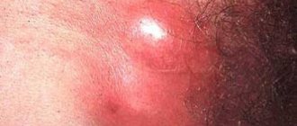
Examples of photos of male cellulite on the stomach
Due to the reasons described in the first paragraph, cellulite on the stomach of men looks different than on women. As a rule, these are ordinary fat deposits, that is, excess fat. The skin does not look flabby and sagging. It is often taut and elastic. Of course, it all depends on the stage of cellulite.
See what cellulite looks like in men in the following photo:
Definition of cellulite
Cellulite accompanies women of all times. 98% of women have cellulite. Many women do not accept it and try to hide areas of the body covered with orange peel.
Orange peel-like changes in the skin are called liposclerosis, fibrosclerotic edema, or female lipodystrophy. The word "cellulite" was first used in 1920. At that time it was believed that skin inflammation was the cause of the formation of fat folds. But this was wrong and it is now known that cellulite is not accompanied by inflammation.
Cellulite is a degeneration of the skin. Marketers decided to make a “disease” out of cellulite in order to force women and girls to buy special cosmetics and cosmetic procedures.
Both medicine and cosmetology can treat cellulite. But there is no doubt that advanced cellulite becomes a serious problem due to the fact that it leads to stagnation of fat or venous clots - blood clots.
How to get rid of the problem
Some treatment methods (to varying degrees) have an effect on cellulite. These include the following:
- Acoustic wave therapy (AWT) is a technique that applies acoustic waves (also called sonic, pressure, or shock waves) to affected areas.
- Laser, light or radiofrequency therapy are procedures that combine laser (or radiofrequency) and massage therapy.
- Undercutting is a procedure that involves inserting a needle under the skin to break up the fibrous connective tissue structures that separate clusters of fat cells. Medical professionals perform the cutting using a device known as a Cellfina. The course can last up to two years or more.
- Vacuum tissue release is another new procedure that can help break down tough fibrous tissue structures.
- Methylxanthines are a group of chemicals that include aminophylline, caffeine and theophylline. These chemicals are present in many anti-cellulite creams due to their ability to break down fat deposits. However, skin creams cannot provide the necessary concentration of these chemicals in the time required to significantly break down fat.
- Creams with retinol. Retinol may help with skin thickening, which may explain the reduction in the appearance of cellulite. The product must be used for six months or longer to see the effect.
- Carboxytherapy is a medical procedure that injects carbon dioxide (CO2) directly under the skin. A small study found that it may be helpful in treating cellulite.
- Laser liposuction adds laser treatment to the typical liposuction fat removal procedure.
- Ultrasonic liposculpture uses sound waves to try to reduce cellulite.
You can continue to enumerate. There are many practices that are used, but without the approval of official medicine. Some practices help some, some don't. The effectiveness of therapy is influenced by many factors.
The most reasonable treatment for men is the desire to get their body in order. Limit your alcohol intake, especially beer, normalize your diet, and pay attention to physical activity.
Hyperventilation of the lungs during physical activity in the fresh air leads to increased oxygenation of blood and tissues. Oxygen is directly involved in oxidative processes, including the breakdown of fat.
Fighting methods
Treatment of cellulite begins with an analysis of the location of foci of destruction on the patient’s skin. This procedure is mandatory - different parts of the body react differently to hardware or manual procedures. Eliminating lipodystrophy is a complex task based on restoring blood and lymph circulation. Patients are prescribed physical exercises, massage, wraps, and electrical stimulation.
Procedures prescribed by a doctor allow you to solve several problems simultaneously:
- break down fats;
- prevent the formation of new fat deposits;
- ensure the outflow of fluids from tissues;
- increase the tone of subcutaneous vessels;
- perform muscle stimulation;
- even out skin texture.
Their solution is facilitated by hardware procedures: pressotherapy, microcurrent electrical stimulation, electrolipolysis. The listed manipulations ensure restoration of blood flow and increase the intensity of lymph movement. Thanks to this, the normal state of the patients’ subcutaneous tissue is restored, and the relief of the skin is normalized.
Liposuction remains a radical method of treating lipodystrophy. Surgical manipulation is prescribed for patients with a genetic predisposition to the accumulation of subcutaneous fat on the thighs or abdomen. Before the operation, biomaterial will be submitted for laboratory tests. Doctors often refer patients for x-rays or computed tomography. Visualization of the focus of lipodystrophy allows you to plan the course of surgical intervention.
Methods for preventing and eliminating cellulite
Since this problem arises due to a whole range of different reasons, the methods for solving it should be multidirectional and include:
- diet;
- physical exercise;
- cosmetic procedures.
You need to start by making an appointment with a body cosmetologist, who will conduct an examination, collect anamnesis, and prescribe a course of treatment, taking into account the stage of cellulite and the characteristics of your health condition.
The main methods for getting rid of “orange peel” are:
- Massage (manual and hardware);
- Wraps;
- Fitness;
- Proper nutrition.
Let's pay more detailed attention to each point.
Massage for cellulite
Let's start with a massage. This is an effective way to eliminate fat deposits, restore skin firmness and elasticity, and shape your figure.
There are many massage techniques. Within the framework of our topic, first of all, it is worth mentioning anti-cellulite manual massage. It is usually intended to smooth and tone the skin on the legs. The technique for performing this massage is as follows: the massage therapist’s hand movements should be fast and include 50% rubbing and 50% squeezing. The impact is superficial. A course of at least 10 sessions is recommended.
Hardware methods for treating cellulite
Hardware methods - LPG and Endosphere - have also proven themselves well.
LPG is a vacuum-roller effect on body tissue. The technique was developed in France in the 80s of the 20th century. With the help of self-propelled rollers located in the handpiece, the skin fold is mechanically kneaded, which is captured by the vacuum. The whole body is massaged: the specialist moves the handpiece horizontally and vertically, and the rollers rotate in one direction or the other. The most problematic areas of the body (hips, butt and others) are treated with special attention.
Result of LPG massage:
- Removing excess fluid;
- Stimulation of lipolysis;
- Activation of blood circulation and lymph flow;
- Production of collagen in tissues.
All this leads to the fact that the skin is evened out, excess volumes and kilograms disappear, and the silhouette becomes more toned. This type of massage requires a special elastic suit that protects the surface of the skin, making the procedure comfortable and ensuring hygiene. The standard course is 10 sessions, the duration of 1 session is 35 minutes in accordance with the protocol of the device manufacturer.
Endosphere therapy was developed by Italian scientists in the early 2000s. This is a method of compression microvibration transmitted to the body using a special device - a cylinder equipped with rotating spheres made of special silicone. The vibration of the spheres causes the deep layer of tissue to “work”, stimulating the work of mechanoreceptors (Merkel particles), thereby activating the process of lipolysis (fat breakdown) and the breakdown of fibrous compounds.
When carrying out the procedure, the cosmetologist changes the speed of movement of the handpiece and thereby sets the parameters of vibration of the spheres and their rotation, providing pressure on the body (“pump effect”). In this case, the specialist varies the intensity of the impact depending on the individual tasks of the patient.
Endosphere therapy is done directly on the skin, onto which massage oil is applied for better gliding of the spheres. Even if cellulite is only on the thighs, the procedure involves not only one or another leg, but also the body as a whole - otherwise the procedure will be ineffective.
This is a course technique, on average 6 to 12 sessions are required, each of which lasts 1 hour 15 minutes in accordance with the recommendations of the creators of the technology.
Endosphere Effect:
- Lymphatic drainage effect;
- Smoothing the skin surface (eliminating cellulite);
- Elimination of local fat deposits;
- Figure correction;
- Weight reduction.
By removing excess water, puffiness is relieved, the skin becomes dense and elastic, more toned.
These devices are recognized leaders in the field of body care.
Many people are interested in:
- Which one is better?
- How is LPG different from Endosphere?
In answering these questions, we must first say that these are two fundamentally different techniques of influence, as can be seen from their description. Secondly, which of these devices will be effective can only be decided by a cosmetologist after examining the patient, taking into account the medical history and the stage of cellulite. Third, a combination of these two methods may be required to achieve the desired effect.
As mentioned above, getting rid of cellulite requires an integrated approach. Therefore, it will be useful to combine massage with wraps, which have a beneficial effect on the quality of the skin. They can be of various types: based on oligo-elements (algae, for example), mud, cream. The temperature regime used is also different - there are hot wraps and cold ones.
At home, such procedures can be carried out only after consultation with a specialist: if cosmetics are used incorrectly, the timing of the procedure and other requirements are not followed, the disease can be aggravated.
How does cellulite wrap work?
A special composition is applied to the skin, then the body is wrapped in a thermal blanket, the desired temperature is set and after the allotted time the procedure ends. The temperature chosen by the cosmetologist should activate the effect of cosmetic products, due to which lymphatic drainage occurs, the functioning of blood vessels is normalized, the skin is nourished and becomes smoother.