Consequences of mole removal
The majority of moles appear in a person during the first twenty years of his life. About one in a hundred newborns are born with moles. Acquired marks are, as a rule, benign formations, congenital ones are one of the types of minor developmental defects. Moles can be located under the skin (subdermal) and on its surface as a pigmented formation consisting mainly of melanocyte cells. Their dark color is due to the presence of too much melanin pigment.
Possible consequences
The consequences of mole removal can be very diverse. They depend not only on the nature of the mole or its shape, but also on the level of professionalism of the physician to whom the patient turns. By the way, the plastic surgeon Elena Vladimirovna Salyamkina is distinguished not only by her professionalism, but also by the quality of the equipment and tools used during the operation. An important factor, by the way, is also the reaction of the patient’s body.
Care after mole removal is one of the most important components in maintaining health after surgery. This is what influences how long the healing will take. If you strictly follow the attending physician’s instructions for caring for the affected area, then the consequences after removing the mole will be absolutely “harmless”.
At the site of a mole removed by a surgeon, a thin black crust most often forms. It must be treated with a solution of potassium permanganate for several days, alternating it with a brilliant green solution (brilliant green). In addition, the wound can also be treated using a variety of ointments with an antiseptic effect, containing antibiotics. The main thing is that these drugs should be prescribed exclusively by the attending physician.
Under no circumstances should you forcibly peel off the scab, as this may cause the wound to reopen, which may slow down the rapid healing process. During the week you should not take baths, visit swimming pools and saunas, and you should also avoid using a variety of decorative cosmetics. You can only apply sunscreen lotions and creams to your skin, if, of course, there is any need for this. These restrictions are due to the fact that if the crust gets wet, it may come off ahead of time. As a result, the wound may open, and some infections or bacteria may enter it. Accordingly, such a nuisance as inflammation may occur.
How long does it take for the skin to heal?
Complete healing of the skin occurs in a person after removal of a mole in three weeks, or maybe two, but discomfort at the operated site can be observed for another six months. The timing, of course, depends solely on the size of the wound - the larger it is, the longer the healing will take. As soon as the crust falls off on its own, a pink patch of fresh skin forms under it, which you should not forget to protect from direct sunlight.
Attention: If the removal procedure was carried out in the summer, during the period of its healing it is necessary to avoid tanning and even more so from visiting a solarium. If the removed mole was on the face or on any other part of the body that cannot be protected from the sun’s rays, it generally requires special care - generously lubricating the area with sunscreen with a protection level of at least 60 when going outside.
Remember that young, new skin is incredibly sensitive to any external factors, especially ultraviolet rays. If ultraviolet radiation does get into these places, persistent pigment spots may appear. It is necessary to protect this area from direct sunlight and treat it with cream until the color of the area with young skin is equal to the color of the rest of the skin.
Important: There is no need to lose vigilance on days when the sun is covered by clouds, since even at this time ultraviolet radiation reaches up to 85%, and when the sun's rays are reflected from sand, water or snow, the absorption of ultraviolet radiation by the skin almost doubles.
Scars
A scar after mole removal is quite normal. You should not expect that the removal procedure will take place without any consequences. In most cases, such a scar will not be able to go away on its own, just like just a small scar after removal of a mole.
So, how can you deal with this problem? For example, you can try lubricating these places with cocoa butter - now you can buy it in one of the pharmacies.
A colloidal scar after mole removal can also be removed using a silicone patch, which must be worn for two or three weeks. However, such a patch should also be prescribed exclusively by a specialist.
But if, after removing a mole, a tubercle appears that does not resolve, you may have to use the services of plastic surgery. In this case, the patient does not have to worry about being left with a rough, pronounced scar. The scars are smoothed out so that they are practically invisible.
If a mole was removed not because it spoiled a person’s appearance, but because it was constantly injured, you must remember to regularly examine your body. Particular attention should be paid to remaining moles and their changes in appearance.
Results after different removal methods
If a person is planning to remove a mole on the body, he needs to be prepared for the fact that the results after removal can be very varied. Everything depends on the complexity of the operation, the state of health of the person, and, of course, on the method of removing the mole.
For example, after using a laser, there are no scars or marks left, and accordingly, even a rehabilitation period is not needed. The procedure itself takes only a few minutes. It is sterile, so it is absolutely impossible to get any infection. So you definitely shouldn’t expect negative consequences after laser removal.
If moles are removed with liquid nitrogen, the result may be somewhat unexpected, because moles can have different depths, and it is not always possible to remove them to the very root with nitrogen. After such removal, a person may also encounter troubles such as a scar or a scar, and most often, a severe burn. The skin takes a very long time to heal after this procedure, almost six months. But removal with nitrogen also has its advantage - it is the most affordable type of mole removal.
However, the most dangerous consequences can arise when you remove a mole yourself, since only a specialist can determine the condition of the mole, its depth and level of danger.
When surgically removing a mole, you must be prepared for the fact that a small scar will remain in its place. Therefore, this method is most suitable for removing moles that are located in areas covered by clothing.
The main thing that a person who has decided to remove a mole needs to remember is that no matter which method he chooses, he must first undergo a series of tests and pass the necessary tests. Specialists must make sure whether this formation is benign or malignant.
vip-hirurg.ru>
A lump appeared in place after the removal of a mole: what to do?
After removing a mole, you need to treat the area and minimize the negative impact from the outside on it.
After removing a mole with a laser or as a result of surgery, one of the decisive periods begins, on the normal course of which the health of the entire body depends. Removal of a nevus is necessarily accompanied by a long rehabilitation period, during which it is necessary to properly handle, care for and monitor all changes at the site of removal.
If this area of the skin is treated correctly, not overheated in the sun and minimizes the influence of chemical and physical factors on it, it will slowly heal and there will be no trace left of the intervention. However, not everything always goes well and often after such operations unpleasant consequences arise, such as:
- A scar appeared at the site of removal. The situation is typical and does not pose any threat. There is no need to do anything, the scar is a natural phenomenon that will disappear on its own after some time. There's no need to worry.
- Redness after mole removal is an adequate reaction of the body to surgery. But if the redness around the mole does not go away over a long period of time, it is important to consult a specialist.
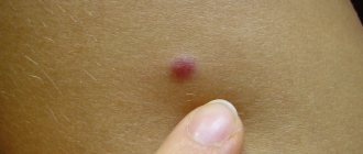
- A lump appeared at the site of the removed mole. If any formations appear, it is recommended to see a doctor immediately. Often, compaction under a mole signals the initial stage of melanoma (skin cancer) and the development of malignant tumors. They may be benign, but it is better to consult a doctor immediately in order to eliminate all health risks.
On average, the wound healing process takes 2-3 weeks.
During this period, it is important to carefully observe all changes in the skin at the site of removal, wipe the wound with products prescribed by specialists, and if it is noticed that something new has formed, immediately consult a doctor. If healing occurs normally, a small scar will remain after a couple of weeks, which will soon disappear.
Nevus removal: the right choice of the safest method
Most nevi do not pose a serious problem for humans, but any changes require urgent consultation with a doctor. He will also be able to determine whether the formation needs to be removed or not.
Is it possible to remove nevi?
Sometimes removal is the only option to prevent a mole from turning into a malignant tumor.
Frequent indications are:
- injured or a potential threat,
- big sizes,
- aesthetic inconsistency,
- change in shape, color, texture.
The latter is the most “alarming bell”.
Especially if swelling appears around the formation, the boundaries become blurred, and itching or burning occurs. This happens under the influence of endogenous or exogenous factors. The latter include visiting a solarium, prolonged exposure to the sun, and injuries.
Types of therapy
Treatment methods are selected depending on the location and type of nevus. Sometimes it is left untouched, but observation by a dermatologist is recommended. If the formation is benign, then hospitalization is not required, but all procedures must take place in a medical facility.
Laser method
This type is carried out in most cases using local anesthesia. The laser is effective for benign formations:
- intradermal,
- epidermal.
The good thing about this method is that it is done very quickly. On average, the procedure takes about five minutes. The doctor cuts off the nevus in layers using a device that has directed laser radiation. It is possible to adjust the depth and intensity of beam penetration.
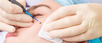
Laser nevus removal is non-contact and one of the safest. It allows you to remove nevi on the face or other areas where the skin is especially sensitive.
Photo of laser removal of nevus on the face
With laser there is no blood loss, pain, or risk of infection. Most moles, when completely removed, never appear in that place again. The wound heals in a few days, in rare cases it takes two weeks.
There are also some negative sides. After removing large birthmarks, a white spot remains on the skin. Since the laser evaporates the formation cells, it is not possible to send them for histological examination. Therefore, such therapy is used when the nevus is carefully examined by a doctor and tests are taken.
Surgical excision
The use of a surgical scalpel is a classic method in which a bordering incision is made. It is done in such a way as to touch healthy skin. After the manipulation, the wound is sutured, and the formation itself is sent for examination.
The method has proven itself well when the nevus:
- penetrated very deeply into the layers of the skin,
- has several broken parts,
- suspected of developing into melanoma.
Doctors do not carry out such treatment for herpes, infectious or inflammatory diseases. A wait-and-see approach is chosen until complete recovery. The procedure is carried out under anesthesia and guarantees the removal of the entire mole.
The disadvantage of removing a nevus surgically is the long healing period of the wound. Often a scar remains in this place. In addition, after surgical excision, you should not stay in the sun for a long time.
The procedure itself takes 30 minutes. The time depends on the location of the nevus and its size. In modern clinics, after surgery, silicone absorbable patches are used, which make it possible to make the scar after surgery less noticeable.
Cryodestruction
This method makes it possible to cope with the formation of cold.
To do this, very low temperatures are used to freeze and immediately destroy organic formations. In most cases, the procedure is prescribed when there is no need to send a mole for histology; it is also used to remove formations in a precancerous state.
The indications are:
- change in nevus size,
- painful sensations,
- bleeding,
- formation of precancerous cells.
Cold is used on tumors located in the upper layers of the dermis. Under its influence, water crystallizes in cells, and the level of electrolytes and metabolites reaches a critical threshold. This destroys the cell membrane.
The procedure is carried out using liquid nitrogen or a cryodestructor. In both cases, the removal itself takes 10 seconds. The procedure is painless, so there is no need for pain relief.
The disadvantage of the method is the lack of ability to control the depth of exposure, so in inept hands, burns and tissue scarring are possible.
Electrocoagulation
The method allows you to deal with a mole using thermal effects obtained by electric current.
During the process, the treated area is cauterized, allowing:
- quickly get rid of nevus,
- after healing, obtain a smooth area of skin without a scar,
- examine cells of formation,
- eliminate even the smallest formations.
This method is used on small nevi or if there is a base. Contraindications include: the appearance of malignant tumors, herpes, skin coagulation disorders, the presence of foci of chronic infection.
Before the procedure, local anesthesia is administered. The mole is then removed using a metal loop that carries a current. A small number of healthy skin cells are also cauterized to “seal” the vessels and prevent blood loss.
After the procedure, high-quality education care will be required throughout your recovery.
Radio knife
Using a radio knife is one of the most gentle techniques. High-frequency waves generate energy, which is used to cut tissue and evaporate cells. This allows you to get an almost invisible mark on the work site.
The main advantage is that healthy tissues are not damaged. The procedure itself takes no more than 20 minutes and is painless. The radioknife is used on all parts of the body, and formations can be examined.
Such removal cannot be carried out if skin formations begin to turn into malignant tumors, there are herpes or inflammatory diseases. It is also contraindicated for those who have heart rate sensors in their bodies.
The device itself removes moles during operation, disinfects and stops bleeding. It is possible to use a radioknife to work with precancerous formations.
Surgitron device
The technique allows you to remove a nevus using a cold radio knife.
The method does not lead to complications and has a low level of trauma. The impact on the mole is carried out using high frequency radio waves. Energy leads to rapid destruction of cellular structures.
The method can be used for single and multiple nevi. However, one of the contraindications is pregnancy. The remaining restrictions are the same as for the radio knife.
A crust remains at the treatment site, which falls off within a week, leaving a mark like an abrasion. A variety of medications are used to heal it. At the same time, solariums and rest in the open sun are prohibited.
What could be the consequences?
Regardless of the method chosen, removal of a nevus is a serious intervention in the functioning of the body. Before the procedure, it is better to learn about the consequences in advance.
Natural changes include:
- the formation of wounds and their covering with a crust,
- formation of a pink spot (young healthy skin).
Depending on the method, discomfort may continue for another 2-3 weeks, and final healing is possible only after several months.
After removal, scars or indentations may appear. This phenomenon is typical for a laser or surgical scalpel. Skin pigmentation is sometimes observed. Cryodestruction is an inexpensive method, but there are enough consequences after it. For example, if the nevus is not completely eliminated, the mole may appear again. The use of liquid nitrogen is a burn that damages healthy tissue. In addition, a tubercle may appear at the site of an incompletely removed formation.
During surgical intervention, the possibility of formation of a keloid scar remains, and further infection of the wound cannot be ruled out. If the nevus is not completely removed, then it is possible not only for a new one to appear, but also for it to degenerate into melanoma. Therefore, any manipulation must be carried out by a doctor.
Patient reviews
Despite the fact that many strive to choose a more gentle method of treatment, it is not always possible to refuse to use a scalpel. Patients note that this method is painless, and the ability to send tissue for analysis is a big plus.
Despite this, most people prefer a laser or radio knife. If the doctor finds a melanoma-dangerous nevus, then they choose a method that allows further examination of the cells. According to reviews, the most painless is the radio knife, which acts quickly.
Price for procedures
The simplest thing is to go to a clinic, where the nevus will be removed for free.
In this case, a surgical scalpel is used. When contacting a clinic, the cost of the procedure will depend on:
- localization,
- sizes,
- selected method.
Of course, the price for the same manipulation will vary depending on the chosen clinic. Some types of formations require several visits to the doctor, for example, when treating a giant pigmented nevus, the operation is performed in 2 or 3 stages.
Included in the price:
- preliminary inspection,
- diagnostics,
- operation,
- use of anesthesia,
- histology.
How to remove a nevus at home?
Extreme types of impact (cutting, tearing off) should be completely excluded, as this is fraught with the development of cancer.
For removal at home use:
- garlic,
- iodine,
- ointment with celandine,
- vinegar, apple juice.
In all these cases, a cotton pad is moistened and used to lubricate the nevus. All folk remedies require a huge amount of time, and the consequences can be unpredictable.
Thus, nevi are neoplasms that can cause harm if handled incorrectly. Therefore, the treatment method is chosen by the doctor who decides on the need for a histological examination.
Is it dangerous to remove moles? The following video will tell you about it:
Consequences after removal of a nevus in this video:
gidmed.com>
Methods for removing moles: from surgical to radio wave
Moles are benign elements formed on the skin due to excess accumulation of melanin. As long as they do not cause discomfort and do not pose a cosmetic problem, they can be tolerated. But if the neoplasms are injured, cause anxiety or spoil the body with their presence, it is better to remove them.
What methods of mole removal exist, which one should you prefer? The choice of the optimal option depends on the condition and structural features of a particular element, as well as the degree of potential health hazard.
How to remove a mole without surgery?
The procedure for removing moles with liquid nitrogen is carried out in the form of cold exposure to the lesion with a low-temperature substance. Cryodestruction is performed quickly and painlessly. Its affordability is attractive to patients.
But a significant drawback is the inability to predict how deeply nitrogen will penetrate into the epidermal layers and whether it will completely destroy nevus cells. If the neoplasm is not frozen out at the root, it is possible that it will reappear in the same place.
If after removal of a mole a bump appears, it means that it was not completely removed. A person can easily feel a lump in the treated area. If the procedure was performed by an inexperienced specialist, his uncertain actions can cause burns to healthy tissue. And this will not only damage the surrounding skin, but will also lengthen the healing period of the treated area.
Removing moles by electrocoagulation involves heat treatment of the element. To perform the manipulation, a specialist can use two types of currents:
- direct current used in galvanocaustics;
- high-frequency current, suitable for performing physiotherapy measures.
The indication for the procedure is the small size of the tumor and its small base. But in the case of chronic infectious pathologies in the body, the presence of viral infections and malignant skin lesions, electrocoagulative treatment is not recommended.
It is also not suitable for patients with altered or impaired blood clotting. Individual intolerance to current procedures is also a reason to refuse such therapy.
Small and medium-sized wounds left from the effects of the coagulator heal quickly and without scarring. But if a large area has been subjected to current treatment, there is a possibility of scar tissue forming.
Video: the principle of the electrocoagulation procedure is shown.
Laser and radio wave therapy
Of all the methods for removing moles, laser evaporation works well. Consequences and complications after the procedure occur in very rare cases. In general, the healing process proceeds without deviations. Patients who come to the clinic with small flat spots, if all medical recommendations are followed, do not leave any traces.
If a convex intradermal neoplasm was removed with a laser beam, an inconspicuous scar may form or the remainder of a small depression at the site of the former nevus. Sometimes laser intervention leaves a trace in the form of pigmentation of the cleansed skin.
Video: laser mole removal.
Radio wave removal of moles is considered the best and more modern. Non-contact intervention carried out using a radio knife is suitable for removing elements from the thin skin of the eyelids and mucous membranes. Radio waves do an excellent job with nevi of different sizes and shapes and remove them from any part of the body.
The advantages of radio wave therapy are:
- speed of the procedure - the whole action takes about 5 minutes;
- absence of swelling and bleeding - the patient immediately goes home;
- impossibility of tissue burns;
- sterility of the procedure;
- not a long recovery period.
The formation of a colloidal scar does not occur after removal of a mole with a radioknife. There may only be a small red mark on the cleared area. Therefore, the use of this technique to remove birthmarks from the face is not always advisable.
Improper care of the treated lesion can lead to the appearance of pigment spots. To avoid this, the crust that forms at the site of the mole should not be wetted for a couple of days. It should also be protected from direct sunlight.
Video: removing a mole with a radio knife.
Features of surgical excision of moles
Today, the surgical method of removing moles has lost its popularity due to its traumatic nature. However, it also has certain advantages:
- excellent result without recurrence of pathology (if performed correctly);
- possibility of transferring material for histological analysis;
- no contraindications;
- the operation is performed with little involvement of healthy tissue, which reduces the risk of secondary elements appearing on the skin;
- affordability.
How are moles removed surgically? The operation is performed under local anesthesia with subsequent transfer of the excised material for histological examination. His answers are needed for differential diagnosis, that is, from them a specialist will be able to judge the nature of the removed tumor and prescribe adequate treatment.
Before cutting out the pathological lesion, the doctor treats the area surrounding it with an antiseptic solution. To avoid relapses during the removal process, the surgeon also excises small areas of surrounding healthy tissue. Next, he applies cosmetic stitches. On the 2nd day, the medical staff bandages and treats the wound. After a week, the stitches are removed.
Video: how moles are removed with a scalpel.
What kind of doctor removes moles? A nevus located in an area of increased trauma requires consultation with a surgeon. After inspecting the item, he will decide whether to remove it.
You should also contact a surgeon if you have bloody birthmarks that appear on the child’s body. Having monitored the dynamics of the hemangioma, he will make a prediction about its spontaneous resorption or, conversely, suggest getting rid of it.
kozhnyi.ru>
Laser removal of moles: reviews, photos
Moles, especially those located on the face, have long been considered evidence of an aristocratic or noble family, and therefore they were often even specially painted. But in the modern world, the attitude towards these spots, regardless of their location, is very ambiguous - some are indifferent to them, some are afraid of their degeneration into a severe tumor - melanoma. The latter often try to remove moles with a laser as quickly as possible to eliminate the cause of their anxiety - but is this always justified?
Moles in dermatology
Moles, or as experts also call them pigmented nevi, are congenital or acquired accumulations of special melanocyte cells that contain the brown pigment melanin. These cells are usually evenly distributed over the skin and protect it from ultraviolet radiation, but in some cases they can collect in clusters that look like dark spots, which, in fact, is a nevus. In most cases, they do not pose any particular danger, but in some situations, the melanocytes of nevi may degenerate into malignant melanoma cells. This is what most concerns about moles are associated with and justifies the desire to remove them.
In addition, despite the fact that most nevi are several millimeters in size, sometimes they can be significant in size - up to several centimeters in diameter. If they are located on the face, then this is a pronounced cosmetic defect. In addition, the larger the size of the nevus, the higher the likelihood of developing various disorders in it, including malignant degeneration. That is why we can say that removing moles with a laser or other instruments, in this case, turns into not just a cosmetic manipulation, but also a method of preventive treatment.
Many people are frightened by the possible consequences of such removal, because it is widely believed that skin cancer often appears after such interventions. In fact, all biological material obtained during laser removal is sent to a histology laboratory, and after the procedure you can visit a specialist at any time if there are any changes. That is, the entire process, from the beginning of nevus removal to complete healing of the postoperative wound, is under the control of specialists and the appearance of any negative changes will be recorded by a specialist. This will allow timely measures to be taken to eliminate and treat pathological changes, especially when the mole is localized on the face.
Indications for the procedure
Laser removal of moles is not performed in every case; there are fairly strict medical indications for this. Removal based only on aesthetic indications is possible only if there is absolute confidence that such an effect will not have negative consequences. The prerequisites for such a cosmetic procedure on the face or other parts of the body are the following factors:
- significant size of the mole, its strong expression, creating significant aesthetic inconvenience;
- redness or other changes in the color, shape, size of the nevus;
- condition after injury or damage to a mole;
- Some experts also recommend removing a mole in cases where it begins to fade, that is, disappear from the surface of the skin.
Removal of a mole with a laser can be carried out only after a histological examination of its tissues - if no signs of malingization were found. In cases where histology shows suspicious changes, the person is referred for examination to an oncologist - it is he who, after conducting repeated studies, makes a final verdict regarding the nature of the nevus. In cases where there is proven malingization of cells, the use of a laser is excluded; only a surgical removal method is used, even when the tumor is localized on the face. In such a situation, the threat of serious consequences is much more priority than possible scars or marks on the skin.
Benefits of laser removal and its types
Fortunately, such a pessimistic scenario for the development of a mole is quite rare, and most removals of this formation occur precisely for cosmetic reasons. At the same time, there are other ways to get rid of nevi on the face and in other noticeable places - the most popular are the diathermocoagulation method and, oddly enough, surgery. However, compared to them, the use of a laser has a number of very important advantages:
- The most important advantage is the absence of marks and scars after the manipulation. According to medical statistics, such consequences occur in approximately 2-3 people per thousand cases of removal.
- The possibility of carrying out the procedure in an outpatient setting - unlike surgical removal, there are fewer requirements for sterility and lighting of the room.
- The procedure is painless - there is no need to use various anesthetics and additionally load the patient’s body with drugs.
- Some types of lasers have the ability, in addition to directly removing tissue, to stimulate cellular regeneration. This speeds up healing and shortens the post-procedure rehabilitation period to 5-7 days.
- The procedure is non-invasive – removal of moles with a laser is not accompanied by the introduction of instruments or suture material into the tissue, which sharply reduces the risk of side effects and infectious complications.
Just like all medicine and cosmetology in general, the method of laser removal of nevi is also progressing, so at the moment there are two main methods of laser exposure to tissue:
- Ablation method - a laser beam removes mole tissue layer by layer down to a layer of healthy underlying tissue. At the same time, for ordinary nevi, one procedure lasting several seconds is spent, while large formations can be removed in several stages. The layer of removed cells is sent for histological examination; CO-2 and erbium lasers are mainly used for this technique.
- The vascular coagulation method is a more subtle and less common technique that requires the cosmetologist to have special skills and knowledge. Its essence lies in the fact that with the help of a neodymium laser, the vessels that feed the nevus are blocked (coagulated), as a result of which its tissues undergo necrosis. This method is perceived quite ambiguously in the scientific community.
In the vast majority of cases, only the ablation technique is currently used - only a few state-of-the-art salons use a combination of these two techniques. In this case, the erbium laser removes nevus tissue, and the neodymium laser closes the vessels, preventing bleeding.
Contraindications and side effects
The laser mole removal technique cannot be used for certain conditions that may affect both the mole itself and the skin around it:
- Inflammatory skin lesions in the immediate vicinity of the location of the mole itself. For example, this may be an exacerbation of a herpes virus infection of the lips - then removal of nevi on the face cannot be done.
- Reduced blood clotting - each such formation has a developed capillary network, which is why injuries to moles are characterized by prolonged and relatively heavy bleeding. Therefore, when the hemostatic system is disrupted, it is better not to remove it.
- Malingization of nevus cells - if histological examination reveals signs of a malignant process - the use of a laser for removal is strictly prohibited.
After removing a mole with a laser, complications or side effects are extremely rare - in thousandths of a percent, scars may appear, as well as slight pigmentation at the site of the nevus. To reduce the risk of complications and speed up regeneration, you need to avoid prolonged exposure to direct sunlight, not visit solariums for about a month, and also intensively moisturize the skin.
Reviews about the procedure
Dmitry 32 years old, Krasnodar Since birth, I had a huge mole on my left cheek - it always stood out in size on my face, due to its black tint it looked like a spot in my photos, and was the cause of complexes in childhood.
I didn’t dare remove it, because according to rumors it could cause cancer, disfigure the face, etc. Purely by chance I came across an advertisement online for a clinic that practiced laser removal of nevi - the reviews about its specialists were good and I also decided to undergo the procedure . The size of the mole by that time was already 1.5 centimeters, so I had several treatments over the course of two months. But now in place of the ugly spot there is only slightly changed skin - I am completely satisfied with the result, I recommend it to everyone. Valeria 25 years old, Ufa From my mother I inherited a whole scattering of small moles on my neck and décolleté - from an oncological point of view they never bothered me, but aesthetically I didn’t like them and I wanted to remove them. After the tests were carried out and their safety was confirmed, I removed several especially pronounced nevi near the chest as a test. In their place a crust appeared, which fell off after a week, and after another 7-10 days there were no traces of the moles left. Now I am completely confident in this method and am looking forward to getting rid of the remaining formations. Arina 37 years old, Mikheevo Five years ago, removing moles saved my life - I decided to get rid of a nevus on my face that I had had since birth. Having passed the necessary smears and tests, the dermatologist informed me that I couldn’t have laser removal and that I needed to see an oncologist as soon as possible. As a result, I had a first-degree melanoma removed - if I had not decided on this procedure, I would have found out about it too late, because there were no external preconditions. Leave your review Laser procedures are increasingly used in all areas of medicine, including cosmetology. Having a number of advantages over traditional techniques, they open up a whole range of possibilities for both the doctor and the patient. This also applies to laser removal of nevi - ten years ago it was almost impossible to remove such a formation without a scar, whereas now a laser does it quickly and without problems.
LadyCleo.ru>
What happens after a mole is removed
Every day in our country hundreds, and maybe thousands of skin lesions are removed using different methods. Unfortunately, healing is not always perfect. Today I will talk about the possible cosmetic results of removing moles and warts. Read this article to the end and you will learn how to avoid problems after such operations ( tips are marked in italics
).
Normal healing after removal
Normally, on the third or fifth day after removal of the mole, a dark, dry crust remains at the site of the operation. The same thing happens when you get scratched or cut. The size of the crust is usually the same as the removed formation or 1 mm larger. Gradually it peels off from the skin and, after about 2-4 weeks, disappears completely. At the site of removal, a small reddish pit remains, no more than 1-2 mm deep. At the end of the fourth to sixth week, the bottom of the fossa rises to the level of the surrounding skin. By the end of the third month, in most cases, the redness disappears and a barely noticeable mark remains on the skin from the removed mole.
BEFORE removal 3 months AFTER
The picture below shows the healing process as a diagram:
normal healing after removal is called a normotrophic scar
Hypertrophic scar (bulge)
A hypertrophic scar is a red lump that rises above the surrounding normal skin. It may be painful or itchy, but these symptoms usually go away over time.
Such a scar is in no way associated with the development of melanoma and skin cancer after removal of a mole, if, according to histological examination, a benign formation was removed
.
When, a few weeks after the removal of a mole or wart, something similar appears in its place, any person will begin to worry. Fortunately, a hypertrophic scar does not pose any danger other than a cosmetic defect. Sometimes, after a few months, less often after a few years, it itself becomes flatter, softer and acquires a lighter color than the surrounding skin. If this does not happen or you want to speed up this process, it makes sense to contact an experienced dermatologist-cosmetologist.
If you notice the development of such a scar, please do not worry - it is not forever and doctors are quite successful in dealing with it. There is only one case where it is better for you to see the doctor who performed the removal - if the mole was removed without histology. To reduce the likelihood of scar formation, strictly follow your doctor’s recommendations and consult him again if you notice the first signs - redness and thickening.
Hypotrophic scar (depression)
Another, quite common, situation is when a mole was removed, and a small depression has formed in this place. This condition is called a hypotrophic scar and means that the skin cells have not worked enough to restore its surface. Unlike a hypertrophic scar, this healing option is not so obvious; as a rule, it does not cause concern, does not require correction and gradually smoothes out after 1-2 years
.
White spot (hypopigmentation)
This condition occurs in two cases. In the first case - if within 3-4 months from the operation you have been sunbathing for a long time. In the second case, if the mole or wart was located in the deep layers of the skin, and the cells that form the pigment were completely removed and were not restored. This condition is also not dangerous to health and can go away on its own without your participation. To prevent hypopigmentation from developing, try to protect the surgical site from the sun for 3-4 months. If you still cannot avoid the appearance of a white spot, remember that it goes away on its own, in most cases, after 1-2 years.
BEFORE removal 3 months AFTER
Suppuration
Sometimes, if the rules of surgery are not followed or the wound is not properly cared for, inflammation and subsequent suppuration may develop at the site where the mole or wart was removed. This unpleasant condition has 4 main symptoms: pus, redness, swelling and tenderness. We can talk about suppuration only when all 4 symptoms are present AT THE SAME TIME.
You should not think that suppuration has occurred when there is only redness around or slight swelling of the wound. If a clear or yellowish liquid sometimes comes out from under the crust, there is also no cause for concern. This is due to the fact that only viscous white !liquid! (pus) is one of the main signs of suppuration. In my practice, there were cases when patients, 3-4 days after removal, noticed a whitish-yellowish bottom of the wound and thought that this indicated suppuration. Fortunately, this impression is also deceptive. This color is nothing other than the normal color of the second layer of skin (dermis).
When superficial removal of skin formations using shave biopsy or radio wave surgery, the shape of the wound eliminates suppuration. This is due to the fact that with this type of removal, the skin is not completely cut, no stitches are applied, and there is simply nowhere for the pus to accumulate, unlike traditional operations.
Reappearance of a mole after removal (recurrence of pigmented nevus)
If the mole is not completely removed and nevus cells remain in the skin, a relapse may occur. In this case, at the site of removal, after the crust falls off or a little later, a dark spot appears, no more than 1-3 mm in size, not protruding above the skin. According to the few studies of this issue, recurrence of a pigmented nevus does not lead to the development of melanoma and skin cancer, if the histology shows a nevus. This condition requires nothing more than observation. If desired, the relapse can be removed again, but you must remember that the likelihood of scarring in this case increases.
I cannot help but note that, in my experience, the reappearance of a mole is possible even if its complete removal is confirmed by histological examination. This question is still awaiting its researchers.
Summary, or briefly about the main thing:
The cosmetic result of mole removal only partially depends on the surgical technique and the doctor’s qualifications. The size and depth of the formation being removed in the skin also have a significant impact. Otherwise, all of the above options for skin restoration depend on the individual characteristics of your body. Modern science, unfortunately, is not able to accurately predict in advance which of them and to what extent will affect the regeneration of your skin.