The article was prepared by a specialist for informational purposes only. We urge you not to self-medicate. When the first symptoms appear, consult a doctor.
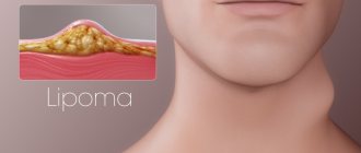
The term “fat” in everyday life usually means lipoma - a tumor of fat cells concentrated in a capsule of connective tissue. The localization of this benign formation is almost any part of the human body, but more often a lipoma occurs where there is a relatively small layer of subcutaneous fatty tissue. Wen can form on the shoulders, arms, elbows, hands, wrists and even fingers. These formations very rarely degenerate into malignant tumors, but this possibility should not be completely ruled out.
Features of wen on the hands and fingers
The appearance of any non-standard formation under the skin is a good reason to consult a doctor. Although most often the tumor turns out to be an ordinary wen, a non-specialist does not have the necessary knowledge to make an independent diagnosis. A fatty tissue is not a tumor in the usual sense; it is a connective tissue capsule filled with fat cells.
Features of wen on the hands:
- In very rare cases, lipid formations on the hands may not have a capsule; fat accumulates under the epidermis layer, penetrating deep into the muscle over time. This pathology is called diffuse lipoma;
- Wen on the hands, reaching impressive sizes, leads to impaired motor functions;
- Saturation of the hands and fingers with nerve endings, contact of the formation with the joints can lead to pain when the wen grows excessively;
- Wen on the hands are more often injured due to the specificity of their localization;
- Even a small formation on the hand is clearly visible, causing psychological discomfort to its owner.
Wen can increase in size, reaching the size of an orange or a tennis ball. The growth of the formation intensifies even more after its mechanical damage. Wen on the fingers are injured especially often, since the hands take part in hundreds of different operations during the day.
Drug treatment
If the size of the lipoma does not exceed 2-3 cm, the doctor uses drug therapy: a medicine is injected inside with a thin needle, triggering the breakdown of adipose tissue. For complete resorption it will take 3-4 procedures and 2 months. The probability of a successful outcome is 80%. The method is contraindicated in a number of chronic diseases.
Unlike many formations, a wen on the arm does not pose a threat to life; if it is small in size, the problem is of an aesthetic nature. The tumor must be treated in the early stages: as it grows, nerve endings and blood vessels are affected.
It is worth remembering: the larger the size of the lipoma, the more difficult it is to remove, and the more difficult the rehabilitation period. In rare cases, a neglected wen becomes a source of mutation processes and degenerates into a malignant formation. If you miss the moment when the lipoma begins to change, you can end up with serious health problems.
Reasons for the appearance of wen
There are several factors, the combination of which leads to the appearance of lipid formation on the hands and fingers. However, there are no specific reasons that cause the development of this particular type of wen. Many aspects can immediately influence the formation of lipomas.
Causes of wen:
- Hereditary predisposition;
- Hypothyroidism, diabetes mellitus and other endocrine pathologies;
- Impaired functioning of the gastrointestinal tract, liver or kidneys;
- Frequent psycho-emotional disorders, stress;
- Pathological addiction to nicotine, alcohol, drugs;
- Unbalanced diet - predominance of fats and carbohydrates in the menu.
Genetically determined predisposition, diseases of the liver and endocrine glands are a common cause of lipomatosis - multiple formation of wen under the skin of the hands and fingers. In this case, the localization of tumors is not limited only to the hands; wen forms on any part of the patient’s body.
What does a wen look like?
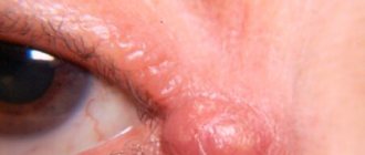
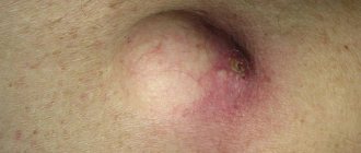
A lump on the elbow or other part of the body has an unfixed condition. Although it has its own clear boundaries, it feels like if you touch and press on it, it is unable to stay in place, it rolls under the skin, and sinks into the muscles. The lipoma can attach itself to the bone, so it is better not to delay it, otherwise the surgical intervention will become more complicated. A wen looks like a lump, a wart, a white mole: a person may not even suspect that it is something serious. The size may depend on the age of formation. There is no pain, inflammation or temperature.
Wen on fingers
At the beginning of its development, such a formation may look like a regular boil. The presence of a soft, painless mound on the finger for a long time is a reason to identify it as a wen. There is a very thin layer of epidermis and muscles on the fingers, so the lipoma does not have the opportunity to grow greatly. However, the growth of wen on the fingers leads to the fact that it begins to interfere with the functioning of the joints, touching and damaging them.
The advantage of this formation is that a wen on the fingers is easily diagnosed, especially if it grows steadily. Frequent injury to a fatty tumor forces the patient to seek medical help. Removing a large formation on a finger requires the surgeon’s exquisite work to preserve the functionality of the limb.
Surgical removal
First of all, the surgeon conducts a visual examination, ultrasound or x-ray. If a malignant tumor is suspected, additional studies are prescribed, but this rarely happens. Next, surgical removal occurs, and the contents of the wen are, by default, sent for histology in order to finally exclude the possibility of oncology.
- With the puncture-aspiration method, the contents of the capsule are sucked out with a long needle. In this case, relapses are possible, since the formation itself remains intact. This option is recommended for use on wen of very large or very small sizes in open and easily accessible areas. Stretched skin does not recover after the intervention, which will require additional cosmetic procedures.
- Small formations are cauterized with liquid nitrogen (cryodestruction) or high-frequency currents (electrocoagulation). The wound after electrocoagulation heals within two weeks, no scar remains. After cauterization with nitrogen, pigment spots may appear.
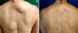
- Wen is regularly removed using the classical method. The operation is performed in a hospital under local anesthesia or under general anesthesia with hospitalization if the wen is large (size exceeds 15 cm). The surgeon makes an incision with a scalpel, removes the resulting capsule along with its contents, the wound is cleaned and sutured. Sutures are removed on the 10th day after surgery. Surgical removal leaves scars that will have to be removed.
- With the endoscopic method, the wen is punctured and a miniature camera, an endoscope, is inserted inside. The capsule is separated from the surrounding tissue and removed. Sometimes vacuum suction of adipose tissue is performed through a puncture, then the capsule itself is cut out. In both cases, the puncture heals quickly and without complications.
- Laser removal implies the exclusion of re-formation of lipoma. There are no scars or scars left, and no relapses occur. Recovery takes place under the supervision of a doctor and lasts one to two weeks.
- Radio wave method - removal of a wen with a radio knife. Under the influence of intense radiation, adipose tissue and capsule are removed. This is an effective procedure that does not leave scars. Recovery after surgery lasts 5 days.
The advantages of the radio wave method and laser removal are that intact tissue is not affected. The operation takes place without blood or pain, excluding infection, suppuration or swelling. The wound heals completely, no scar is formed. The rehabilitation period is short without loss of performance.
Regardless of the method of removal, after surgery the patient is prescribed medications to prevent inflammation. The resulting scars and scars are removed separately using laser resurfacing, dermabrasion and other cosmetic procedures.
Wen on the palms
Separately, we should consider xanthomatosis, a metabolic disease in which multiple lumpy growths with soft, loose, yellow contents form on the palms. It is represented by a mixture of cholesterol and cholestanol. Xanthomas are localized not only on the hands, they can affect the eyelids, corners of the eyes and, in general, any parts of the body of a person suffering from a serious disorder of lipid metabolism. Treatment of xanthomatosis involves a combination of drug therapy with surgical or cosmetic removal of yellow plaques and growths on the skin.
The main misconceptions regarding the treatment of wen on the hands
Self-medication of a fatty tumor at home is very dangerous, no matter where it is and no matter what its minimum size.
There are several other misconceptions regarding the treatment of wen on the hands:
- You can get rid of wen with the help of medicinal herbs and compresses. Relieving minor symptoms with such methods is quite possible, but it will not be possible to completely cure the wen and eliminate its cause.
- Returning to a healthy lifestyle will help cure the wen. To prevent education, you can stick to a diet and give up bad habits, but the process that has begun cannot be stopped by such measures.
- There are special diets designed to get rid of fatty tumors. Body weight and the size of the wen are in no way related to each other, so losing weight will not help get rid of the problem or even stop the growth of the formation.
Opening the tumor capsule on your own will not help get rid of it, since the fragments of connective tissue remaining in the wound will become the basis for relapse.
Is it possible to delete
The decision about whether it is necessary to remove a wen in the wrist or elbow area should be made only by a doctor. Sometimes the doctor advises not to touch the tumor at all, but only recommends observation with regular conducting of the necessary studies. This happens with small tumors that do not have a visible tendency to grow and develop and do not show any symptoms. And surgical removal is prescribed for the following manifestations:
- inflammation;
- sudden growth;
- the appearance of pain;
- change in skin color.
In other cases, the surgeon, assessing each case separately, selects the most suitable treatment method.
Under no circumstances should you remove a lipoma on your arm yourself. This action will not give the expected results and will worsen her condition. Although the wen is a benign formation, trauma can lead to reorganization into a malignant tumor.
Differentiation of a wen on the arm from atheroma
A sebaceous cyst (atheroma) can have symptoms similar to a lipoma, so distinguishing these formations from each other can be very difficult.
Differentiation of wen from atheroma - main signs:
- Localization of atheroma is the sebaceous gland, localization of the wen is its own capsule of connective tissue;
- The atheroma is tightly attached to the skin and is crowned by a clogged mouth of the gland, and the lipoma moves quite freely under the epidermis;
- Pain and discomfort are frequent companions in the initial stages of atheroma development, and lipoma is painless, its main symptom is constant growth;
- The localization of atheroma is the upper layers of the epidermis, its contents can be visible outward, and the lipoma is located deeper;
- Atheroma is denser and more elastic to the touch than lipoma;
- The skin over the surface of the lipoma can be gathered into a fold, which cannot be said about a sebaceous gland cyst;
- Lipoma has a more impressive growth potential, while atheroma rarely reaches the size of a quail egg.
When treating small lipomas (up to 3 cm in diameter), it is possible to use drugs that absorb adipose tissue. Atheroma cannot be treated with such methods, especially if the cyst is inflamed.
Methods for diagnosing formations on joints
When contacting a specialist, a clinical examination is initially performed. The doctor studies the symptoms and palpates the elbow or knee, determining:
- consistency (soft, dense);
- presence of pain;
- mobility of internal nodes.
If the data obtained indicates the presence of a lipoma on the knee or elbow, treatment is prescribed. If an inflammatory process or tree growth is suspected, additional diagnostics are carried out:
- X-ray excludes the possibility of fracture or dislocation;
- Ultrasound examination allows you to recognize the internal state of the joint (presence of pus) and periarticular tissues;
- cytological analysis allows you to see the shape of the formation;
- A puncture is prescribed to determine the type of bursitis based on fluid analysis.