Introduction
Dermatitis herpetiformis is an inflammatory skin disease with a chronic relapsing course, pruritic polymorphic lesions and typical histopathological and immunopathological features. Based on a number of evidence, dermatitis herpetiformis is considered a cutaneous manifestation of celiac disease.
Both diseases are observed in gluten-sensitive individuals, exhibiting the same HLA haplotypes (Dq2 and DQ8), and also improve with the introduction of a gluten-free diet. In addition, in patients with dermatitis herpetiformis, almost all cases of intestinal biopsy reveal changes typical for celiac disease in the form of villous atrophy with intraepithelial lymphocytosis, as well as the generation of circulating autoantibodies to the enzyme tissue transglutaminase. Dermatitis herpetiformis is more common in Caucasians, although an increasing number of cases are being reported in the Japanese population. The incidence per 100,000 population in Scotland is 11, in Sweden 19.6-39.2. The prevalence of dermatitis herpetiformis in Finland is 75.3 per 100,000 population (8 times lower than the prevalence of celiac disease in this country) with an annual incidence rate of up to 3.5 per 100,000 population for the period 1980-2009, showing a decreasing trend in recent years.
As a rule, dermatitis herpetiformis develops in the fourth or fifth decades of life, although people of any age can suffer from dermatosis. In a recent study conducted in a group of 159 patients with dermatitis herpetiformis, approximately 27% of patients were 10 years of age and 36% were under 20 years of age, showing that, at least in Italy, childhood dermatitis herpetiformis is more common. than in other countries.
In 2009, our group published guidelines for the treatment of patients with dermatitis herpetiformis. However, according to recent publications, several new data have been obtained regarding the clinical and immunopathological features of dermatitis herpetiformis. In addition, new guidelines for celiac disease were developed by the European Society of Pediatric Gastroenterology, Hepatology and Nutrition (ESPGHAN) in 2012.
Clinical features
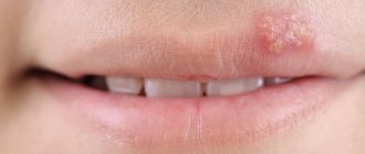
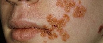
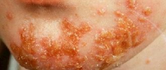
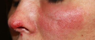
Dermatitis herpetiformis typically presents as symmetrical, grouped polymorphic lesions consisting of erythematous areas, urticarial plaques and papules involving the extensor surfaces of the knees, elbows, shoulders, buttocks, sacrum, neck, face and scalp. The rash is accompanied by itching and burning of varying intensity. Subjective disorders precede the development of lesions.
Along with these manifestations, several atypical presentations have been reported in patients with dermatitis herpetiformis, including purpura, petechiae on the hands and feet, leukocytoclastic vasculitis, plantar keratosis, blisters mimicking chronic urticaria, and lesions mimicking pruritus pigmentosa. Interestingly, in some cases the dermatosis may present with only erythema or only severe itching, making diagnosis challenging.
Differential diagnosis in children is carried out with atopic dermatitis, scabies, papular urticaria, and in adults - with eczema, other bullous autoimmune diseases of the blisters (especially IgA linear dermatitis and bullous pemphigoid), nodular prurigo, urticaria, erythema multiforme.
Forms
Dermatitis herpetiformis has several types and forms. In determining what type of pathology it is, visual analysis of the skin rash plays an important role. The most common types are:
- vesicular. In this condition, small vesicle-type bubbles form on the skin;
- papular. This variety is characterized by the formation of multiple small nodules with no voids;
- bullous With this type of pathology, a large number of blisters filled with liquid contents are formed;
- urticariform. This type of pathology is characterized by the formation of multiple blisters due to detachment of the upper layer of the epithelium.
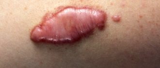
This is what Dühring's dermatitis on the hands may look like.
In the case of Dühring's dermatitis herpetiformis, in which the rash merges with each other and transforms into erosion, we can say that the defect has acquired an atypical form of dermatitis. Such negative transformations are also classified and among them the most common are:
- vegetative. With this complication, complicated foci of villous type are formed;
- localized. Symptoms are expressed only in a small area of the skin;
- pemphigoid. In this case, Dühring's dermatosis herpetiformis is characterized by a rash of large compacted blisters that are difficult to rupture;
- strophuloid. The formation of blisters and papules occurs. Moreover, nodules form first, and small blisters appear on top of them;
- trichophytoid. With such an exacerbation, the rash will have wavy edges and peel off. These symptoms are in many ways similar to fungal infections. For this reason, doctors give clinical recommendations to undergo a study in order to establish the root cause of the negative symptoms that appear;
- eczematoid. At the site of the lesion, multiple rashes of papules and blisters are observed, which literally transform into weeping erosions within a few days.
There is another form called senile dermatitis. As a rule, Dühring's senile dermatitis occurs against the background of age-related changes in the body, which cause metabolic disorders, problems in the liver, and inhibition of blood flow activity. With such a pathology, a rash of different sizes appears on the body, which forms an atypical form of the defect.
Dühring's disease in old age is dangerous due to its course. Therefore, at the first symptom, you should immediately consult a doctor for advice.
Histopathology
The typical histological picture of the affected skin of patients with dermatitis herpetiformis is characterized by subepidermal vesicles and blisters associated with the accumulation of neutrophils at the apices of the dermal papillae. Skin histopathology in dermatitis herpetiformis is not a reliable diagnostic criterion, as it can also occur in other bullous diseases, including linear dermatitis, congenital epidermolysis bullosa. In addition, as shown by Warren and Cockerell, the histopathological appearance is nonspecific in approximately 35% to 40% of cases. Therefore, to make a correct diagnosis, histological examination should always be performed in conjunction with direct immunofluorescence (DIF), which is the gold standard for diagnosing dermatitis herpetiformis.
Preventive measures for DD
Due to the unclear etiology of the disease, primary preventive measures have not been developed. Therefore, when we talk about preventive measures, we mean preventing or minimizing relapses. After all, the chronic form is usually accompanied by periodic exacerbations, which are unpredictable and can occur at any time throughout life. Prevention lies precisely in not provoking them, the main aspects of which include:
- Compliance with recommendations for gluten-free dietary nutrition (food containing iodine and certain types of grains is strictly prohibited).
- Regular examination by a dermatologist (registration at a dispensary).
- Maintaining personal hygiene will significantly reduce the likelihood of infection.
General preventive measures, of course, include a healthy lifestyle. After all, bad habits aggravate the physical condition of the body and have a destructive effect on the immune system, which can lead to another exacerbation of dermatosis herpetiformis.
Direct immunofluorescence
Direct immunofluorescence reveals two patterns: 1) granular IgA deposits in the dermal papillae and 2) granular IgA deposits along the basement membrane. Sometimes a combination of both options may occur. A third variant has recently been described, consisting of fibrillar IgA deposits primarily located at the tips of the dermal papillae. This pattern is often observed in up to 50% of Japanese patients with dermatitis herpetiformis.
Other types of immune deposits that can be detected by direct immunofluorescence include the presence of perivascular IgA deposits in the upper dermis, as well as granular IgM C3 deposits at the dermal-epidermal junction and/or dermal papillae.
The sensitivity and specificity of direct immunofluorescence in the diagnosis of dermatitis herpetiformis is about 100%. Additionally, according to ESPGHAN guidelines for celiac disease, positive direct immunofluorescence in patients with suspected dermatitis herpetiformis eliminates the need for duodenal biopsy to diagnose celiac disease. Direct immunofluorescence should be performed on intact skin, since in damaged skin IgA may be eliminated by inflammatory cells. In addition, patients should be on a normal diet during the examination period because IgA deposits may disappear from the skin in patients on a gluten-free diet. If the patient is on a gluten-free diet, then he should be switched to a diet with normal gluten content at least a month before the biopsy.
In case of negative results of direct immunofluorescence in patients with a high clinical suspicion of dermatitis herpetiformis, a repeat biopsy from another unaffected skin area should be repeated. Negative results of direct immunofluorescence in dermatitis herpetiformis are very rare. In such cases, a combination of clinical, histological and serological data helps to establish the diagnosis.
Serological tests
Patients with dermatitis herpetiformis usually have specific antibodies that may be found in patients with celiac disease. Among them, the IgA antibody test to tissue transglutaminase is considered the most sensitive and specific diagnostic method for first-line serological testing in patients with suspected dermatitis herpetiformis. Some patients may have a deficiency of IgA antibodies to tissue transglutaminase. Specific and sensitive serological markers of dermatitis herpetiformis are also IgA+IgG total antibodies to endomysium (EMAS), IgA+IgG total antigliadin (anti-DGP) and IgA antibodies to epidermal tissue transglutaminase (anti-TGe IgA).
Anti-TSH antibodies (IgA antibodies to tissue transglutaminase)
Anti-TSH antibodies belong to the IgA1 subclass and represent a marker of intestinal damage and a test of adherence to a gluten-free diet in patients with dermatitis herpetiformis/celiac disease. Commercially available enzyme-linked immunosorbent assay (ELISA) kits have a sensitivity ranging from 47% to 95% and a specificity of more than 90% for the diagnosis of dermatitis herpetiformis. The ELISA method for determining the listed serological markers of celiac disease is currently quite sensitive, cheap and simple.
EMAS (IgA+IgG total antibodies to endomysium)
These antibodies are directed against the connective tissue surrounding smooth muscle cells. Antibody detection is based on indirect immunofluorescence in monkey esophageal tissue. EMA testing showed specificity close to 100%, and sensitivity of the order of 52%-100% in diagnosing dermatitis herpetiformis. Since these antibodies are absent in patients on a gluten-free diet, this test is a marker of diet compliance. However, as more expensive, time-consuming, and technician-dependent, testing should only be performed in cases of doubt.
Anti-DGP antibodies (Total Ig A + IgG antibodies to deamidated gliadin peptides)
In patients with celiac disease, DGP anti-antibodies show lower sensitivity and specificity than anti-TSH and EMAS. In clinical practice, the DGP anti-antibody test should be used only in doubtful cases.
Anti-ETG antibodies
(IgA antibodies to epidermal tissue transglutaminase)
Recent data have shown that patients with dermatitis herpetiformis have antibodies directed against epidermal tissue transglutaminase, which is considered an autoantigen of dermatitis herpetiformis. For dermatitis herpetiformis, the sensitivity of the method is in the range of 52%-100%, and the specificity is above 90%, which is similar to the test for anti-TSH antibodies. This test is not widely available in all laboratories and is currently used only for research purposes and not for clinical management of patients.
Other antibodies
Other antibodies that are currently being tested as markers for celiac disease and/or dermatitis herpetiformis include the anti-TSH neoepitope antibody and the anti-GAF3X antibody. Although they may be good markers of dermatitis herpetiformis, further research is needed to confirm their usefulness in diagnosing the disease.
Traditional therapy
Representatives of traditional medicine have a positive attitude towards the use of folk remedies based on medicinal herbs. Although in each individual case you need to consult a doctor.
Using plants such as knotweed, sea buckthorn, and knotweed to prepare decoctions, tinctures or ointments, you can significantly alleviate the patient’s condition.
Equally effective for relieving symptoms of DD are mistletoe, calendula, tricolor violet, as well as licorice and juniper.
An infusion of Leuzea and Aralia and a decoction of Eleutherococcus perfectly strengthens the immune system.
Infusions are prepared using vodka or water in the traditional way. Alcohol is infused for about 10 days, proportions: 1 tbsp. l of one plant or dry collection per 500 ml of vodka. Decoctions: 7 tablespoons of table herbs per 1 liter of boiling water, leave for 2-2.5 hours.
Belladonna herb can become the basis for preparing a medicinal ointment at home. It is enough to take 2 parts of rendered lard (internal) and 1 part of the dried, ground leaves of the plant. The mixture is placed in the oven (90 degrees) for 5-6 hours. The finished medicine is passed through a filter and the sore spots are treated.
HLA haplotype testing
As with celiac disease, virtually all patients with dermatitis herpetiformis carry either the HLA-DQ2 (DQA1*05, DQB1*02) or HLADQ8 (DQB1*0302) variant alleles. Thus, the presence of these alleles provides sensitivity close to 100% for dermatitis herpetiformis. A negative HLA test may be helpful in ruling out the diagnosis of dermatitis herpetiformis. It may also be useful as a screening tool for patients at high risk of celiac disease, including first-degree relatives of people with celiac disease.
Small intestinal biopsy
As with celiac disease, patients with dermatitis herpetiformis histologically in most cases reveal villous atrophy, elongation (hyperplasia) of the crypts, shortening and thickening of the villi, infiltration of plasma cells, lymphocytes, mast cells, eosinophils and basophils. However, in general, the histopathological changes in patients with dermatitis herpetiformis are milder than those in patients with celiac disease.
The histological picture of celiac disease is classified according to the modified Marsh system into 3 stages, starting from the first with normal mucosa and ending with the third, with villous atrophy (Marsh III).
Dermatitis herpetiformis can be considered as celiac disease of the skin and, according to the latest guidelines, in patients with an established diagnosis of dermatitis herpetiformis, confirmation of the diagnosis by duodenal biopsy is not required. However, in doubtful cases of dermatitis herpetiformis (eg, with atypical clinical or immunopathological features), all necessary measures should be taken to diagnose celiac disease, including a duodenal biopsy. In addition, a biopsy should be performed in case of gastrointestinal complications and if lymphoma is suspected.
Causes of Dühring's dermatitis herpetiformis
The exact reasons that provoke the development of the disease have not been fully established, but a number of factors have been identified that, according to experts, can become a catalyst for the development of this type of dermatitis, these include internal factors:
- autoimmune diseases;
- disruptions in the functioning of the endocrine system;
- hormonal surges - pregnancy, menopause;
- allergies to protein/gluten/drugs/vaccines;
- gastrointestinal diseases;
- low level of immunity;
- oncology;
- severe fatigue/stress/mental illnesses;
- viral invasions;
- hereditary factor.
In addition to allergies to proteins or gluten, increased sensitivity to iodine has been noted in these patients.
Find out more
Diagnostic algorithm
In patients with clinical and/or histological features suspicious for dermatitis herpetiformis, an additional skin biopsy for direct immunofluorescence and a serological test for IgA antibodies to tissue transglutaminase should be performed. Then the diagnostic algorithm should be as follows:
1. In the case of typical findings from direct immunofluorescence (ie, granular IgA deposits at the cutaneous-epidermal junction or in the dermal papillae) and a positive serological test for antibodies to transglutaminase, diagnoses of dermatitis herpetiformis and, accordingly, celiac disease may be eligible.
- In case of typical results of direct immunofluorescence, but a negative serological test for transglutaminase antibodies, testing for HLA DQ2/DQ8 is necessary. If HLA DQ2/DQ8 testing is negative, the diagnosis of dermatitis herpetiformis can be excluded. If positive, patients should be further examined. In particular, to exclude a previous false-negative test result for antibodies to transglutaminase, it is necessary to determine total antibodies to endomysium (EMAS) and total antibodies to deamidated gliadin peptides (DGP). If the EMAS antibody or anti-DGP test is positive, the diagnosis of dermatitis herpetiformis is valid. If the results are negative, guidelines for diagnosing celiac disease should be followed, including performing a duodenal biopsy to confirm intestinal involvement before starting a gluten-free diet.
- In case of negative direct immunofluorescence and the presence of antibodies to transglutaminase, testing for HLA DQ2/DQ8 is indicated. If the HLA DQ2/DQ8 test result is negative, then dermatitis herpetiformis can be excluded, but if positive, then patients should be further evaluated. First, a repeat biopsy from a different skin site is necessary to rule out false-negative direct immunofluorescence results due to improper sample collection from a previous skin biopsy. If repeated direct immunofluorescence shows typical findings for dermatitis herpetiformis, then the diagnosis can be confirmed. If direct immunofluorescence results are again negative, then guidelines for diagnosing celiac disease should be followed, including performing a duodenal biopsy to confirm intestinal involvement before starting a gluten-free diet.
- In the case of negative results of direct immunofluorescence and transglutaminase antibody testing, the diagnosis of dermatitis herpetiformis can be excluded, and the clinical and histological data of the patients should be reconsidered to establish a different diagnosis.
What therapy is indicated for DD?
Before deciding on a medicinal course of treatment, the patient must be sent for a thorough examination of the digestive organs to rule out chronic, infectious and oncological diseases.
The main medication is Dapsone, it relieves symptoms after the first dose according to the following regimen: 100 g (2 times a day) – 5 days; then a break of 2 days and again the same dosage. A total of 3 to 5 visits.
This potent drug should be prescribed with extreme caution. After relief of pronounced symptoms, the dosage is reduced to 5 mg after 1 or 2 days. The medication is taken under the supervision of a doctor, because it can cause a number of undesirable effects: anemia, allergies, nausea with vomiting, and puts excessive stress on the liver. The doctor warns the patient about the incompatibility of Dapsone with Amidopyrine and drugs containing barbiturates.
If the medicine mentioned above is not suitable for a particular patient, he may be prescribed:
- Etebenecid;
- lipoic acid;
- Methionine;
- antihistamines;
- sodium dimercoptopropanesulfonate;
- vitamin B complexes, rutoside, ascorbic acid.
Diucifon is also an effective remedy; it is taken for about 2 months; an individual regimen is developed for each patient, taking into account a 5-day break.
The sulfonic group also includes: Avlosulfone, Sulfapyridine (according to a special scheme).
For severe itching - Claritin, Zyrtec, Erius.
Severe forms of DD are not always treatable with the medications listed above. In such cases, doctors are forced to prescribe hormonal drugs: Triamcinolone, Prednisolone, Dexamethasone. These medications can come in different forms: ointments, tablets, intramuscular injections.
Dermatol ointment (5%) - used to treat opened vesicles.
A solution of salicylic, boric acid, fucorcin, brilliant green - for unbroken blisters.
It is recommended to treat light wounds and erosions with aniline dyes. In case of secondary infection, aerosols containing glucocorticosteroids and antibiotics are effective.
When DD appears on the oral mucosa, rinsing with antiseptic solutions is indicated.
If outpatient treatment does not give the expected result, the patient is hospitalized, in particular, for the following indications:
- Spread of the rash over larger areas of the body.
- The lesion is infected for the second time.
The patient's dose of Dapsone is increased or Sulfasalazine is prescribed.
Treatment
As noted earlier, dermatitis herpetiformis is a cutaneous manifestation of celiac disease. Therefore, the first condition in treatment is the patient’s lifelong adherence to a gluten-free diet. However, in the first month after diagnosis or in the inflammatory stages of the disease, a gluten-free diet alone will not be enough to control symptoms and additional medications may be used, including dapsone, sulfones or steroids.
Gluten-free diet
A strict gluten-free diet is the basis for the treatment of dermatitis herpetiformis/celiac disease. The permissible gluten level is no more than 20 parts per million. However, some countries allow products with gluten levels up to 100 ppm (very low levels).
A gluten-free diet may resolve gastrointestinal and cutaneous manifestations, as well as prevent the development of lymphomas and other diseases associated with gluten-induced enteropathy and malabsorption.
A gluten-free diet resolves gastrointestinal symptoms in an average of 3-6 months, which is significantly faster than rashes, which require an average of 1-2 years of gluten-free dieting to resolve. The rash invariably returns within 12 weeks of stopping the diet. IgA antibodies may disappear from the cutaneous epidermal junction after years of a strict gluten-free diet.
Gluten is present in wheat, rye, and barley. Although in the past the mainstay of a gluten-free diet was the avoidance of all gluten-containing grains, including wheat, barley, rye and oats, recently some authors have shown that oats can be safely consumed by patients with celiac disease. However, oats should not be contaminated with wheat, barley or rye, which is the case with most oats.
Several recent studies have shown that 20% of cases of dermatitis herpetiformis may go into remission and, therefore, doctors should constantly reconsider the need for a gluten-free diet for their patients.
Despite the obvious benefits of a gluten-free diet, in practice many patients with dermatitis herpetiformis are reluctant to follow it. In reality, it requires meticulous monitoring of all foods consumed, is time-consuming, and socially restrictive for patients. Gluten-free products are not widely available and are more expensive than their gluten-containing counterparts.
Dapsone
Although there are no randomized controlled trials in the literature on the effectiveness of dapsone, it is recognized as a therapeutic option for patients with dermatitis herpetiformis for a period of 6 to 24 months until the results of a gluten-free diet appear. The initial dose should be 50 mg/day in order to minimize possible side effects. The dose may then be increased to 200 mg/day and given until disease control is achieved. In the maintenance phase, at a dose of 0.5-1 mg/kg/day, itching and the development of new skin lesions can generally be controlled.
Side effects associated with the use of dapsone are dose dependent and are more likely to occur in patients with underlying medical conditions such as anemia, cardiovascular disease, or glucose-6-phosphate dehydrogenase deficiency.
These include hemolytic anemia, usually developing within the first 2 weeks) and methemoglobinemia. Hypersensitivity to dapsone is considered the most severe complication and develops within 2-6 weeks in approximately 5% of patients with fever, photosensitivity, rash, malaise, lymphadenopathy, neurological effects, nephropathy, hypothyroidism, gastrointestinal symptoms and liver damage in some cases up to to liver failure.
Therefore, patients using dapsone should be closely monitored. Before starting therapy, a general blood and urine test should be performed, the level of glucose-6-phosphate dehydrogenase should be determined, methemoglobinemia should be excluded, and liver and kidney function should be examined. Thereafter, patients should be monitored weekly for the first month for anemia, methemoglobinemia, and symptoms of neuropathy. After the first months, a complete blood count should be performed twice a month for the next 2 months and then every 3 months (along with liver and kidney function testing.
Sulfasalazine, sulfapyridine and sulfamethoxypyridazine.
If dapsone is ineffective or in case of its adverse effects, sulfasalazine (1-2 g / day), sulfapyridine (2-4 g / g) and sulfamethoxypyridazine (0.25-1.5 g / day) are indicated.
All three drugs have similar side effects, including gastrointestinal disorders (nausea, anorexia, vomiting), hypersensitivity reactions, hemolytic anemia, proteinuria and crystalluria. Therefore, a general blood and urine test is recommended before treatment. Tests should be repeated monthly after the first 3 months, and every 6 months thereafter.
Enteric-coated formulations of the drugs currently available may prevent symptoms associated with gastrointestinal disorders.
Other drugs
Among other drugs, in cases of localized forms of the disease, powerful (betamethasone valerate or dipropionate) or very strong (clobetasol propionate) topical steroids can be used to reduce itching and prevent the appearance of new lesions. Systemic steroids or antihistamines, although they can reduce itching and burning, but their effectiveness is considered quite low.
Other drugs that have been anecdotally reported to be effective include topical dapsone, cyclosporine A or azathioprine, colchicine, heparin, tetracyclines, nicotinamide, mycophenolate mofetil, and rituximab.
Several new experimental approaches are currently being investigated for the treatment of celiac disease, including the use of engineered grains and gliadin inhibitory peptides, immunomodulatory strategies to prevent the development of an immune response against gluten, correction of intestinal barrier defects, and others (reviewed by Fasano et al).
Patients with dermatitis herpetiformis should undergo regular follow-up (6 months after diagnosis and annually thereafter) by a multidisciplinary team with at least a dermatologist and a nutritionist. The purpose of these visits is to evaluate adherence to the gluten-free diet and the presence of dyslipidemia, and to rule out the possible development of intestinal malabsorption and/or celiac disease-related conditions, including the development of other autoimmune diseases and complications such as resistant celiac disease, ulcerative ileitis, celiac sprue, or lymphoma. Among the autoimmune or immune-mediated diseases associated with dermatitis herpetiformis, the most frequently described in the literature are Hashimoto's thyroiditis, insulin-dependent diabetes mellitus, pernicious anemia, multiple sclerosis, Sjögren's syndrome, lupus erythematosus, rheumatoid arthritis, vitiligo, and psoriasis.
Prevention of Dühring's dermatitis herpetiformis
Since Dühring's dermatitis is recognized as a chronic disease, the main task is to minimize relapses that occur from time to time. For prevention purposes it is necessary:
- Follow the diet recommended by your doctor.
- Take examinations according to the schedule agreed with your attending physician.
- Complete the course of treatment completely, even if the symptoms subside.
- Observe personal hygiene rules.
- Get tested to identify other diseases.
- Treat acute and chronic diseases of internal organs.
- Eliminate allergens.
It is important to remember that this article is for informational purposes only and is in no way a guide to action. If you experience any symptoms similar to dermatitis herpetiformis, you should immediately visit a doctor.