The article was prepared by a specialist for informational purposes only. We urge you not to self-medicate. When the first symptoms appear, consult a doctor.
The development of bedsores on the body complicates the treatment of patients in intensive care wards, geriatric departments, as well as during the period of rehabilitation at home, after heart attacks, strokes, with spinal paralysis, complex fractures of the limbs, spinal injuries, comatose states, and other pathologies, with a person forced to be in a monotonous pose.
What are bedsores?
Bedsores are a pathological change in the skin, subcutaneous tissue, muscles, bones, and other body tissues, developing as a neurotrophic disorder, the causes of which are disturbances in the innervation, blood and lymph circulation of a local area of the body, with prolonged contact with a hard surface.
Brief description of pathological changes on the body:
- develop on the side of the body adjacent to the hard surface;
- characterized by stages of pathogenesis, beginning with stagnation of blood circulation, in the absence of treatment, ending with neurotrophic necrosis of the wet or dry type, sepsis or gas gangrene;
- develop most quickly, within 24 hours, in exhausted patients with congestion in cardiovascular failure;
- localized on protruding areas of the body, the most typical areas of damage are:
- when the patient is positioned on his back, the area is affected (sacrum and coccyx, buttocks, spinous processes of the spine, area of the shoulder blades, heels);
- when the patient is positioned on his stomach, the area affected is (knee joints, iliac crests, protruding surface of the chest);
- when the patient is positioned on his side or half-sitting, the area (ischial tuberosities) is affected;
- rarely localized on the back of the head and folds of the mammary glands.
Specific localization of bedsores on the skin: under plaster casts, in places of tight fit of materials that do not penetrate moisture (oilcloth diapers, rubber tubes), folds of bed linen, bandages, etc.
Specific localization of bedsores on the mucous membranes: under dentures, with long-term drainage of the urethra - on the urethra, with long-term catheterization of blood vessels - on the mucous membrane of blood vessels.
Bedsores rarely develop in young, conscious people without a history of chronic diseases. Typically, in this category of patients, if bedsores occur and develop gradually, there is a high probability of missing the impending pathology.
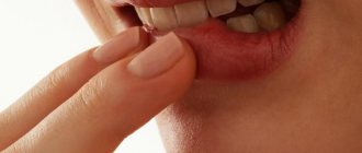
The first signs of bedsores
- Subjective sensations that the patient can communicate to caregivers when he is conscious and has preserved pain sensitivity in parts of the body:
- tingling on the skin in places where bedsores are likely to develop, is associated with stagnation of biological fluids (blood, lymph) feeding the nerve endings;
- loss of sensation (numbness) after about 2-3 hours in this area of the body.
- Visible signs of an incipient bedsore that caregivers must know:
- stagnation of peripheral blood and lymph, initially in the form of venous erythema of a bluish-red color, without clear boundaries, localized at the point of contact of the bone and muscle protrusions of the body with the bed, intensity of skin coloring: from barely noticeable to rich;
- desquamation of the skin epidermis with or without preliminary formation of purulent blisters.
These are signs of a developing bedsore. It is urgent to take measures to prevent further deterioration of the pathology.
What to do to eliminate the first symptoms of a bedsore?
To do this you need:
- change the patient’s position every two hours; if there are no contraindications, it is recommended to use special pillows to change the position of the limbs and body relative to the surface of the bed, forming gaps between the skin and the bed;
- monitor the level of the head of the patient’s bed; the head of the bed should be lower or level with it;
- regulate the humidity of the patient’s skin with hygiene products (washing cream, foam, solution, spray, you can warm baths (it is forbidden to use hot water), do these procedures at least twice a day, in case of uncontrolled bowel movements, remove contaminants as quickly as possible;
- remove excess moisture from the skin and skin folds (water, remains of liquid food, urine, wound exudate, sweat) using special absorbent pads, diapers, napkins, towels, films;
- regularly remake the bed or change bed linen at least once a day;
- do not perform intense massage; light stroking of skin areas with signs of stagnation is allowed; carry out this procedure carefully, without friction, especially in areas with close bones;
- use anti-bedsore mattresses of balloon or cellular type, equipped with special silent compressors to maintain and change the rigidity of its base, with adjustable and programmable inflation of different areas.
- use, for patients in wheelchairs, pillows filled with gel foam, air, monitor changes in body position in the chair at least once an hour.
Why are bedsores dangerous?
Bedsores are pathologies whose treatment is best avoided. If this could not be done, then when foci of skin maceration form, pathogenesis develops very quickly, with the formation of foci of tissue necrosis and is characterized by long-term treatment of a purulent wound. The outcomes of bedsores are dangerous. In some cases, bedsores are caused by:
- extensive excisions of soft tissues and the formation of defects with impaired innervation and blood circulation of the underlying areas of the body,
- lower limb amputations;
- necrotic damage to the periosteum and bone tissue in the form of osteomyelitis, periostitis;
- depletion of the body's defenses, complicating the treatment of the underlying disease;
With the development of bedsores of the wet necrosis type, the wound becomes infected, with the development of purulent processes (phlegmon, sepsis, gas gangrene).
With the development of bedsores of the type of dry necrosis, a protracted pathogenesis develops with long periods of healing of the defect.
Complications
The very development of bedsores to the last stage is already a serious complication. But if you don’t know how to treat bedsores and how to help the patient, the situation will worsen. Fistulas and abscesses will begin to form, and the likelihood of sepsis increases. The patient may catch an infection - and then the consequences will be completely unpredictable.
If bedsores appear, treatment should be started as quickly as possible - preferably at the first stage. But in case of missed situations, you also cannot give up. Now there are many remedies for bedsores for bedridden patients - some of them make the course of the disease easier, some of them allow the tissue to heal.
Causes of bedsores
The cause of bedsores is as follows. Our body is completely riddled with small blood vessels. Through these vessels - capillaries - blood flows to various organs of the body. If blood vessels are compressed, blood stops flowing to the tissues, as a result of which the tissues become necrosis.
If a person remains motionless for two hours, his blood vessels are compressed and blood stops flowing to certain areas of the body tissue. Therefore, bedsores form. Remember that it is very dangerous to sit or lie still for a long time.
Bedsores also form if a wet sheet is often pulled out from under a sick person. In this case, blood vessels rupture. This is completely invisible to the human eye. But after the blood vessels rupture, blood stops flowing to the tissues. Bedsores form.
Also, blood vessels can rupture if a person cannot, for example, walk and constantly slides down to take a different position.
When to see a doctor
You should consult a doctor as soon as the patient shows the first signs of bedsores. The doctor will examine the patient and tell you how to treat bedsores: treatment and treatment are carried out according to an individual plan - depending on the patient’s illness, the characteristics of his situation, taking other medications, concomitant diseases, etc. A lot of data needs to be taken into account in order to find a treatment for pressure ulcers that will not be useless.
You can contact our clinic for help. We are located in the center of Moscow, near the Mayakovskaya, Belorusskaya, Novoslobodskaya, and Tverskaya metro stations.
Risk factors for developing pressure ulcers
It has been observed that bedsores develop in bedridden patients at different times. In medical institutions, to systematize the assessment of risk factors for the development of pressure ulcers, the Norton, Braden or Waterlow scales are used. At home they don't matter. Based on these criteria, risk factors associated with care errors and individual characteristics of the patient, suitable for home use, have been formulated.
Factors associated with errors in the organization of patient care:
- untidy bed, changed less than once a day;
- rare change of underwear to dry and clean ones;
- neglect of hygiene procedures (treating the body with special solutions, drying, massaging areas of the body where possible without additional trauma to the bedsore);
- hard, uneven bed surface.
Factors associated with the individual characteristics of the patient’s condition:
- elderly age;
- exhaustion or, conversely, obesity of the patient;
- diseases of the cardiovascular system;
- diseases associated with impaired innervation of the body (including strokes);
- disorders associated with changes in metabolic processes in the body (diabetes mellitus, impaired water-salt metabolism or normal drinking restrictions);
- unbalanced nutrition or lack of protein in the diet, protein dystrophies (protein metabolism disorders);
- the patient's condition (coma, dementia, etc.) in which he does not control bowel movements or urination.
In addition, factors that provoke the occurrence of bedsores include smoking, diabetes, lack of water and little nutrition, excess or, on the contrary, very low weight, urinary and fecal incontinence, dirty skin, crumbs and small objects in the bed, allergic reaction to products skin care, folds, seams, buttons on underwear, as well as injuries and diseases of the spinal cord and brain, sweating at elevated temperatures.
Preparing to visit the doctor
Before visiting a doctor, do not treat wounds with ointments. To know how to treat bedsores, the doctor will also need information about all the patient’s illnesses and his condition. When accompanying a patient to an appointment, be prepared to answer all these questions: about the bedsores themselves, the reasons for the bedridden condition, the patient’s behavior and other points.
If the situation is advanced and the patient cannot be transported to the clinic in a wheelchair, then a doctor’s visit is required. The preparation is the same: information about the condition, as well as hygiene procedures without additional treatment of wounds.
Stages and degrees of bedsores
The pathogenesis of bedsores is characterized by stages of development. There are four stages of pathogenesis.
Stage I bedsores
Visually determined by venous erythema at the site of skin contact with a foreign surface. Venous erythema is the result of obstructed blood outflow from a local area.
How to distinguish venous erythema from arterial hyperemia and bruise?
- Difference from arterial hyperemia:
- the color of venous erythema is red-bluish, the color of arterial erythema is bright red;
- the local temperature of venous erythema corresponds to the skin temperature or slightly lower, the local temperature of arterial hyperemia is a warm area of the skin at the site where the temperature is determined.
- Difference from a bruise:
- the red-bluish color of the skin at the site of finger pressure does not change (bruise)
- the same color of the skin at the site of pressure turns pale (venous hyperemia).
Venous hyperemia (erythema) of protruding bony areas of the human body adjacent to foreign surfaces, without violating the integrity of the skin, is the most important sign of a pressure ulcer of the first stage.
Stage II bedsores
Visually determined by thinning of the epidermis - the upper layer of the skin, followed by peeling and the formation of blisters. Pathogenesis develops as follows: venous congestion provokes disruption of tissue nutrition, innervation of a body area, excess fluid in the skin, causes swelling (maceration) and rupture of epidermal cells.
Superficial damage to the skin in the form of thinning and peeling of the epidermis, loss of integrity, maceration (moistening) is the most important sign of the second stage of a bedsore.
Stage III bedsores
Visually defined as a wound with suppuration (contamination with microflora) or without suppuration.
Involvement in the pathogenesis of deep layers of skin tissue, subcutaneous tissue, muscles with a purulent type of inflammation and the beginning processes of necrosis (tissue death) is the most important sign of the third stage of a bedsore.
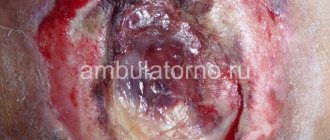
Stage IV bedsores
Visually it is defined as a local cavity or defect formed as a result of decay (necrosis), along the edges of the cavity there are walls of the defect on which purulent inflammation continues.
A necrotic cavity and its expansion due to inflammation of the walls is the most important sign of the fourth stage of a bedsore.
In different parts of the body, there may be different stages of bedsores.
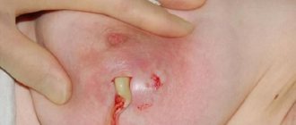
Purulent bedsores
Suppuration of the bedsore area begins in the second stage, develops in the third and fourth, after the wound is contaminated with staphylococci, streptococci, and other pyogenic microorganisms of the area.
A common way of developing a purulent bedsore is erysipelas and phlegmon. In severe cases, a bedsore develops into sepsis or gas gangrene.
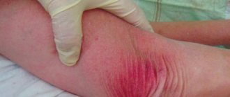
Development of purulent bedsores similar to erysipelas
Erysipelas is a local purulent inflammation in a local area of the skin. The causative agent is hemolytic staphylococcus and other pyogenic microorganisms. Erysipelas occurs in people with weakened immune systems, in the older age group, and is accompanied by toxicosis and hyperthermia.
Symptoms of erysipelas with bedsores:
- the skin at the site of the bedsore is bright red with a noticeable dense cold swelling - the main symptom;
- increase in body temperature up to 39 °C;
- headache, weakness, nausea;
- a purulent wound develops;
- exudate from the wound, without treatment, enters the bloodstream.
Development of purulent bedsores according to the type of phlegmonous inflammation
Phlegmon is a diffuse purulent inflammation without clear boundaries. The causative agent is staphylococcus, other pyogenic microorganisms, E. coli. Phlegmon can be localized under: skin, fascia, in the intermuscular space.
Symptoms of phlegmonous inflammation with bedsores:
- a glossy swelling without clear boundaries, red in color, hot to the touch - the main symptom of the onset of phlegmonous inflammation;
- body temperature up to 40 °C;
- rapid development of a fistula with purulent or putrefactive contents.
Development of bedsores according to the type of septic inflammation
Sepsis or purulent blood poisoning is a continuation of erysipelas or phlegmonous inflammation. The extreme stage of sepsis is septic shock, which often results in the death of the patient.
Symptoms of purulent septic inflammation in bedsores
Symptoms can be identified such as:
- atypicality – a variety of symptoms, the absence of the main one (pathognomonic).
- high temperature at the beginning of the process and low at the end;
- rapid development of the process.
The symptoms of sepsis begin to be combated in the early stages of a bedsore with the help of antibiotic therapy. Currently, bringing a patient to a septic state rarely happens, perhaps due to the patient’s immunodeficiency state and insensitivity to antibiotics.
Development of bedsores like gas gangrene
Gas gangrene is a severe purulent, putrefactive pathology. The causative agent is the microorganism Clostridium, most often Clostridium perfringens (soil microorganism). Infection occurs when contaminated soil comes into contact with the macerated surface of a bedsore.
Symptoms of gas gangrene in bedsores:
- sounds of crepitation (crunching) when passed over a damaged area of skin is a pathognomonic symptom.
- rapid development within six to seven hours;
- skin color gray-blue;
- the wound is dry;
- strong putrid odor.
Levomekol
One of the most popular ointments for bedsores. Levomekol is inexpensive and has a complex effect. The ointment relieves inflammation, removes pus, and cleans the wound. The antibacterial effect of the drug helps get rid of the infection that often develops in wounds with bedsores. Reviews of Levomekol are mostly positive, especially since the product has a very reasonable price. "Levomekol" helps at the prevention stage and with any stages of bedsores (wounds need to be treated two to three times a day). Among the disadvantages are an inconvenient tube and a too liquid texture when heated.
Levomekol
OJSC "Nizhpharm", Russia
An ointment that has an antimicrobial, anti-inflammatory, drying effect and is used for: Purulent wounds (including those infected with mixed microflora) in the first (purulent-necrotic) phase of the wound process.
from 38
1236
- Like
- Write a review
Skin treatment for bedsores
There are three areas of skin treatment for bedsores.
- Hygienic treatment of the skin to maintain the physiological properties of the skin (humidity, skin pH, softening the integument, increasing elasticity), removing physiological secretions of the body (sweat, sebum, epidermal scales), aggressive environments of feces (urine and feces) and deodorization of the skin;
- Preventive treatment of the skin to stimulate local blood circulation, restore sensitivity, and prevent skin cracking.
- Therapeutic treatment, use of medicines in preparative forms (ointment, cream, solution, gel, spray, powder, powder, solution for external use, etc.),
Solution for treating bedsores
In modern surgery, the use of solutions in the treatment of bedsores has somewhat lost its former importance. Meanwhile, simple solutions have been successfully used for many decades in military field surgery to treat wounds and their complications.
Solutions in any concentrations are not recommended for prevention and treatment: iodine, KMnO4 - potassium permanganate, brilliant green, hydrogen peroxide.
It is important to monitor the patient's diet. Your diet should include foods that contain zinc, iron and other beneficial microelements. Bedsores are often caused by iron deficiency. Dairy products, fish, poultry, and chicken eggs are rich in it. The patient should eat more green vegetables and fresh fruits. For those patients whose digestive system does not cope well with meat, broths are suitable as an alternative.
On this topic:
Prevention of bedsores
Bedsores on the heels, buttocks and tailbone
The usual position is for the patient to lie on his back. The most vulnerable places for the development of bedsores are the buttocks, tailbone, heels, and sometimes the area of the shoulder blades. Therefore, first of all, when performing hygienic procedures on a patient, you should pay attention to these areas of the body.
Bedsores on the heels
An unusual localization at first glance, but it occurs frequently. There are several heel diseases that, although not associated with bedsores, are evidence of the vulnerability of this area of the body, namely: heel necrosis (Haglund-Schinz disease), bursitis, heel epiphysitis, and so on.
Causes of pressure sores on the heel:
- load when lying on your back;
- thick skin, it is difficult to notice the beginning of the pathology;
- cracks in the skin of the heels, increased likelihood of microbial contamination;
- congestion of the lower extremities is a common companion for older people and occurs in some diseases (diabetes mellitus, etc.).
Symptoms of bedsores on the heels
Many sources indicate the absence of warning signs of bedsores on the heels. The appearance of a bedsore may be preceded by a white spot, tingling, or loss of sensation in the heel area.
Preventive actions
Prevention of pressure sores on the heels includes:
- use heel relief devices (wedge-shaped pillows, sheepskin, special mattresses);
- stimulate blood circulation (light massage of the calves), rub in 2% camphor alcohol, change the position of the legs every two hours;
- protect the skin of your heels from microflora, for example, using a colloidal butterfly bandage (Comfil Plus), which creates the effect of a moist, sterile chamber. The materials from which the bandage is made and the impregnating composition allow the bandage to be left on the wound for two to five days.
Treatment of bedsores on the heels
The remedies and methods for treating bedsores on the heels do not differ from the treatment of bedsores on other parts of the body. The anatomical features of the location of the bedsore are taken into account.
Bedsores on the buttocks
Bedsores on the buttocks have dangerous consequences. This area is located close to important organs (rectum, hip joint, pelvic organs, nerve nodes and blood vessels innervating the lower extremities), disruption of which significantly affects the quality of life, and in some cases, the preservation of life.
The buttocks have a powerful muscular system that should resist the formation of bedsores. However, regular contamination of the skin with urine and feces, without proper hygienic treatment, stimulates the development of bedsores. When a necrotic focus forms, extensive soft tissue defects are formed that are difficult to treat. The warning signs and symptoms of bedsores on the buttocks are typical.
Preventive actions:
Prevention of pressure sores on the buttocks includes:
- carry out regular hygiene procedures for the buttocks and perineum using washing creams, liquids, sprays; to simplify the procedures, use special latex-coated gloves that isolate the hands of the person caring for the patient and do not injure damaged areas of the patient’s skin;
- to prevent diaper rash on the skin of the buttocks and perineum, use absorbent panties, sheets, diapers, diapers, and neutral absorbent powders;
- Regularly, every two hours, change the patient's position, use anti-bedsore mattresses with programmable changes in the hardness of the bed and its different areas, use pillows, bolsters and other devices to prevent bedsores on the buttocks.
Treatment of bedsores on the buttocks
In the early stages, Russian-made multi-truss wipes or their analogues are recommended. The use of multifarm wipes is indicated for purulent processes with weak to moderate exudation. The exposure time and course frequency are indicated on the packaging.
Treatment of advanced forms of bedsores on the buttocks is carried out taking into account the anatomical features of this part of the body, similar to the treatment of purulent wounds.
Bedsore on tailbone
This area of the body protrudes somewhat and when lying down, the bones of the tailbone are in close contact with the bed of the bed. Bedsores on the tailbone are dangerous due to the small muscle layer and the presence of important nerve plexuses in it. Melting of tissues during necrosis damages nerve endings and provokes disruption of the innervation of the lower part of the body.
Symptoms of a bedsore on the tailbone
Precursors and symptoms of a pressure sore on the coccyx correspond to the classical ideas of the development of its pathogenesis. Due to the anatomical proximity of the buttocks and tailbone, preventive measures and treatment of the early stages are identical to those on the buttocks.
Dermazin
This ointment effectively destroys almost all bacteria that cause wound infection. At the same time, Dermazin has excellent penetrating ability; it treats bedsores well even with tissue necrosis. It contains silver sulfadiazine, which acts effectively and delicately at the same time. For maximum effect, the ointment is applied under the bandage once or twice a day (just before doing this, you need to thoroughly clean the wound). Sometimes an allergic reaction to Dermazin is possible, but this is rare. Reviews about the drug are mostly good, the cost is quite affordable.
Dermazin
Lek d.d., Slovenia
— treatment and prevention of burn infections (including before autodermoplasty);
- treatment and prevention of infection of trophic ulcers and wounds. from 180
465
- Like
- Write a review
How to treat bedsores?
Treatment of pressure ulcers of the second, especially the third and fourth stages should correspond to the treatment of purulent wounds. Purulent wounds are difficult to treat, but over many years of field surgery, a standard treatment algorithm has been developed. Of course, additions and improvements to treatment are regularly made, but the goal and objectives of treatment have remained unchanged.
I. In the first phase of pathogenesis
In the first phase, when the bedsore wound is filled with pus and necrotic tissue, you should:
- ensure the drainage of pus from the wound;
- relieve swelling;
- suppress microbial contamination of the wound.
Inspection of the wound and cleaning of the edges from necrotic tissue is performed in a surgical department. To drain the pus, drainage tubes are made and they are regularly inspected.
Passive drainage can be done at home:
- To do this, the wound is filled with special napkins soaked in compounds that promote the outflow of pus. The napkins are changed periodically. You can use regular bandages as napkins, the edges of which do not fall apart into threads. Solutions and ointments are used to impregnate napkins.
Outdated medicines:
hypertonic solutions 10% sodium chloride, 3-5% boric acid solution and others. Currently, the use of such solutions is limited due to the low suction force from 4 to 8 hours.
Hydrophobic ointments (liniments, emulsions) on a petroleum jelly basis (Liniment according to Vishnevsky, synthomycin emulsion, tetracycline, neomycin and others). Their disadvantage is that they do not absorb pus; the antibiotics in their composition do not work at full strength.
Modern medicines:
hydrophilic (water-soluble ointments) - Levomekol, Levosin and other water-soluble compounds. They remove pus from the wound well, within about 20-24 hours.
Attention! Use hydrophilic ointments only if there is pus in the wound; in another situation (no pus), these ointments are not effective.
- Enzyme therapy is the next method of surgical treatment of purulent wounds (therapy with pus-removing enzymes).
Proteolytic enzymes (trypsin, chymotrypsin, others). To enhance their action, use a combination of these or other enzymes with ointments, for example, a combination of enzymes and Iruksol ointment.
- Antiseptic solutions for external use. furatsilin, hydrogen peroxide, boric acid (currently used to a limited extent). Modern formulations are indicated for use - 0.5% solution of iodopyrone, 1% solution of dioxidine.
- Physical methods of treatment. They use traditional methods (UHF, ultrasonic cavitation, oxygenation, vibrophonation, laser therapy and other similar methods)
II. In the second phase
In the second phase, after cleaning the bedsore from pus, healthy tissue appears. A healthy scab is a thin layer of dried granulations. A purulent scab is a thick crust consisting of dried pus. Recovery under a purulent scab is impossible!
When healthy granulations appear, treatment is prescribed:
- relieving inflammation;
- protecting healthy granulations (healthy tissues) from accidental damage;
- stimulating tissue repair processes.
Oflomelid
This ointment for bedsores contains anti-inflammatory, disinfectant and analgesic substances - all this together helps to quickly cope with the problem. Oflomelid usually needs to be applied once a day to reduce the risk of infection of the wound and clean it. Significant relief occurs already in the first days of using the drug, and bedsores can heal completely after two weeks of constant use. Reviews about Oflomelid ointment are only positive, despite its cost (although the tube of the product is large). The only disadvantage of Oflomelid is that the ointment is not available in all pharmacies.
Oflomelid
Novopharm-Biosintez LLC, Ukraine
Infected purulent wounds of various localization and etiology in the first (purulent-necrotic) phase of the wound process, incl.
accompanied by severe pain: - infected burns of II-IV degree; - bedsores; - trophic ulcers; — postoperative and post-traumatic wounds and fistulas; - wounds after opening abscesses, phlegmons, after surgical treatment of abscessed boils, carbuncles, hidradenitis, suppurating atheromas, lipomas. from 93
5.0 2 reviews
365
- Like
- Write a review