Complications after otoplasty
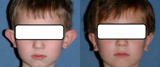
Aesthetic otoplasty is a frequently performed operation. There are many different ways to correct protruding ears, and each can have complications. The plastic surgeon performing otoplasty must be aware of possible complications, their cause and how to eliminate them.
Asymmetry
Asymmetry may exist before otoplasty. If asymmetry occurs after surgery, correction can be avoided if the asymmetry is minor.
Surgical correction will depend on the cause of the asymmetry (i.e., too narrow or too weak a suture on one side, too much or little skin excision on one side, etc.).
Bleeding and hematoma
Bleeding after otoplasty may be caused by inadequate hemostasis, hypertension, vasodilation due to the use of local anesthetics, postoperative trauma, or existing coagulopathy.
Surgery should be performed as early as possible to stop the source of bleeding and evacuate clots. Untimely removal of a clotted hematoma can lead to tissue necrosis or chondritis.
Cartilage relief disorders
Palpable and visible cartilage edges or irregularities may be caused by cutting the cartilage along the anterior surface. To avoid this, it is necessary to use cartilage-sparing techniques.
Cartilage necrosis
This may occur due to a hematoma causing excessive pressure on the cartilage. The necrotic area must be removed. And this in turn can lead to deformation that will require correction.
Chondritis and perichondritis
Typically, clinical signs of infection (pain increasingly resistant to analgesics, swelling, redness of the ear) begin 3–5 days after surgery.
Chondritis may require drainage and debridement of the affected cartilage. The use of antibiotics during and after surgery and the placement of a drain can prevent the development of chondritis.
Granulomas
Braided nonabsorbable sutures are more likely to cause granulomas than monofilament nonabsorbable sutures.
To eliminate a granuloma, it is necessary to remove the sutures and the granuloma itself. Non-absorbable sutures, such as nylon or polypropylene, can cut through the skin, causing fistulas or granuloma.
Hypersensitivity
Postoperative hypersensitivity is the result of irritation of the nerve endings of the cutaneous nerves.
It usually subsides when there is no pressure on the ear and is quickly eliminated with mild painkillers.
Infection and suppuration
Infection is usually diagnosed by the presence of excessive pain, erythema, fever, or purulent discharge.
The festering wound must be opened, sanitized and thoroughly washed with an antiseptic solution. Oral antibiotics are also prescribed for at least 7 days. Failure to treat the infection at an early stage can lead to perichondritis, chondritis and cartilage necrosis.
Hypercorrection
Excessive correction of the middle third of the ear leads to a “telephone handset” deformity.
Excessive correction of the upper and lower concha leads to a “reverse telephone ear” deformity.
Narrowing of the external auditory canal
Surgical deformity resulting from anterior displacement of the pinna can cause obliteration (narrowing) of the ear canal.
This can be prevented by careful preoperative planning and adherence to a protruding ear plan.
Pain
Excessive postoperative pain may be a sign of a developing hematoma. The dressing should be removed and the wound examined for swelling indicating a hematoma.
Treatment consists of finding the source of bleeding, electrocoagulating it and evacuating the hematoma.
Recurrence of deformity (protruding ears)
Recurrence of the initial ear deformity can be caused by trauma, insufficient weakening of the cartilage, dehiscence or cutting of the suture.
Rough scars
Hypertrophic or keloid scars may occur in approximately 2-5% of patients.
The earring hole can be a source of keloid scars. Excessive tension in the suture area can cause the scar to expand.
Careful and conservative skin resection can reduce the formation of rough scars.
About 8% of hypertrophic scars resolve without treatment, but 80% of keloid scars recur.
Treatment may include
- use of hormonal drugs (triamcinolone), alone or in combination with 5-fluorouracil or bleomycin,
- removal with or without radiation,
- cryotherapy with or without steroid injection and
- compression therapy.
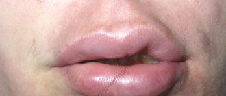
Skin necrosis
Skin necrosis is usually the result of postoperative compression bandages being too tight or it may be caused by excessive electrocoagulation of the skin. This can lead to rough scars.
Seam extrusion (cutting seams)
Suture cutting can occur as a result of permanent sutures being placed too close to the skin and placing excessive tension on the cartilage. Also, the development of infection can lead to cutting of stitches.
Telephone ear deformity
This deformity is created by the uneven distance of the pinna, where the upper and lower poles of the ear are still convex in relation to the middle third of the ear.
The reason is due to inadequate correction of the middle pole, which is excessively tightened by the suture, as well as due to excessive removal of skin in the postauricular area, soft tissue or cartilage in the middle third of the ear.
Prevention includes
- tightening the middle seam last to avoid potential overcorrection,
- avoiding excessive cartilage removal.
Treatment requires removal of incorrectly placed sutures and application of new ones.
Insufficient correction
Insufficient correction of the upper helix and lobule results in a relatively noticeable upper third of the ear called a “telephone deformity.”
Insufficient correction of an overdeveloped bowl can result in “reverse telephone ear.”
Unrealistic patient expectations
The surgeon must detect the patient's unrealistic expectations before ear surgery. In such cases, it is better to refuse the operation.
Early postoperative period
The most crucial moment is rehabilitation after otoplasty, because the extent to which the desired result is achieved depends on how it proceeds. After this operation, rehabilitation is divided into two types: early and late. The duration of the early rehabilitation period after otoplasty takes about 10 days and is accompanied by pain and swelling. Immediately after the operation is completed, a compression bandage is applied to the head. It is very important to follow the recommendations for wearing this bandage, because it performs such important functions as:
- fixing the ears in the desired position;
- protection of ears from accidental injuries;
- containment of swelling and spread of hematoma.
The success of otoplasty and the rehabilitation period largely depend on wearing a compression bandage, which is removed only on the recommendation of the plastic surgeon.
How to cope with pain?
If your ears hurt badly after otoplasty, you can use various methods to relieve discomfort. Of course, the very first and most effective way to get rid of pain is through medications with an analgesic effect. An injection of an anesthetic drug is usually given in a plastic surgery clinic shortly before the patient is discharged from the hospital.
Taking analgesics at home is not always necessary. The surgeon is guided by the current situation. Mild painkillers may be prescribed for home use to relieve moderate pain. If the pain becomes too noticeable and current medications are not helping to cope with it, the patient can contact his surgeon and ask to be prescribed medications with a more pronounced effect.
Quick Recovery Tips
Mostly, the period for which ears hurt after otoplasty is about 3-7 days, depending on individual characteristics. To speed up tissue healing and, accordingly, relieve discomfort, the patient must follow all the rules for caring for the injured area during the recovery period, prohibitions and recommendations of his doctor.
Heat treatments, such as a steam bath, sauna, or even a hot bath, promote blood flow to damaged areas, which delays tissue healing. Sports and physical activity also negatively affect the healing rate. Bad habits, an abundance of spicy and salty foods also increase the recovery time.
Suture sites must be treated daily with disinfectants. At first, you need to wear a fixing bandage. You cannot remove or move it without your doctor's permission. It is advisable to sleep on your back.
Indications
Otoplasty is performed if the patient wants to eliminate protruding ears, asymmetry of the shells or other external defects. Today, young people often turn to surgeons who want to sharpen the upper tip of the shell or change any parameters of the lobe.
The main indications also include:
- Small and large ears
- Birth defects of various types (protruding ears, protruding ear, “macaque ear”, etc.)
- Acquired defects and consequences of injuries (ruptures and deformations, wearing tunnels, etc.)