Warts in the vagina appear against the background of human papillomavirus infection, which is spread, among other things, through unprotected sexual intercourse. The pathology is based on the human papillomavirus (HPV). It is the main cause of genital warts in women. There are about 100 different HPVs in nature, 60 develop on soft tissues and cause warts, and another 40 cause infections on the mucous membranes of the human body. According to statistics, women are more often affected by HPV. Research has shown that about 50% of sexually active people become infected with the human papillomavirus in their lives during the first years of sexual activity. It is very important to detect the disease at an early stage, as the disease can lead to cervical cancer. It is condylomas inside the vagina and in the anus that most often have a high oncogenic risk.
What are condylomas in the vagina
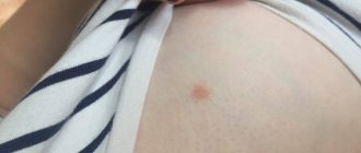
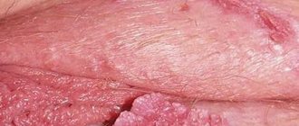
Genital warts in the vagina are neoplasms that resemble warts in appearance. They are localized mainly on the skin and mucous membrane of the genitals, and tend to merge. The causative agent is the human papillomavirus (HPV), which is transmitted through unprotected sex with a carrier partner.
Often, HPV carriage can be asymptomatic for many years, and only when a woman’s immunity decreases, the disease begins to manifest itself as warts and condylomas. Externally, such neoplasms resemble cauliflower; growths can be around the anus, on the vulva or in the vagina.
Warts can grow and cause burning or itching. They are benign tumors, but if left untreated, HPV causes mutations in human cells, which also lead to cervical cancer. Therefore, it is so important to surgically remove condylomas in the vagina in a timely manner.
Types of condylomas
There are two main types of condylomas:
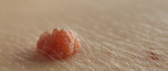
1. Pointed.
When examining the vagina, you can see growths that resemble papillae.
This type of condylomas is called genital warts. They look like a neoplasm on a leg, most often they are flesh-colored. They can appear either singly or in groups. Usually located on the walls of the vagina. 2. Flat.
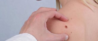
These growths are difficult to notice. The shape of condylomas can be round or oval. The growths are not distinguished by their shade and merge with the mucous membrane. Neoplasms have a slow growth rate and do not cause discomfort to the woman. Diagnosis of condylomas When suspicious growths are detected, consultation with a gynecologist is a mandatory procedure. The examination involves the following activities:
- examination in a gynecological chair;
- examination of a smear from the cervical canal;
- colposcopy;
- blood analysis;
- histology;
- PCR diagnosis.
There is a guaranteed way to diagnose condylomas in the vagina - using a vinegar solution. The mucous membrane of the labia should be treated with vinegar. As a result, a spasm of the vessel occurs, which feeds the skin tumor. Over time, the growths will lighten and stand out, due to this, their exact number and diameter can be determined. It is even possible to see subcutaneous tumors that are located in the deep layers.
Treatment options
Methods for getting rid of condylomas in the vagina involve radical removal of growths and conservative treatment, supplemented with traditional medicine. Only the doctor decides how to treat condylomas. Conservative therapy There are many conservative ways to treat condylomas of the anal area, vulva, and vagina. This is the use of antiviral drugs, cytostatic agents, drugs with a general strengthening effect, and immunostimulants. If the growths are located in the area of the vulva, and their coverage area is no more than 10 cm2, you can use cauterizing agents (Condilin, Podophyllin, Verrucatsid, Collomac, Solcoderm). These are drugs that cause necrosis of the treated surface. Therefore, medications are not recommended for the treatment of the anal area and urethra. Prohibited when carrying a child. Removal of condylomas in the vagina is usually done surgically, sometimes supplemented by the use of antiviral drugs. This group includes Panavir, Interferon, Isoprinosine. Therapy is often supplemented by the use of drugs that increase immunity, for example, Polyoxidonium, Aldara, Immunal. Radical treatment Today, to get rid of condylomas, electrocoagulation, cryodestruction, surgery, laser vaporization, and a radio wave knife are used. But not all methods are suitable for treating condylomas. The use of nitrogen in this case is more traumatic and not as effective as other methods. As for the surgical removal option, this process is invasive and is prescribed only in very severe cases. When treating, the choice is generally made in favor of an electrocoagulator, a radio wave knife and a laser. All these methods are used under anesthesia, so they do not cause discomfort to the woman during surgery. They are practically non-traumatic, unlike surgical intervention, they have a fast healing time. The difference lies in the operating principle of the device. A radio wave knife and laser evaporate infected tissue layer by layer. The electrocoagulator cauterizes the growths by applying current at high frequencies. Any of the methods has certain contraindications, but, in general, these treatment methods can be used to remove tumors in women on the clitoris, labia minora, cervix, and vagina. Folk methods Turning to folk methods, you can find many recipes that are recommended for getting rid of growths. The most popular remedies include celandine juice. It is used to get rid of various skin growths. But during the processing process, you need to act carefully so that the composition does not get on healthy tissue. Iodine can help cure condylomas, as it has a drying effect. The product can only be used on the surface of the skin; if applied to the mucous membrane, a burn will occur. Traditional recipes also have certain contraindications. In addition, none of them are suitable for getting rid of condylomas in the vagina. Therefore, folk treatment can only complement the main one. Before use, you should definitely consult your doctor. Condylomas during pregnancy Growths during pregnancy are also common. In this case, of course, there are certain features that need to be taken into account both during conservative treatment and when removing the tumor using various methods. The formation of growths provokes, first of all, the appearance of hormonal imbalance, as well as a weakening of the immune system during gestation. Large condylomas will affect the birth process and complicate it. When diagnosing condylomas, doctors recommend giving birth to a child by caesarean section.
How do condylomas appear in the vagina?
The favorite place for the appearance of genital warts is the external genitalia. In women, they appear on the cervix, anus and labia. In men - on the head of the penis, the skin of the foreskin and around the anus. Once a doctor discovers genital warts, treatment should begin as early as possible because some types of human papillomavirus have increased oncogenic properties.
The disease may be asymptomatic. When condylomas are injured by clothing, during defecation or during sexual intercourse, short-term bleeding often appears. Inflamed tumors can itch and cause discomfort. There are strains of HPV that cause unusual vaginal bleeding. This often happens when warts caused by HPV become cancerous.
It is important to visit a gynecologist in a timely manner, and if you notice genital warts, it is better to get examined immediately. In most cases, a smear will help detect the virus in a timely manner and help prescribe correct and timely treatment.
In our clinic you can undergo examination and treatment of vaginal condylomas. The technical equipment of the medical center allows for highly reliable examination and removal of genital warts using minimally invasive methods. For any severity of condylomatosis, you should be examined by proctologists or venereologists, since there are frequent cases of squamous cell carcinoma developing against the background of HPV.
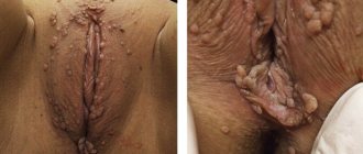
Genital warts, genital warts (AC), exophytic warts, venereal warts, Genital warts in the English literature - benign formations caused by the human papillomavirus (HPV) 6, 11, 30, 42, 43, 44, 45, 51, 52 types, but in 90% of cases types 6 and 11 are found. Genital condylomas have the appearance of warts, papillomavidous or broad-based formations. Histological examination reveals a picture typical of the cytopathogenic effect of HPV - thickening of the epidermis, acanthosis, thickening and elongation of interpapillary wedges, koilocytosis of superficial keratinocytes [4]. Most people who become infected with HPV, including the low-oncogenic type, do not develop symptoms of the infection. The source of infection, of course, is a sick person. In 65% of people, clinical manifestations occur within 90 days after contact with a sexual partner, although the exact timing is unknown, since it is impossible to reproduce the disease in experimental animals. Sexual transmission is not the only route for genital warts; the household route is equally dangerous - through contaminated underwear, hands; infection of a child during childbirth is well known. The established fact of positive HPV tests in pubic hair follicles is very important. It is known that in healthy men, HPV is detected in 17-21% of cases; in patients with genital warts, pubic hair follicles are infected in 32-55%. Thus, regardless of the type of sexual contact, skin-to-skin contact is dangerous [6]. This “unpretentiousness” of the virus in choosing its habitat determines the extreme prevalence of both the pathogens themselves and the diseases they cause. The lowest frequency of OC lesions was recorded in Russia - 34.7 per 100 thousand population (2009), while in Germany (2006) the frequency of exophytic condylomas in young women was 462 per 100 thousand, in England and Wales (2000) - 715 per 100 thousand population (cited from 2). There is probably an underestimation of this pathology in Russia. The most important feature of low-oncogenic HPV types is their ability to associate with highly oncogenic ones. Every fourth patient with AC is diagnosed with cervical intraepithelial neoplasia of varying severity [7], and removal of genital warts does not eliminate viruses of high oncogenic risk. This is what determines the development of cervical or vaginal cancer in women who have OK. It should be noted that genital warts can also be accompanied by a rare type of cancer - verrucous (giant cell condyloma of Buschke-Levenshtein). It is associated with viruses of types 6, 11 and has a tendency to local invasion.
Factors that contribute to the persistence of HPV infection are well known: age under 21 years at first sexual intercourse, 4 or more sexual partners in a lifetime, immunodeficiency conditions, and the presence of other STIs. From a practical point of view, atopy is a very important suppressive factor, no matter what clinical forms it has, which affects the therapeutic effect and the high probability of relapse in patients with genital warts. There are many myths about genital warts: for example, warts only occur during unprotected intercourse. But, unfortunately, even “protected sexual intercourse” does not guarantee safety, since condoms are not able to protect the skin of the pubis and perineum, where the infection also persists.
Diagnosis of OK is simple; genital warts are well identified visually and when examined with a colposcope. Condylomas are localized on the cervix, vaginal walls, perineum, perianal area, and the external opening of the urethra and clitoris are often affected. Colposcopically, condylomas may appear as pointed growths, have a rounded papule-like shape, or resemble papillary formations. The size of the growths varies - from small single ones to massive ones, blocking the entrance and vaginal cavity. A biopsy of condylomas is carried out if the diagnosis is unclear, there are signs of atypia, warts are stained, their increased density and fixation on the underlying tissues, bleeding or ulceration.
Clinical symptoms do not always depend on the degree of damage. There are often situations when, in the presence of single OCs, itching, burning in the vagina, constant discomfort or discomfort during sexual intercourse, and even pain are bothered, while extensive lesions do not cause any complaints. If OK is obvious, HPV testing is not recommended because it will not lead to a change in treatment.
Genital condylomas should be differentiated from molluscum contagiosum, squamous cell papilloma, condyloma lata, fibroepithelial polyp, and much less often from dermatitis and psoriasis.
Molluscum contagiosum is caused by a DNA pox virus. Typical for this disease are single or grouped small, smooth papules 3-6 mm in diameter with a pinpoint depression in the center. The rashes, even if they are multiple, never stick together, although they can form plaque-like foci.
Squamous cell papilloma is a multiple papillary formation located in the vestibule of the vagina below the hymen ring. Sometimes papillomas are localized on the medial surface of the labia minora. Papillomas are covered with stratified squamous epithelium and are often an accidental finding in the absence of complaints. Their connection with HPV has not been proven, the origin is unclear.
Fibroepithelial polyp is a benign papillomatous formation without vascular atypia, histologically it has characteristics characteristic of a polyp.
Condylomas lata with syphilis are flat, dense, pink-red formations on a wide base.
If you suspect dermatitis, psoriasis, or in unclear cases, consult a dermatologist. It is worth remembering about vulvar and vaginal cancer, where only a comprehensive assessment will allow you to avoid mistakes.
There are two approaches to the treatment of genital warts: traditional - complete elimination of all lesions or removal of condylomas as a cosmetic nuisance. There is no consensus regarding male HPV carriers without clinical manifestations. Currently, preventive treatment for men is not accepted, since there is no reliable data on its effectiveness. Spontaneous recovery is known - the reverse development of genital warts in 29-39% of patients. Often, children in whom the appearance of condylomas at the age of 2-3 years is associated with intrapartum infection are spontaneously cured. The effectiveness of various methods of treatment of OK, even taking into account repeated courses, is 60-80%. Relapses in 25-50% of cases occur within the first 3 months after treatment and are most often caused by reactivation of the virus [5]. Destructive physical treatments are the most popular, and the first choice of method may be electroexcision, laser vaporization, cryosurgery, radio wave excision or photodynamic therapy. Electroexcision frees viruses within a radius of 20 mm from the center of the lesion. Conservative treatment of small lesions is carried out with podophyllin, trichloroacetic acid or solcoderm. In English-speaking countries, 5% imiquimod cream (Aldara) or 15% synecatechin ointment is used, treatment is carried out for 16 weeks. There is currently no evidence that local interferon injections lead to regression or reduction in the recurrence rate of genital warts.
Material and methods
We tested various types of therapy (medicinal and destructive) in 397 women with genital warts of the vagina and vulva; in some we used the domestic drug Panavir, which is available in various forms: injection solution, suppository and gel. The mechanism of action of the drug is multifaceted and, as experience shows, its use is effective for various viral diseases. The drug prevents adhesion and inhibits the penetration of the virus, stops the opening of the viral capsid, disrupts the transcription and replication of viral DNA, the synthesis of capsid proteins, stops the assembly of viral particles and their exit from the cell, and has a complex immunomodulatory effect [1, 3]. The basis of the drug is a biologically active heteroglycoside - panavir.
The age of the patients ranged from 16 to 40 years, all women complained: 119 (30.1) experienced itching and discomfort in the vagina, 90 (22.8) had chronic leucorrhoea, 88 (21.8) had discomfort during sexual intercourse. 5%), recurrent candidiasis - 5 (11.3%), cosmetic defect bothered 55 (13.9%). Viruses 6, 11 types were isolated from 327 (82.2%) women, of which 48 were in combination with HPV types 16, 18, 25 were isolated from types 42, 44, 51, 45 were not infected with HPV was found. Two women had massive lesions, the rest had condylomas of the vagina or vulva with an area of 5-8 cm2. Among concomitant infections, chlamydia was found - in 35 (19%), herpes virus - in 19 (4.8%), ureaplasma - in 97 (24.4%), Candida
- in 49 (12.3%), trichomonas - in 9 (2.2%). The remaining patients had nonspecific bacterial microflora in various titers. Chronic cervicitis was noted in every third woman with OK, bacterial vaginosis - in every fourth, colpitis - in every fifth. Stage I cervical epithelial dysplasia was diagnosed in 48 (12.1%) patients.
At the first stage of treatment, the concomitant infection was treated in accordance with the identified pathogen, followed by antiviral therapy with Panavir in 140 women (group 1). The comparison groups included 165 women (group 2) who received only electrical excision of condylomas and 92 patients (group 3) in whom electrical excision was supplemented with the use of α-interferon in suppositories vaginally for 2 weeks after surgery.
The treatment regimen with panavir of the 1st group consisted of using a combination of two forms of the drug: rectal suppositories (1 suppository every other day, N10) and vaginal gel 2 times a day for 14 days. After antiviral therapy, condylomas were removed by electroexcision.
Results and discussion
The results were assessed by the frequency of relapses, elimination of viruses and regression of complaints (see table).
As follows from the table, the most effective method was the use of Panavir and subsequent electroexcision of condylomas compared with the results in groups of women who received only electroexcision or its combination with the use of α-interferon. It should be noted that immunocorrection in the treatment of HPV is necessary and this fact is generally recognized. Local therapy is more effective if accompanied by the use of systemic immunomodulatory drugs, as this leads to a more lasting anti-relapse effect. This was confirmed in our study by the fact of elimination of viruses, including highly oncogenic types. Of the 28 women of the 1st group with combined HPV types 6, 11, 16, 18, negative results of the polymerase chain reaction test were found in all of them; moreover, herpes viruses were not found during repeated analysis. Subjective sensations of discomfort in the vagina also disappeared in all patients. Among patients in group 2, 18 women had combined HPV infection caused by viruses of low- and high-oncogenic types. The performed electroexcision had no effect on their presence, and the persistence of these types of virus remained. Viruses 6 and 11 types were isolated from 59 (35.7%) of 165 women in this group, which probably determined the high rate of relapses 3 and 6 months after treatment, as well as the persistence of complaints in every third patient.
The use of electroexcision and α-interferon in the postoperative period led to the elimination of viruses in 76.2% of women. Highly oncogenic types of the virus identified in 2 women in this group persisted after treatment. Symptoms such as leucorrhoea continued to bother me (in 16 women) and itching in the vagina persisted (in 12). It should be emphasized that our proposed treatment regimen with Panavir differs somewhat from that recommended by the manufacturer, but purely empirically this option was accepted as optimal. Only local use of Panavir gel in combination with destructive methods is equal in effectiveness to the use of a combination of α-interferon and the destruction method, while the combination of a fairly long-term systemic treatment with Panavir (rectal suppositories) and local therapy (vaginal gel) in combination with the destruction of condylomas gives a good lasting effect .
The cost of treating genital warts is high worldwide. Relapses of the disease, repeated courses of therapy and, at the same time, insufficient effectiveness lead to an increase in the cost of treatment so much that it can be compared with the treatment of AIDS. The use of Panavir in the proposed version is a one-time cost, but it is highly effective, amounting, according to our data, to 99.3% at 6-month follow-up and 92.5% at 2-year follow-up. The ability of the drug to provide an immunomodulatory effect, proven in numerous studies, and an antiviral effect in various diseases is encouraging in the treatment of infections caused not only by low-oncogenic HPV types, but also by highly oncogenic ones, which can help prevent cervical cancer.
conclusions
1. The proposed treatment regimen for patients with genital condylomas with a combination of fairly long-term systemic and local use of Panavir in combination with subsequent electrical excision of condylomas gives a good and lasting effect.
2. The ability of Panavir to have an immunomodulatory and antiviral effect is encouraging in the treatment of infections caused not only by low-oncogenic but also highly oncogenic types of HPV.
Causes of condylomas in the vagina
The main method of transmission of HPV, which leads to the formation of condylomas, is genital. But experts believe that the human papillomavirus is present in the body of many people, but it does not cause the development of diseases in everyone.
HPV is transmitted through any type of sexual intercourse, be it anal, vaginal or oral contact. Wearing a condom during sexual intercourse can help prevent transmission of the virus, but does not provide 100% protection. HPV is often transmitted through wounds, scratches and cuts - these are the main places of entry of the papillomavirus into the body. Transmission from mother to child is possible.
A mother can transmit the virus to her child during childbirth if the baby has microcracks in the skin and mucous membranes. If a child is infected with the strain of HPV that causes genital warts, he or she may also develop airway papillomatosis (growths on the vocal cords).
Anxiety, stress and depression can affect the secretion of hormones and thus cause hormonal imbalance, which can activate the existing papillomavirus. When the production of hormones is disrupted, a person’s immunity is noticeably reduced, and this creates an ideal environment for the proliferation of the human papillomavirus. The use of birth control pills can also disrupt the balance of estrogen and progesterone and thus contribute to the spread of infection.
Here are the factors that provoke the appearance of genital warts on the genitals of men and women:
- sexually transmitted diseases, genitourinary infections (ureaplasmosis, trichomoniasis);
- decreased immunity;
- injuries to the vaginal mucosa, penis;
- promiscuity;
- changes in vaginal microflora;
- intestinal dysbiosis.
Most often, condylomas appear during unprotected intimate contacts in combination with a weakening of the body’s natural defenses.
Diagnostic methods
A qualified gynecologist can make an accurate diagnosis for condylomas and viral warts in the vagina. Based on the examination, the specialist draws conclusions about the development of papillomavirus infection. Additionally, the woman is prescribed an extended colposcopy and PCR test.
A cytological examination of a smear from the cervical canal area allows one to identify atypical cells and confirm or exclude the presence of dysplastic processes in the cervix. Immunological studies help determine antibodies to papillomavirus in the blood. Additionally, it is necessary to conduct screening for common sexually transmitted diseases and HIV infection.
Diagnosis of a vaginal tumor
The method for identifying a benign tumor depends on the type of tumor and its location or localization.
- Examination by a gynecologist.
The doctor's use of mirrors makes it possible to recognize the formation node and determine the wide base or pedicle. You can see a tumor up to the size of a chicken egg. The doctor may notice a raised red or bluish spot above the swollen mucosa. At the same time, pay attention to thickened folds and loose structure, diagnosing hemangioma. With the help of palpation, a formed formation of a dense structure located on the vaginal wall is determined. - Colposcopy.
A widely available way to examine a woman’s vagina using a colposcope. Allows you to detect varicose changes in blood vessels if a hemangioma has formed. Using this method, you can correctly determine the site for collecting material for a smear for cytology. - Transvaginal ultrasound.
Able to detect any vaginal tumor and determine its structure, taking into account the echogenicity index.
Taking a smear for cytological examination can provide the most accurate results regarding the presence of atypical cells.
PCR diagnostics are carried out to detect a viral infection associated with the presence of papillomas. After all, HPV has a high oncogenic risk.
When removing benign tumors, a histological examination is required.
Treatment methods for condylomas
Most often, patients come to us with genital perianal condylomas, the removal of which can be carried out in various ways:
- radio wave therapy (removal of condylomas with a radio knife);
- cryodestruction (cauterization of condylomas with low-temperature liquid nitrogen);
- laser coagulation (evaporation of pathological neoplasms with a laser);
- electrocoagulation (use of high-frequency current).
To prevent recurrence of condylomatosis, antiviral therapy is prescribed. Patients are also advised to periodically visit specialists and get tested. Women who have had condylomas removed should have smears twice a year and undergo histological diagnosis.
Therapy for pregnant women
Genital warts in the vagina can appear during pregnancy due to a weakening of the body's natural defenses. In this case, it is necessary to contact a gynecologist as soon as possible so that the specialist can conduct a comprehensive examination and identify possible concomitant diseases, such as thrush. Surgical removal of genital warts is not performed during pregnancy, since any surgical interventions are contraindicated for the expectant mother.
Specialists limit themselves to local therapy, which eliminates itching, inflammation, and prevents the occurrence of infectious complications and frequent relapses of the disease. Topical antiviral drugs should be used only after the permission of the attending physician.
But sometimes local treatment of vaginal condylomas does not bring the expected result. This is all due to a natural decrease in immunity, which allows you to maintain the viability of the fetus and avoid miscarriage. Also, an exacerbation of human papillomavirus infection is caused by hormonal changes in a woman’s body.
If local medications do not help get rid of vaginal condylomas, it is recommended to wait a while, using daily antiseptics and other safe means that will inhibit the growth of the virus. After childbirth, in most women, vaginal warts completely disappear, which indicates a connection between human papillomavirus infection and natural changes in the body of the expectant mother.
Prevention
It is almost impossible to avoid the initial entry of papillomavirus into the human body. Experts have come to the conclusion that HPV first enters the bloodstream during puberty and the active beginning of intimate life. For a long time, the virus may not manifest itself in any way, but with a decrease in immunity, hormonal imbalance, damage to the mucous membranes of the genitals and the action of other predisposing factors, papillomavirus infection develops. Unprotected intimate contacts and frequent changes of sexual partners predispose to the onset of the disease. As a result of contact with a man who has viral warts on the genitals, a woman can become infected even if she has a strong immune system.
Prevention of the appearance of genital warts of the vagina and external genitalia includes, first of all, the prevention of accidental intimate contacts without the use of barrier contraceptives. It is recommended to adhere to the principles of a healthy lifestyle, carefully monitor the quality of intimate hygiene, and if any signs of gynecological pathologies and external inflammatory processes appear, contact qualified specialists.
Types of Benign Tumors
Benign neoplasms of the vagina are represented by various types. They are usually detected during a routine examination by a gynecologist, if they do not make themselves felt. If discomfort occurs, the woman consults a doctor herself.
- Myoma, fibroma (or fibromyoma).
These types of benign neoplasms usually have a round or oval shape. The relief of the tumors is smooth. The consistency is particularly dense and soft. Tumors form from cellular growths of various tissues (smooth muscle, connective tissue, etc.). Neoplasms have limited mobility. Their connection with the submucous membrane of the vagina or skin fold (labia majora) is completely painless. - Hemangioma.
It is a soft neoplasm of blood vessels. The tumor has a red or bluish tint. The mucous membrane around the formation is swollen, thickening of the folds and looseness of the epithelium are observed. This type of tumor can expand and cover the cervical canal and uterine cavity. Complications (ulceration and tumor necrosis) are often possible. - Lipoma.
The neoplasm is formed from adipose tissue. Localized in the pubic area and labia majora. A fat capsule or wen occurs as a result of blockage of the sebaceous ducts. The consistency of the formation is soft, mobile, and has a clearly defined shape. - Myxoma.
It is formed by the remains of embryonic connective tissue. The tumor is typical for elderly patients. The localization site is the subcutaneous fatty tissue of the pubis and labia majora. It grows slowly. The neoplasm is not inclined to change its nature and does not pose any particular danger to women. - Papilloma (condyloma).
The neoplasm is formed from epithelial tissues. Represents papillae on legs or on a broad base. Usually found on the walls of the vagina. The cause is HPV. - Lymphangioma.
The tumor has a finely lumpy structure. Formed from lymphatic vessels. It is a formation of a pale bluish hue, consisting of many nodules. There is lymph inside them. The mild swelling may be painful and especially tender. Sometimes dense inclusions appear. There are known cases of suppuration. - Cysts.
These are specific formations or lumps filled with fluid or epithelial tissue. Especially often, Bartholin gland cysts are found in a woman’s vagina. Their occurrence is associated with the presence of a bacterial infection in the body.
Retention cysts can occur in the upper part or on the walls of the vagina. Their appearance is related to the intrauterine development of the embryo. At an early stage of development, two ducts are born: Gartner's (Wolf's) and Müller's. When the fetus develops, the formation of the male genital organs occurs from the right and left Gartner's ducts. The right and left Müllerian ducts initiate the development of the female genitalia. The duct, left out of use, gradually dies off. Sometimes the remnants of both passage systems degenerate. As a result, the woman develops cysts. Most often this is the fate of the young. Sometimes the cause of the appearance of cysts in the vagina can be atypical focal inclusions from the epithelial layer or former hematomas.