Fistulas at the site of a surgical suture are a rare but unpleasant complication associated with non-compliance with the rules of asepsis and antisepsis and the use of non-absorbable threads. If a patient develops a ligature fistula after surgery, it should be treated by a highly qualified surgeon in a specialized center to prevent relapse. It should be noted that strict adherence to the sanitary and epidemiological regime and the use of modern suture material during operations at the Igor Medvedev Center reduces complications such as ligature fistulas to zero. Our specialists also successfully treat complications of operations performed in other medical institutions.
Ligature fistulas
A ligature is a thread used to tie a vessel, restoring the integrity of organs and skin. A fistula is a reaction of the tissues around the thread, leading to their melting, failure of the suture, divergence of the edges of the wound, bleeding, etc.
Causes
Any abscess is a localized inflammatory process limited to the capsule.
The capsule may consist of an omentum, inflammatory adhesions and adjacent tissues. The abscess cavity most often contains aerobic and anaerobic bacteria that have migrated to this area from the gastrointestinal tract. The immediate sources of abscess formation are infectious agents and inflammatory factors that affect the condition of tissues. Bacteria can penetrate into the structures of the abdominal organs from the external environment during surgery, the gastrointestinal tract and other areas. Often we are talking about secondary peritonitis due to rupture of the intestinal walls or pancreas.
Main reasons:
- Inflammation of the appendix of the cecum (appendicitis). In this case, purulent exudate is formed in the closed intestinal cavity. If treatment is not carried out in a timely manner, perforation of the intestinal wall with penetration of pus is possible.
- Inflammation of pancreatic tissue followed by necrosis. Exudate can also penetrate into the free abdominal cavity and form an abscess.
- Rupture of the wall of the duodenum due to peptic ulcer disease of the organ.
- Inflammation of the gallbladder and its complications, such as gangrenous cholecystitis.
- Mesenteric ischemia and development of tissue necrosis.
- Purulent inflammation of the female genital organs.
- Complication of surgical intervention in the gastrointestinal tract, severe injury.
- Other sources of exudate include diverticulum rupture, leaky intestinal anastomosis, and hematoma rupture.
The composition of the pathogenic microflora of the abscess depends on the source of inflammation. Most often, E. coli is found in pus. In patients who have been taking antibiotics for a long time, inflammation can be caused by pathogenic and opportunistic fungal microorganisms.
Why are ligature fistulas dangerous?
Since the thread in the case of a fistula becomes a source of chronic infection, the fistula leads to intoxication of the body, impaired immune reactions, and, in the early period, to suppuration of the wound and failure of the sutures. Treatment of ligature fistulas
- conservative
- Washing the wound with antiseptics, taking antibiotics, vitamins, immunomodulators, and anti-inflammatory drugs helps cope with fresh ligature fistulas. Minimally invasive techniques are also used: cauterization of excess granulations with an electrocoagulator and laser, ultrasound treatment of fistulas.
- surgical
If a patient has a long-existing ligature fistula, it is treated surgically, the same can be said in case of complications from the wound (suppuration, suture dehiscence). In this case, the ligatures are removed, the edges of the wound are refreshed, fibrous tissues and excess granulations are removed and new sutures are applied using sterile atraumatic synthetic material with different resorption times.
Classification
Doctors describe different forms of the disease based on the source of the inflammatory process. It is very important to establish the localization of the abscess in the early stages of diagnosis for effective surgical treatment.
Basic forms:
- Metastatic abscess is the formation of a secondary pyogenic capsule containing pus as a result of the spread of infection from distant sites. Bacteria and fungal microorganisms can spread through the blood and lymph flow.
- Postoperative ulcers are pathological areas formed as a result of surgical treatment of diseases. In this case, the pathology may be caused by a failed interintestinal anastomosis, infection, or an error during removal of the appendix.
- Perforated abscess. The inflammatory focus is formed as a result of rupture of the walls of the inflamed anatomical structure. This could be a rupture of the walls of the appendix, pancreas due to pancreatic necrosis, or another organ.
- Post-traumatic abscess resulting from trauma to the abdominal organs.
Imaging and diagnostic procedures can help determine the source of inflammation.
What is an abscess and what causes it?
An abscess is a painful pustule surrounded by inflamed tissue. Often it can be easily felt. Such a formation can appear on any part of the body, but most often it affects:
- armpits;
- area around the anus and vagina (Bartholin gland abscess);
- tissue around the tooth;
- skin in the groin area;
- hair follicles.
During their development, abscesses are filled with necrotic masses and can open on their own. However, it is best to consult a doctor in case of such a disease, who will open the purulent focus and clean (drain) it.
Causes of abscesses:
- as an independent disease, abscesses of the skin and soft tissues most often occur, caused by blockage of the openings of the sebaceous and sweat glands, the formation of cysts and the proliferation of pathogenic microorganisms there;
- purulent soft tissue abscess can be a complication of skin damage;
- a post-injection abscess develops when the drug is administered with a non-sterile syringe; in severe cases, a purulent process – phlegmon – may spread;
- such a process often complicates the course of any infectious diseases of a bacterial nature, for example, a peritonsillar abscess develops as a complication of tonsillitis;
- Abdominal abscesses can develop when microbes are transferred through blood vessels;
- in some cases, the cause of the disease is not bacteria, but protozoan microorganisms, for example, amoebic liver abscess;
- pathology may arise primarily due to the penetration of a large number of pathogens with high virulence (damaging ability) into the tissues, and a lung abscess may form.
Signs of an abscess occur more often in people with weakened immune systems. Risk factors for developing pathology:
- long-term treatment with glucocorticoids and chemotherapy drugs;
- diabetes mellitus, malignant tumors;
- diseases of the blood and hematopoiesis – sickle cell anemia, leukemia, HIV infection;
- Crohn's disease, ulcerative colitis;
- severe injuries or burns;
- alcoholism, drug addiction.
Other risk factors are exposure to a polluted environment, contact with dust, hydrocarbons, poor skin hygiene, atherosclerosis of peripheral arteries or severe varicose veins.
Symptoms
In the early stages, this inflammatory disease can be identified by symptoms. This may be an exacerbation of the symptoms of a previously diagnosed pathology or the spontaneous appearance of discomfort in the abdominal area.
Possible signs:
- severe pain in the abdominal area;
- abdominal muscle tension;
- nausea and vomiting;
- lack of appetite;
- dizziness and weakness.
- increase in body temperature;
- cardiopalmus;
- chills;
- intestinal obstruction.
The nature of the symptoms depends on the location of the abscess. When a subdiaphragmatic abscess forms, the patient may complain of cough, fever and pain that appears during inspiration.
Causes of ligature fistulas:
- non-absorbable threads (silk)
- infection of suture material before and during surgery (intestinal contents)
- suture rejection reaction
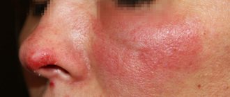
The most common cause of fistula formation is still infection; without it, the thread is covered with a connective tissue capsule, inflammation and fistula formation do not occur. What is a ligature fistula?
- granulation and swelling around the thread
- separation of pus from the hole where the thread passes and from the wound
- redness, tissue hardening and pain around the threads
- increase in body temperature
Fistulas do not always form immediately; when using non-absorbable threads, they can appear several months and years after the intervention!
Diagnostics
If purulent inflammation is suspected, the patient will need the help of a surgeon. The doctor will ask the patient in detail about the complaints and study the anamnestic data. A physical examination, including palpation of the abdomen, helps determine the location of the inflammation. The required treatment method is selected after receiving the results of instrumental and laboratory diagnostics.
Possible examination methods
- Blood analysis. The inflammatory process is indicated by an increase in the number of leukocytes.
- Visualization of organs using contrast radiography or computed tomography. The resulting image allows the specialist to see the location of the purulent capsule.
- Ultrasound imaging.
- Examination using minimally invasive intervention (diagnostic laparoscopy).
The data obtained through visual and laboratory examinations is used to perform surgery and prescribe medications.
Risk factors
Favorable background for the appearance of abscesses are also:
- long-term diseases of the gastrointestinal tract (gastroenteritis, enteritis, colitis);
- peripheral circulatory disorders (atherosclerosis, varicose veins, postthrombophlebitis disease);
- metabolic disorders (obesity, hypothyroidism, vitamin deficiency).
Diabetes mellitus with severe damage to the blood vessels plays a particularly significant role in the development and progression of the purulent process.
Treatment
The patient requires hospitalization in a clinic for timely treatment and careful medical supervision. The abscess can only be removed through surgery, but in addition to surgical treatment, patients are prescribed medications.
Treatment methods
- Opening and draining the abscess. The type of surgical access depends on the location of the abscess. If several foci of inflammation are detected, a wide dissection may be required.
- Intravenous administration of broad-spectrum antimicrobial agents to suppress the infectious process.
- Percutaneous drainage of small ulcers.
After surgery, continued antimicrobial therapy is necessary. If, as a result of inflammation and subsequent treatment, the functions of one or another organ are impaired, the patient will require additional rehabilitation. If surgery is performed in a timely manner, the prognosis is positive.
Lung abscess - symptoms and treatment
Treatment of a lung abscess can only be conservative; if necessary, it is supplemented with minimally invasive procedures. If these methods are ineffective or complications arise, surgical treatment is performed.
General treatment
- Therapeutic and hygienic exercises (for example, morning exercises) to improve physical health.
- Sleep 9–10 hours.
- High-calorie foods rich in proteins: meat, poultry, fish, dairy products, eggs, nuts, legumes, etc.
- Vitamin therapy: vitamins C, B1, B6, B12, etc.
- Infusion therapy is the introduction of glucose-saline solutions to restore the level of fluid and salts in the body, normalize metabolic processes, remove toxins and replenish energy.
- Breathing exercises to improve air exchange in the lungs. The doctor may advise you to blow air through a tube lowered into water and breathe through your nose: take a deep breath and exhale quickly, or take a slow breath, hold your breath for a few seconds and exhale slowly, etc.
- Parenteral nutrition is the intravenous administration of amino acids, albumin, protein, plasma or glucose solutions with insulin to compensate for the lack of vitamins and microelements.
- Blood transfusion to restore hemoglobin levels in severe anemia.
- Desensitizing and bronchodilator therapy to reduce the sensitivity of the bronchi to inflammatory factors, as well as improve their patency. May include: Eufillin, Pipolfen in the form of tablets, intravenous or intramuscular injections; Berodual in the form of a solution for inhalation; expectorants; vibration massage.
- Stimulation of the cough reflex to clear the bronchi of pus. The easiest way is to press on the cricoid cartilage or, after taking a deep breath, while exhaling, sharply squeeze the lower parts of the chest with your hands. You can also use inhalations with hypertonic sodium chloride or 10% Acetylcysteine solution. Stimulation can be performed every two hours.
- Inhalations with humidified oxygen.
- Ultrasonic inhalation of Terrylitin, Terrydecase, antibiotics, interferon to liquefy pus and necrotic masses in the abscess cavity and locally influence microorganisms and viruses [1].
Etiotropic therapy
Etiotropic treatment is aimed at the cause of the disease. In this case, the cause is an infection caused by bacteria, protozoa or fungi. Therefore, for lung abscess, etiotropic therapy involves the prescription of antibiotics that destroy pathogens.
This is the leading treatment method. As a rule, it is effective in the early stages of abscess formation. If an abscess has already formed, antibiotics will not be as effective.
Before an analysis of the pathogen and its sensitivity to antibiotics is ready, it is recommended to treat a lung abscess with broad-spectrum antibiotics. The effect on antibiotic therapy can be seen after 3-4 days, general condition improves after 4-7 days, and improvements on X-ray or CT can be seen after two months.
If the patient's general condition does not improve, this may indicate that the wrong antibiotic has been chosen. The drug is changed, and if necessary, antiviral and antifungal therapy is added.
The duration of treatment will depend on the general condition of the patient and the results of radiological and laboratory monitoring [2][3][12]. A general blood test should be done upon admission and after 3–4 days, a chest x-ray should be done upon admission and after 10–14 days.
Drainage of a lung abscess
Postural drainage. It is used when an abscess ruptures into the bronchus. The patient takes a certain position in which the affected bronchus is directed vertically downwards. This position allows purulent sputum to exit the bronchus on its own. With this technique, the patient's condition improves within 1–2 weeks [1][9].
Sanitation bronchoscopy. This is lavage of the bronchi followed by suction of the contents. It is used when sputum cannot come out on its own due to high viscosity, obstruction of the bronchial tube or a narrow connection between the abscess and the bronchial tube. Depending on the patient’s condition, the procedure is repeated daily or every 1–2 days. If the condition improves, the interval is increased [1][6][9].
Segmental catheterization of the draining focus of the bronchus. Using an installed catheter, pus is removed and medications (proteolytic drugs, antibiotics) are administered, this improves the natural cleansing of the bronchial tube. The catheter is inserted through the nose or through a puncture in the trachea under local or general anesthesia [1][9].
Transthoracic drainage (according to Monaldi). A puncture is made in the place where the abscess is closest to the chest wall. Through it, the abscess cavity is cleaned and antiseptic solutions are injected into it [1][9]. After the procedure, a drainage tube is left in the abscess cavity to ensure constant drainage of the contents of the cavity and for repeated rinsing if necessary.
Temporary endobronchial occlusion. With endobronchial occlusion, the affected bronchus is specially blocked. This is necessary in several cases: when it is necessary to limit the diseased area of the lung from healthy bronchi; stop pulmonary bleeding; close a bronchopleural fistula, in which air from the bronchus enters the pleural cavity and compresses the lung (pneumothorax). To block the desired bronchus, special plugs (occluders) are used, which are installed using a bronchoscope. When the task is completed, the occluders are removed [1].
Surgery
Pneumotomy. In this method, the lung tissue is cut to open the abscess and remove its contents. Pneumotomy is used in severe cases when it is impossible to perform resection (removal of part of the lung) due to the severity of the patient's condition.
First, the location of the abscess is determined, then the chest wall above the bottom of the abscess cavity is cut along the ribs, and if necessary, a rib fragment is removed. Pus is removed from the abscess, the cavity is washed with antiseptic solutions and a gauze swab and drainage tubes are installed in it to completely remove necrotic tissue. The dressing is done the next day, the cavity is again washed with antiseptic solutions. Drainage tubes are removed only when the cavity is completely cleared of necrotic masses and inflammation subsides [1].
Thoracostomy. This is the creation of permanent access (an opening in the chest wall) into the pleural cavity. It is performed to remove pus and air from the pleural cavity when conventional drainage techniques are not effective. The operation is technically simple and can be used in seriously ill patients.
The chest wall in the projection of the accumulation of pus is cut along the rib (incision up to 12–15 cm). Pus is removed from the pleural cavity, the formed bridges are separated, and washed with antiseptic solutions. After this, 10–12 cm of the ribs are removed. Along the edge of the thoracostoma (hole), the skin, muscles, and fascia are sutured to the pleura. The size of the formed thoracostoma is 4×8 cm. Several gauze swabs are placed into the cavity through the formed hole [1].
From the next day, bandages are made and the cavity is treated with antiseptics. In the first three days, tampons are changed, then tampons are not installed. Gradually, the cavity decreases and heals completely without additional intervention. Sometimes it is necessary to close the wound quickly. On average, all procedures take from 1 to 8 weeks.
Ligation of the pulmonary artery. It is not used as an independent method in the treatment of lung abscess. It is used in the development of a complication—arterial bleeding—when other methods are impossible [1].
Lung resection. The operation involves complete removal of the affected lung tissue. This is the most traumatic method. It is used most often, but is not used in severe conditions: sepsis, severe respiratory failure, renal failure, heart failure, etc. The operation is performed under general anesthesia.
Indications for resection:
- hemoptysis;
- prolonged intoxication and fever (about two months);
- bronchopleural fistula;
- rupture of an abscess in the pleural cavity with pyopneumothorax or empyema;
- lung abscess that persists for more than six weeks despite treatment;
- suspected cancer;
- cavity more than 6 cm;
- leukocytosis (increased white blood cell count) for 3–4 weeks despite adequate antibiotic therapy.
The extent of resection may vary:
- atypical resection (resection in which the segmental structure of the lung is not taken into account) - preferable if the abscess with necrotic tissue is completely removed;
- lobectomy (removal of a lobe of the lung) - performed for large and/or centrally located abscesses;
- pneumonectomy (removal of the entire lung) is carried out only with complete confidence that the entire lung is subject to purulent melting [1][9].
Complications arising from the appearance of a postoperative fistula
- An abscess is a cavity filled with pus;
- Cellulitis – inclusion of subcutaneous fat in the inflammatory process;
- Eventration – loss of internal organs due to purulent melting of tissues;
- Sepsis - the spread of purulent contents in the cavity of the chest, skull, and abdominal cavity;
- Toxic-resorptive fever is pronounced hyperthermia as a reaction of the body.
Why is quality diagnostics important?
The disease's symptoms often resemble diseases such as postoperative hernias, suture granulomas, and ligature abscesses. Unpleasant sensations with a hematoma and even signs with metastatic cancer are also similar to the symptoms of endometriosis in a postoperative scar. Therefore, high-quality diagnosis is important. In addition to the standard examination, diagnostic procedures are recommended that can provide more complete information.
The most effective method is ultrasound - with endometriosis of a postoperative scar, you can detect solid hypoechoic formations with unclear contours and a fibrous component or small hyperechoic zones that form as a result of the inflammatory process during monthly hemorrhage into the surrounding tissues. Ultrasonography can also detect the presence of bridges, a capsule of varying thickness, evaluate the characteristics of the blood supply to the pathological focus, etc.
MRI is also considered informative - during the examination it is possible to detect a solid formation and assess the extent of the pathological process.
Also, to clarify the extent of the spread of endometriosis, colonoscopy, cystoscopy, and urography can be prescribed, during which the presence of endometrioid heterotopias in other organs can be excluded/confirmed, since even one lesion remaining after surgery will subsequently cause a relapse. In our clinic, it is also possible to examine blood for tumor markers CA-125, CA 19-9, CEA, HE 4 - these are auxiliary methods that allow us to determine the severity of the disease and evaluate the effectiveness of treatment for endometriosis in a postoperative scar. However, the final diagnostic method is considered to be a morphological study of biological material taken during a biopsy or surgery.
Causes of postoperative scar damage by endometriosis
There are several theories about the origin of the disease, for example, implantation of endometrial particles during surgery, spread through the blood or lymphatic vessels. In most cases, endometriosis of the postoperative scar occurs after delivery by cesarean section, after various transabdominal surgical interventions: appendectomy, cholecystectomy, hernia repair, etc.
There are also a number of factors, the presence of which increases the likelihood of disease: metabolic and hormonal disorders, late pregnancy, unfavorable environmental conditions, prolonged and frequent stressful situations, etc.