Moles on the hands, if they reach large sizes, often cause some discomfort to the patient.
They may also carry health risks.
This is due to the peculiarity of the location.
- A man works with his hands.
Accordingly, if a mole is located on the right hand, it is constantly exposed to mechanical stress.
On the one hand, this leads to trauma, inflammation, and pain.
That is, a mole simply prevents a person from doing physical work.
On the other hand, a traumatic factor carries a risk of malignant degeneration.
- Hands are constantly irritated.
Even if the mole is located on the back of the hand, risks and inconveniences are still present.
Education is constantly exposed to chemical, mechanical, thermal influences.
A person immerses his hands either in cold or hot water.
It uses various chemicals (for example, for washing dishes).
Mechanical irritation continues constantly.
All this increases the risk of malignancy.
- Hands open.
Even in the warm season, most parts of the human body are protected from sunlight.
But this does not apply to the hands.
They are always open.
Therefore, as a result of exposure to the sun, they receive a dose of ultraviolet radiation.
At the same time, insolation is one of the most significant risk factors for the transformation of a mole on the left hand into a malignant tumor.
Types of moles
Traditionally, any formations on the skin that differ in color or texture from the surrounding tissues are called moles.
But in medicine this term is not used.
Pigmented nevi are generally called moles.
These are small benign skin formations, which are a cluster of cells with a high content of melanin pigment.
There are other varieties:
- red moles - formed from blood vessels;
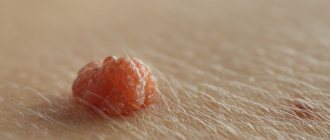
- a hanging mole usually turns out to be a papilloma (a formation of viral origin);
- keratomas - foci of hyperkeratinization of the skin, often occur in older people;
- Freckles are pigment spots that appear or increase pigmentation under the influence of insolation.
Prevention of melanoma
- Since melanoma often develops from moles, it is recommended to conduct regular self-examination of the skin to check for changes in existing nevi and monitor the appearance of new ones.
- If you have a large number of moles, it is recommended to avoid tanning and exposure to direct sunlight.
- When tanning, you must use sunscreen.
- Avoid exposing unprotected skin to direct sunlight.
| Read more about dermatological research at Euroonko | |
| Consultation with a dermatologist-oncologist | from 5,100 rub. |
| Skin examination using the German FotoFinder device | RUB 13,400 |
| Diagnosis of melanoma | from 5,100 rub. |
Book a consultation 24 hours a day
+7+7+78
When are moles on the hands dangerous?
Often people want to get rid of moles on their hands.
There may be several reasons for this:
- aesthetics – often moles look unattractive, especially if there are a lot of them;
- physical discomfort - itching or pain in the mole is possible if it is constantly irritated by mechanical or other factors;
- psychological discomfort - a person may experience fear of the malignant degeneration of a mole (the disadvantage of its location on the hand is that it is constantly in front of the eyes);
- risk of malignancy - if the shape, size, color, structure of a mole changes, its malignant degeneration is possible.
The greatest danger is the transformation of education into cancer.
The most dangerous skin tumor is melanoma.
It doesn't happen that often.
Skin cancer accounts for only about 10% of the total structure.
But this neoplasm has a high degree of malignancy.
Its mortality rate is very high.
The median survival rate is low.
This means that a person's life expectancy after diagnosis is minimal.
Despite the low incidence of the disease, melanoma is responsible for 80% of all deaths due to skin cancer.
How does melanoma manifest?
The symptoms of melanoma are very varied. It can have different sizes, shapes, colors and surfaces. Dimensions vary from 1–2 mm to several centimeters, and the shape can be very different. As for color, there are options here too. Melanoma is most often black, but can be brown, purple, or even pink. There are often cases when several colors are combined in one neoplasm.
The surface of the melanoma may be unchanged, but over time it may ulcerate, bleed, or become weepy. At the initial stage, the symptom of patent leather is characteristic, when there is no skin pattern on the tumor.
From the point of view of clinical manifestations, the following forms of melanoma are distinguished:
- Superficial spreading melanoma. At first it looks like a brown or black spot or plaque; if it rises above the surface of the skin, it is only slightly. She can remain in this state for up to 7 years. As it progresses, it thickens and turns into a node, and its color may change.
- Nodular melanoma. It has the shape of a node, polyp or mushroom, blue-red or black in color. Gradually its surface ulcerates and begins to bleed.
- Lentigo melanoma. It is the result of malignant degeneration of Durrey's melanosis. For a long time (up to 20 years) it exists in the form of a spot or plaque, and then its vertical growth begins. At the same time, the lesion takes on an irregular shape and uneven coloration.
- Acral melanoma. It develops on the fingers or toes in the nail bed and appears as a dark spot under the nail.
When should a mole be removed?
Every person should constantly monitor their moles.
This especially applies to the following categories of people:
- many moles on the body;
- cases of skin cancer among relatives;
- frequent or prolonged sun exposure;
- light skin type.
Any change in the appearance of the nevus should cause concern.
For example:
- the appearance of inclusions or dots inside the mole;
- the appearance of uneven contours;
- the appearance of pain or other subjective sensations;
- presence of discharge;
- uneven terrain;
- heterogeneity of structure and color;
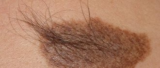
- hair loss in the area where the mole is located;
- ulcerations on the surface.
If you detect any changes, you should consult a doctor as soon as possible.
If melanoma is detected at the horizontal growth stage, the prognosis is favorable.
Timely removal of the formation allows one to achieve relapse-free ten-year survival in 90% of patients, which actually means a complete cure of the disease.
Unfortunately, most people delay too much in contacting a specialist.
If a mole on the hand becomes malignant, this can cause death, since melanoma metastasizes very early.
Interesting Facts
When a mole is located in the center of the palm, a person has strong family and friendly relationships. The sign promises a happy marriage, a large number of friends and career advancement.
Birthmarks vary in shape. Round ones indicate a person’s benevolent character and the presence of positive traits. Markings in the shape of a line are rare: they portend danger. Oval ones indicate an unhindered path in life.
There are crescent-shaped birthmarks. Bearers of such signs have a tendency to gamble, spend money, and are often fans of extreme sports. These people cannot imagine their life without adventure; danger awaits them at every step.
If you see a triangular shape of a mole, then the person is trying to please everyone around him; for them, creating a good impression on strangers is their goal. And square pigment spots can sometimes be seen in sexual maniacs; they indicate the presence of sexual anomalies.
Moles on the hands: what will the doctor do?
The doctor will examine the formation.
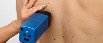
He will perform a dermatoscopy.
This is a basic test to detect suspicious skin growths.
It does not make it possible to accurately state malignant degeneration, nor to exclude it.
But signs of malignancy can be identified, after which a decision may be made about the need for additional diagnostic procedures or removal of the nevus.
Dermatoscopy is the study of an area of skin using a special device.
It provides magnification.
Modern dermatoscopes are also equipped with digital cameras that take pictures.
They allow you to document research results and evaluate changes in a suspicious formation over time.
Benefits of the study:
- non-invasive (does not cause skin trauma);
- painless;
- safe (no risk of infection or other complications);
- It is carried out quickly - about 10 minutes.
For each suspicious mole on the hand, the likelihood of malignancy is assessed.
Patients usually seek consultation with a dermatologist and undergo dermatoscopy for the following reasons:
- many moles on the body;
- changing an existing mole on the hand;
- the appearance of a new mole.
Some patients have no signs of pathology.
They are treated solely for preventive purposes.
It happens that dermatoscopy reveals malignant neoplasms.
Most often it is basal cell skin cancer.
It also happens that melanomas are detected.
During a preventive examination, approximately 2 out of 3 people are diagnosed with a neoplasm that was not noticed by the patient himself.
Treatment of melanoma
The choice of treatment tactics is determined by the stage of the disease. For stages 1–3, the main method is surgery, which, if indicated, can be supplemented with chemotherapy. For 4 common stages, immunotherapy is indicated taking into account the molecular profile of the tumor. If such treatment is not possible, chemotherapy is performed.
Treatment of stages 1–2
The main point in the treatment of stage 1–2 melanoma is radical surgical removal of the tumor. It must be removed within healthy tissues, and the amount of indentation is determined by the data of a morphological study:
- For melanoma in situ, 0.5 cm recede.
- If, according to histological examination, the tumor is more than 2 mm, it is recommended to retreat from its edge by at least 1 cm.
- If the melanoma thickness is more than 2 mm, the indentation should be at least 2 cm.
It is allowed to reduce the indentation when removing melanoma on the fingers in order to maintain their functionality. If an excisional biopsy was used and the diagnosis was confirmed, the scar is excised with the above indentations within 4–8 weeks.
Routine prophylactic removal of regional lymph nodes or their irradiation is not indicated. Instead, a sentinel node biopsy is performed. Only if its results are positive, lymphadenectomy is performed.
Treatment of common forms of the disease
Treatment of patients with advanced forms of melanoma will depend on the possibility of surgical removal of the tumor.
If melanoma is in a resectable state, its radical removal with regional lymphadenectomy is recommended. If metastases were detected during a biopsy of the sentinel node, complete removal of the lymph nodes of the axillary region is carried out with the most complete removal of the fatty tissue in which these nodes were located.
After surgery, the patient is offered immunotherapy and radiation therapy to the area of regional metastasis. RT reduces the likelihood of regional relapse in patients with high-risk factors:
- Damage to 4 or more lymph nodes.
- If the metastasis has gone beyond the lymph node capsule.
Irradiation is carried out in fractionation mode at a total focal dose of 48 Gy.
Patients with high and intermediate risks of melanoma progression are offered adjuvant (postoperative) immunotherapy. The risk group includes patients with the following characteristics of melanoma:
- The thickness of the tumor is from 2 to 4 mm with an ulcerated surface of the tumor.
- The thickness of the tumor is more than 4 mm with any surface.
As part of adjuvant immunotherapy, recombinant interferon preparations and CTLA4 receptor blockers (ipilimumab) are used. This treatment increases the median disease-free survival and overall life expectancy. Adjuvant immunotherapy begins no later than 9 weeks after surgery and continues for up to 12 months.
Treatment of unresectable and metastatic melanoma
Treatment of unresectable and metastatic forms of melanoma is determined by the molecular genetic characteristics of the tumor. The presence of a mutation in the BRAF gene is of primary importance. If the test result is negative (no mutation), a test is performed for a mutation in the CKIT gene.
Treatment of melanoma with a BRAF mutation
First-line therapy uses one of the following treatments:
- Monotherapy with inhibitors
- Monotherapy anti-
- Combined use of BRAF+ inhibitors anti-PD
- Combined use of BRAF+ MEK inhibitors (for patients with rapid progression or large volume of tumor mass).
Treatment is carried out for a long time, until progression or development of severe complications requiring discontinuation of drugs. If the molecular genetic status of the tumor is not specified, these drugs are not prescribed, since there is a possibility of paradoxical acceleration and progression of melanoma growth
These drugs can also cause the development of skin cancer. Therefore, it is necessary to regularly examine the skin for the presence of suspicious growths. If there are any, they are subjected to surgical treatment. Canceling treatment or reducing the dose of drugs is not carried out.
Treatment for the presence of a CKIT mutation
First-line treatment includes anti-PD1 therapy or imatinib (a CKIT inhibitor). Therapy is used until progression or development of complications requiring discontinuation of the drug.
As the disease progresses and BRAF and CKIT mutations are present, nivolumab or pembrolizumab is used as second-line therapy. For slow progression, the CTLA4 inhibitor ipilimumab is used. If immunotherapy is not possible, cytostatics are used.
It should be noted that chemotherapy is less effective in treating melanoma and causes more complications. Therefore, it is not recommended for first-line therapy and is used only when all resources have been exhausted or other treatment is not available.
Treatment in the absence of BRAF and CKIT mutations
In the absence of these mutations, PD1 inhibitors are indicated. If the tumor turns out to be insensitive to them, or progression occurs, they switch to CTLA4 inhibitors (ipilimumab). As part of 3rd line therapy, cytostatics are used.
Hyperthermic perfusion with melphalan can be used to treat unresectable arm melanoma. This method is used as part of palliative therapy for limb preservation in patients who have not responded to immunotherapy and cytotoxic therapy.
Dysplastic moles or nevi
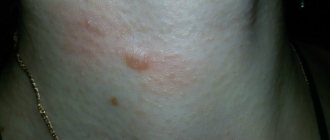
Often, the doctor discovers dysplastic nevi on the skin of the hands.
These are moles whose risk of degeneration is much higher.
Their clinical criteria:
- significantly larger sizes, usually half a centimeter or more;
- irregular or oval shape (while most common nevi are round);
- uneven edges;
- lack of clear boundaries (nevus gradually passes into unchanged skin);
- flat surface;
- heterogeneous color (in one area the mole may be brown, in another – reddish or blue-black).
The hands are one of the common locations of dysplastic nevi.
If they are detected during dermatoscopy, additional examination of the patient is required.
It is carried out to identify laboratory signs of malignancy.
Subsequently, a decision is made on the need to remove the formation.
If it is not removed, the patient is monitored dynamically.
Dysplastic nevus is a precursor to melanoma.
Therefore, tests are taken from the patient: a scraping for cytological examination or a biopsy followed by histological examination.
It has been established that women have a much greater risk of developing melanoma from dysplastic nevi.
Their likelihood of getting sick increases 30 times.
While men with dysplastic nevi suffer from melanoma only 2 times more often than patients who do not have such formations on their bodies.
Stages
- Stage 0, or melanoma in situ - there is dysplasia or non-invasive damage by malignant cells.
- Stage 1 – the thickness of the melanoma is no more than 1 mm, or up to 2 mm but without ulceration of the surface.
- The second stage is assigned if the thickness of the ulcerated tumor is more than 2 mm, or less than 2 mm in the absence of ulceration of its surface.
- Stage 3 is assigned if melanoma metastasizes to regional lymph nodes.
- Stage 4 – any melanoma with metastases to internal organs.
Indications for mole removal
Removing moles on the hands is a simple, harmless and painless procedure.
Therefore, it can be performed both for medical reasons and at the request of the patient.
Such a desire may arise if:
- the mole looks ugly;
- interferes with a person;
- causes him physical or psychological discomfort.
For medical reasons, a nevus can be removed if there are signs of malignancy or its risk is assessed as high.
This may be the case if:
- the mole is constantly injured and inflamed;
- it changes its color, size, shape;
- there are cases of skin cancer among relatives;
- a person often sunbathes in the sun or visits a solarium, so the nevus constantly receives a large dose of ultraviolet radiation.
It should be taken into account that a mole on the hands often receives a lot of ultraviolet radiation, even if the person is not a fan of beach holidays.
If you are often in an open space, your hands are always exposed to sunlight.
To avoid future medical problems, it is better to undergo prophylactic nevus removal.
Including in cases where there are no signs of a pathological process, and atypical cells were not detected during laboratory testing.
The listed situations require removal of the mole as planned.
There are also indications for immediate excision of the formation.
These are:
- increasing the area of an element or its height;
- the appearance of erosions, bleeding;
- inflammation;
- itching;
- regression (reduction in size, lightening);
- the appearance of a corolla around the mole;
- the appearance of smaller moles nearby.
Nevi must be completely removed.
Otherwise, they may recur.
Reason for appearance
If moles appear, then this is either a sign of a certain disease, or it is the influence of environmental factors on health. There is no need to panic if the nevus does not bother you, but for your own peace of mind, you can contact a dermatologist. There are several reasons for the development of birthmarks, namely:
- hormonal imbalance that occurs when the body undergoes changes during adolescence or pregnancy;
- minor injuries, abrasions, mechanical damage to the skin;
- chemical or thermal burns;
- predisposition to the appearance of moles. Usually transmitted through the female line;
- exposure to ultraviolet radiation.