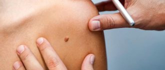
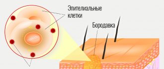
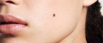
Keratoma is one of the types of benign formations that appear on human skin. Most of all they resemble freckles or age spots. Dermatologists consider abuse of sunbathing as one of the main causes of keratomas. Under the influence of ultraviolet radiation, epithelial cells become keratinized, which contributes to the formation of neoplasms.
The color of keratomas varies from flesh-colored to dark brown. Most often, these tumors appear on the arms, neck and back, much less often they appear on the legs.
Sometimes keratomas disappear on their own over time, but in some cases it is necessary to resort to removal.
Types of keratomas
Dermatologists distinguish several types of keratomas. For example, after forty years, so-called senile formations may appear - spots of a white or grayish tint, the diameter of which may increase over time. They occur most often on the neck, face and on the back of the hands. Sometimes these spots can become crusty and inflamed.
Content:
- Types of keratomas
- In what cases should a keratoma be removed?
- The essence of the radio wave method
- Advantages and disadvantages of radio wave removal of keratomas
- Contraindications to radio wave removal
- How is radio wave removal performed?
- How to behave after radio wave removal
- Possible complications
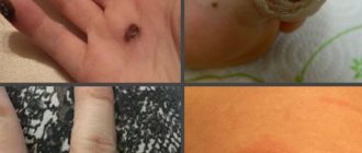
Solar keratomas appear most often in men with fair skin. They occur in those areas of the body that are most often exposed to sunlight. These neoplasms can be considered as harbingers of skin cancer, and therefore require mandatory medical consultation.
One of the most dangerous is seborrheic keratoma - a yellowish-brown spot that over time begins to peel and bleed, causing inflammation. These formations also require mandatory medical monitoring, as do horny keratomas, which are most prone to transform into malignant tumors. Follicular keratomas are the least common. Women are more susceptible to them than men. These neoplasms usually appear on the upper lip and scalp and are pinkish-gray nodules.
How dangerous is a keratoma?
For the most part, keratomas do not pose an oncological risk.
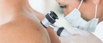
As mentioned above, keratoma keratoma and solar keratoma have an increased risk of malignancy. But what else is the danger of these skin formations? The fact is that even an experienced doctor is not always able to distinguish a keratoma from a malignant neoplasm using clinical methods.
Many malignant tumors at various stages of their development are similar to keratomas. Therefore, in all cases it is necessary to contact a dermatologist-oncologist; only a specialist can correctly diagnose and determine treatment tactics.
Also, keratomas can be dangerous due to complicated course. In the early stages of development, they cause only aesthetic inconvenience (which is important for women or when multiple keratomas are located in open areas of the body). But after the appearance and intensification of keratinization on the surface of the formations, additional complaints may occur - itching, pain, tingling in the area of the keratoma, with deep cracks and injury - the addition of an infection and the occurrence of inflammation, suppuration, with trauma and part of the crusts coming off the surface - severe bleeding.
To summarize: treatment of keratoma is necessary if malignant degeneration is suspected, with a complicated course, with rapid growth, as well as with aesthetic problems.
In what cases should a keratoma be removed?
The decision to remove a keratoma should be made only after consultation with a doctor. In the event that the formation is located on an area of the skin hidden from view and does not cause either aesthetic or physical discomfort, removal can be delayed. However, in some cases, the formation must be removed without fail. For example, doctors recommend removing keratomas that rise above the skin, as a result of which they are often injured and subject to friction. A keratoma should also be removed if it begins to rapidly increase in size, begins to bleed, or becomes covered with cracks. People who spend a lot of time in the sun are advised to get rid of tumors.
The aesthetic aspect is also important. Often, keratomas located on open areas of the skin cause cosmetic inconvenience, causing complexes and self-doubt. In this case, the tumor can also be removed by first consulting a dermatologist.
There are several ways to remove keratomas. The tumor can be removed using a laser, liquid nitrogen, electric current, or surgical excision or radio wave removal. The final verdict regarding which removal method to use in each specific case is made by the doctor after conducting a visual examination and collecting an anamnesis.
Thus, small formations can be removed using cryodestruction with liquid nitrogen - the keratoma simply dies under the influence of low temperatures. The disadvantage of this method is the rather high probability of scarring and scar formation, as well as the inability to use it with reduced blood clotting. In turn, laser removal is quick and painless, eliminates bleeding, but also has a fairly wide range of contraindications.
Electrocoagulation, in other words, cauterization of neoplasm tissue with electric current, is a very effective method of getting rid of keratomas, but in most cases it leaves scars and is contraindicated for people with hypertension.
The most radical method of removing keratoma is surgical excision of the tumor with a scalpel. However, in this case, the appearance of scars of varying severity is inevitable, and in addition, the procedure is painful, although it is performed under local anesthesia.
Recently, the radio wave method of removing keratomas has become the most popular. It is considered the most advanced, very effective and at the same time almost painless.
Removal of skin tumors at the Alla Khazova Clinic
Papillomas, raised moles, warts, keratomas, atheromas and other skin tumors literally haunt a person. The cause of their appearance may be viruses, injuries, bad habits, decreased immunity, exposure to ultraviolet radiation and other negative factors. New growths have different shapes, sizes and colors. They look repulsive and are potentially dangerous - there is a risk of degeneration of a benign tumor into a malignant tumor.
Removing tumors with Surgitron will help get rid of the problem - the most physiological medical procedure. The high efficiency of the method is based on the ability of skin cells to absorb the energy of radio waves and instantly evaporate under its influence. By adjusting the power of energy and the shape of radio waves, we can carefully remove absolutely any tumor without affecting adjacent healthy tissue.
Indications
- Papillomas.
- Warts.
- Moles.
- Molluscum contagiosum.
- Keratomas.
- Hemangiomas.
- Atheromas.
- Lipomas.
- Fibroids.
Contraindications to the procedure
- The patient has metal implants or a pacemaker.
- Herpes is in the acute stage.
- Pregnancy.
- Inflammation, lesions, injury to the skin at the site of exposure.
- Epilepsy.
It is important to know! If your benign tumor suddenly begins to behave uncharacteristically (it hurts, itches, bleeds, changes shape, color, or increases in size), then this may indicate its degeneration into a malignant tumor. Make an appointment with a dermatologist immediately!
The essence of the radio wave method
The radio wave surgery technique is based on the use of an “invisible scalpel” - high-frequency waves. This procedure is also known as radioknife. This method began to be used in dermatology and aesthetic medicine relatively recently and is considered one of the most gentle and at the same time most effective.
Thanks to its configuration, the “radio knife” concentrates high-frequency electromagnetic radiation in a small area, as a result of which it becomes possible to cut tissue with it, as if with an ordinary scalpel. Moreover, if the device has been correctly configured, the radiation does not affect healthy tissue, affecting only the area that has undergone pathological changes. Under the influence of radio waves, several processes simultaneously occur in the epidermis. First of all, microscopic cuts appear, and the pathologically changed cells themselves, under the influence of the knife, begin to generate heat and evaporate on their own. It is noteworthy that wounds under the influence of the same radio waves are immediately disinfected and coagulated.
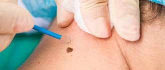
Radio wave removal is most often used to remove single skin tumors with a small area. The radioknife makes it possible to remove keratomas in one procedure. Moreover, the procedure is non-contact and therefore sterile. Radio waves cut tissues and simultaneously cauterize them, which eliminates the development of bleeding. In total, removal of one keratoma using the radio wave method takes no more than three minutes, and the wound heals within a week.
Advantages and disadvantages of radio wave removal of keratomas
The method of radio wave surgery has recently become increasingly popular. This is quite natural, since this procedure for getting rid of tumors on the skin boasts a huge number of advantages compared to other methods.
First of all, the radioknife allows the surgeon to regulate the radiation power, thereby controlling the depth of penetration of the waves. As a result, skin formations are removed very carefully, without damaging surrounding tissues. As a result, unaesthetic scars do not appear at the site of removed tumors.
A tangible advantage of the radio wave method of removing keratomas is the absence of pain. Since the radiation is directed, it does not irritate nerve endings and does not provoke muscle contraction. Therefore, during the procedure the patient practically does not feel any discomfort.
Since the radioknife not only cuts, but also coagulates tissue, the removal process is bloodless. This eliminates the development of inflammatory processes and infection. In addition, the likelihood of developing postoperative edema and other complications is minimized.
Another undeniable advantage of this method is the ability to remove tumors located in hard-to-reach places. Also, the removed tissue can be sent for histological analysis to exclude the possibility that malignant degeneration has begun.
In fact, the radio wave method of removing keratomas has only one drawback: it is used only in cases where it is necessary to remove a formation of small diameter. If the area of the keratoma is large enough, other methods are usually used.
Contraindications to radio wave removal
Like any surgical intervention, radio wave removal of keratomas also has its contraindications, in which the use of this method is highly undesirable:
- You should refrain from using a radioknife if the patient has been diagnosed with diabetes.
- Radio wave removal of tumors for pregnant and lactating women is excluded.
- The procedure should be approached with extreme caution if the patient is diagnosed with cardiovascular failure.
- Removal of the keratoma using the radio wave method should be postponed to a later date if the patient has recently suffered from ARVI, influenza or other similar diseases.
Our advantages over competitors
The high-tech medical center has been providing multidisciplinary medical services to Moscow residents since 2010. Our patients receive the following benefits from keratoma removal:
- Innovative equipment: allows us to offer patients advanced methods of diagnosis and treatment of any pathologies.
- Our own laboratory: our patients receive research results without leaving the clinic, with a guarantee of their accuracy.
- Professional approach: narrow specialists and junior medical staff are trained to work with modern equipment and constantly improve their skills.
- Great offers: follow the information about discounts and promotions on the official website of Medicine LLC - we are pleased to offer you the opportunity to save.
- Convenient location: the medical center is located in the ZAO area, where you can easily reach from anywhere in Moscow.
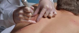
How is radio wave removal performed?
No preliminary preparation is required from the patient before removing the keratoma using the radio wave method. All preparatory work is carried out by the doctor. He must carefully examine the neoplasm, study its boundaries and depth of penetration into the skin. It is on the basis of this data that he makes a decision as to which radio waves need to be used in a given situation and adjusts the radio knife to the desired frequency.
The patient lies down on the couch. The area of skin that will be exposed is disinfected and anesthetized. It should be borne in mind that pain relief in this case is more of a “reinsurance” nature, because the procedure itself is practically painless. The frequency of the current used for manipulation is so high that it does not cause muscle contraction and does not stimulate nerve endings, as a result of which the patient does not experience any unpleasant sensations.
As anesthesia, either an injection of lidocaine or novocaine, or the application of special creams that are used to treat the area of treatment can be used. After five to ten minutes, the anesthesia begins to take effect, after which the doctor begins the procedure.
Externally, the radio knife looks like a thin metal rod connected to the device with a wire loop at the end. It is through this loop that radio waves influence the cells of formation. Also, in addition to the main loop electrode, a ball electrode can be used, which is usually used to level the wound and coagulate blood vessels to prevent the risk of bleeding.
Radio wave surgery is a non-contact method. In other words, there is no physical pressure on the tissue. As noted above, under the influence of a powerful radiation flux, a surge of thermal energy occurs and tissue heating occurs, as a result of which the cells simply evaporate.
Many patients who are not too knowledgeable about how keratoma removal is carried out using the radio wave method are afraid that they can get burned or receive an electric shock during the procedure. However, such worries are completely groundless. During surgery, the electrode does not come into contact with the skin; the doctor simply places it near the surgical field.
After removing the tumor, the doctor treats the wound with an antiseptic and covers it with a sterile gauze bandage.
How to behave after radio wave removal
Removal of keratoma using the radio wave method is considered a simple operation that causes minimal harm to the skin. However, a small wound will appear at the site where the tumor was previously located.
She will heal in stages. First, a thin crust forms, which will fall off on its own over time. Under no circumstances should it be scratched. Also, for at least five days after surgery, the affected area of skin should not be kept wet.
Best materials of the month
- Coronaviruses: SARS-CoV-2 (COVID-19)
- Antibiotics for the prevention and treatment of COVID-19: how effective are they?
- The most common "office" diseases
- Does vodka kill coronavirus?
- How to stay alive on our roads?
Dermatologists recommend not using decorative or care cosmetics based on alcohol or other aggressive substances to care for the area of skin exposed to radio waves for at least six months after removal of the keratoma.
Also, you should not overuse sunbathing or visit a solarium.
Benefits not related to removal technique
- Honest prices.
No hidden fees or intrusive services. Either consultation or removal is paid, strictly according to the price list. You can always check the price with me on VKontakte by sending a photo with a ruler in the background. - Free information support before and after surgery.
If you have any doubts before your visit, you can easily and free of charge by mail or VKontakte. After the operation, you will have my personal phone number, by which you can ask me at any time about what is bothering you. - Fast and convenient.
Removal is carried out within a few minutes, in one visit. - Online appointment booking and removal without calls or waiting
- No preliminary tests, dressings, or suture removal required
- Histology results by email
. You can read them on these sites: docplanner.ru (more than 40 reviews), napopravku.ru (24 reviews), vk.com (more than 150 reviews)