Dermatovenerologist
Khasanova
Alina Rashidovna
9 years experience
Make an appointment
A mole is a benign formation that appears on the skin. Its key feature is pronounced pigmentation against the general background of the skin.
These pigments begin to appear at an early age. Children and adults have them. On the one hand, their presence does not cause any concern. On the other hand, moles from a benign formation can “grow” into a malignant one. Therefore, at the first signs of changes and severe symptoms, you should consult a specialist.
In professional medicine, a mole has another name - nevus. In general, this is not a strict medical concept; the word is used to unite various neoplasms that differ in nature.
Symptoms and signs of moles in adults
In medical practice, sometimes dangerous moles come across that need to be quickly removed. If this is not done, the disease will begin to progress, which can lead to quite unpleasant consequences. Therefore, it is extremely important to monitor the condition of pigmentation on the skin. Especially if you have large moles on your body or those that have begun to grow and visually change.
The symptoms of “bad” moles are as follows:
- moles of a different shape appear, symmetry is lost, the neoplasm grows in one direction;
- the edges become “torn”;
- Over time, the color changes and becomes uneven. This is the first sign that malignant moles are appearing on the body;
- quick resizing;
- change in texture - smooth moles become rough and vice versa;
- if there are hairs on the surface, their sudden loss should cause a person to worry;
- peeling, itching and burning in the place where the mole is.
Do you have any suspicious moles?
Only a doctor can accurately diagnose the disease. Don't delay your consultation - call
Acquired melanocytic nevus (mole) - symptoms and treatment
Depending on the location of melanocytes, three groups of acquired nevi are distinguished:
- Transitional nevi are formed by nests of melanocytes located at the border of the dermis and epidermis. They may be in the form of a spot or slightly raised above the skin. The color varies from light brown to black, and the center of the mole is darker than its edges.
- Complex nevi consist of nests of melanocytes located both at the border of the dermis and epidermis, and in the dermis itself. They are pigmented nodules that rise above the skin at different heights. Their surface can be smooth or spherical, color - from light to dark brown.
- Intradermal nevi occur when nests of melanocytes are located in the dermis. Nevus cells often stop producing melanin, so moles practically do not differ in color from the surrounding skin, but rise above it. They have the shape of a papule, dome or papilloma, their texture is soft and elastic [42].
Acquired nevi can be classified into typical and atypical. Some atypical variants are divided into separate forms: blue nevus, halo nevus, Spitz nevus, eclipse nevus and nevus of the sebaceous glands.
Atypical, or dysplastic nevi are benign acquired melanocytic nevi that have the characteristics of melanoma: asymmetry, irregular borders, heterogeneity of color and a diameter greater than 6 mm.
Atypical nevi are more common when there are a lot of nevi on the body as a whole. Among white-skinned people, atypical nevi occur in 2–10% of the population [22]. As a rule, they appear after puberty [23]. Typically, there are more nevi in areas exposed to sunlight, such as on the torso and legs, and fewer on the chest and buttocks.
Based on their origin, atypical nevi can be divided into two groups:
- Sporadic nevi appear spontaneously under the influence of various external factors leading to mutations in cells. Relatives of patients usually do not have atypical nevi or melanoma.
- Genetically determined nevi (dysplastic nevus syndrome) - occur due to hereditary predisposition. Relatives also have atypical nevi, and there may be cases of melanoma in the family.
Main characteristics of atypical nevi:
- diameter from 0.6 cm;
- irregular outlines with lighter edges;
- the shape is flat, the mole is flush with the surrounding skin;
- the borders are uneven, smoothly blending into the skin;
- the color is heterogeneous, the color intensity can vary from pale brown to black.
An eclipse nevus is a type of complex nevus that is brown in color with a light center and a darker, star-shaped edge. In children, it often develops on the scalp. Despite the heterogeneous color and uneven borders, such nevi are benign. If there are no other warning signs, such as induration and ulceration, then biopsy or removal of such nevi is not necessary [24].
Halo nevus (Sutton's nevus) is a melanocytic nevus surrounded by a symmetrical round or oval halo of depigmentation, i.e., a lighter area of skin. Haloing usually occurs in acquired melanocytic nevi, but can also occur in congenital nevi, blue nevi, Spitz nevus, and melanoma.
Halo nevi occur in almost 5% of white-skinned children aged 6–15 years, most often located on the back [26]. Common in people with vitiligo or relatives with vitiligo. In approximately half of the cases there are multiple nevi (more than 30) [27].
There are four stages of halo nevus development:
I. Pigmented nevus surrounded by a halo of depigmentation.
II. Pink nevus with a halo of depigmentation.
III. Round area of depigmentation without nevus.
IV. Skin of normal color after repigmentation of the halo.
Any of the four clinical stages can last from several months to several years [28]. Up to 10 years can pass between stages I–II and IV.
If the halo nevus has a typical appearance, then a biopsy is not required [29]. Since children with halo nevi usually have many nevi on their entire body, the entire body is examined. This will help identify atypical nevi and determine whether they need to be monitored or biopsied and removed. If there are atypical or concerning signs, a biopsy of the central portion of the nevus is performed. There is no need to remove the halo.
Multiple halo nevi are common among adolescents and young adults. They are rare in middle-aged and older people and may be a manifestation of an immune reaction to cutaneous or ocular melanoma. In this case, a biopsy may be required.
Blue (blue) nevus is a benign growth of dendritic skin melanocytes that actively produce melanin. The blue color is produced when short wavelengths of light are scattered by melanin in the skin, a phenomenon known as the Tyndall effect. Most often, blue nevi appear on the scalp, neck, back of the hands and feet, and in the sacral region.
There are several variants of blue nevi:
- A common blue nevus is a single, dome-shaped, blue or black-blue papule less than 1 cm in diameter. Such nevi often appear during adolescence and are found on the dorsum of the hands and feet.
- A honeycombed blue nevus is a more raised and larger nodule or plaque, measuring more than 1 cm with a smooth or slightly uneven surface. Such nevi can be congenital or acquired; they are most often found on the scalp, face, sacrum and buttocks.
If the blue nevus is stable, then it does not need to be removed. But if it appears suddenly and grows quickly, or a pre-existing nevus increases sharply, then a biopsy is necessary.
Spitz nevi are benign, usually acquired nevi, which are a collection of melanocytes. They most often appear in childhood and are located on the face and legs. Such nevi grow quickly, which often worries patients and parents.
A Spitz nevus is a papule or nodule that is uniformly pink, brown, red, or red-brown in color. Usually symmetrical with a clear outline, less than 1 cm in diameter. The surface may be smooth or warty. Sometimes the nevus includes dark pigment in the form of a star or peripheral stripes.
Pigmented spindle cell nevus of Reed is a variant of Spitz nevus that is common among adolescents and young adults. It often appears on the thighs and has the appearance of a thin papule ranging from dark brown to black in color [30].
A small, stable Spitz nevus with typical features can be observed [31]. Such nevi may shrink or disappear over time [32]. But if the Spitz nevus has atypical signs (size more than 1 cm, asymmetry or ulceration), then it is removed after a biopsy.
A sebaceous nevus is a localized lesion, predominantly consisting of the sebaceous glands. It occurs with equal frequency in men and women, regardless of skin color. It is observed in 0.3% of newborns [15]. Often at birth or in early childhood it appears as a single linear or rounded area with a smooth or velvety surface. It comes in pink, yellow, orange or light brown. Typically found on the scalp or face, extensive lesions are rare.
During adolescence, it becomes warty or nodular and may darken and enlarge. In approximately 1% of cases, such a nevus degenerates into cancer. This almost always occurs after puberty [15].
Causes
Why do moles appear on the skin? Every experienced mole doctor will say that there are several reasons for their appearance on the body:
- ultraviolet radiation;
- traumatic damage to the epidermis;
- systematic violation of the integrity of the skin;
- negative influence of radioactive background;
- Constant consumption of unhealthy foods, smoking, drinking alcoholic beverages. Because of this, moles on the body constantly grow;
- problems in the functioning of the endocrine system, changes in hormonal levels;
- hereditary predisposition.
What types of “moles” are there?
When pronouncing the word “mole,” the patient means a permanent spot or nodule on the skin. In fact, there are hundreds of types of benign skin formations that can be called this. Here are the most common ones.
Hemangioma or “red mole”
This is a benign formation consisting of vascular tissue cells. At least one hemangioma can be found in every adult. They look like red/cherry dots or appear as spider veins (spider hemangioma). They can appear spontaneously throughout life, sometimes at the site of injury or inflammation (for example, at the site of a pimple). This is an absolutely benign formation, it does not turn into cancer. To be removed for cosmetic purposes only.
Keratoma
A benign skin formation consisting of epidermal cells. In the old books they were called “senile”, but this definition is not used now, since they can also occur in young people. A keratoma looks like a gray or brown spot protruding above the skin with a rough surface. Their typical location is in areas exposed to solar radiation (face, chest, upper limbs) or places of constant trauma (under laundry straps and elastic bands). Keratomas almost never become malignant, but they can actively grow, itch and bleed. Therefore, dynamic monitoring of these formations is necessary. Can be removed for cosmetic purposes.
Acrochordon (fibroepithelial polyp or “papilloma”)
A benign skin formation, representing a soft polyp on a flesh-colored stalk. Usually occurs in places of mechanical friction and in skin folds, the elements can be located in groups. Doctors often associate these tumors with the papilloma virus. Acrochordons are recommended to be removed as they tend to grow and spread and can become a serious cosmetic problem. They do not pose a danger to life or health.
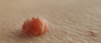
Melanocytic nevus (mole, birthmark)
A benign congenital or acquired skin formation consisting of melanocytes (pigment cells of the skin). This is a flat spot or plaque raised above the skin, most often brown in color. More than 10 types of nevi have been described. On average, an adult has 20-30 such moles on his body. They appear and grow under the influence of ultraviolet radiation. Requires dynamic monitoring, the risk of degeneration into melanoma is 1:200,000. They can be removed by a dermatologist, dermato-oncologist or cosmetologist with subsequent referral for histological examination.
When to see a doctor
There are the following types of moles:
- pigmented;
- vascular.
Usually, red moles on the body are harmless, but only if they have not changed color. If you find a mole, what should you do in this case? First, we advise you to carefully examine it. You need to go to the clinic with a mole if you have:
- bleeding or peeling;
- itching;
- dark areola or rim;
- rapid increase in size;
- sudden color change.
Where to run?
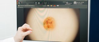
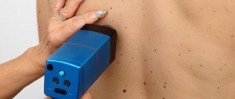
If you notice an unusual mole on your body, be sure to go to a dermato-oncologist. If this doctor is not available, you can consult a dermatologist, surgeon or oncologist. An experienced specialist can visually determine the nature of the nevus. A special device called a dermatoscope helps him with this. Essentially, this is a powerful magnifying glass; by examining a mole through it, the doctor can notice the smallest details that are simply impossible to see with the naked eye. If there is doubt about the diagnosis, the doctor will conduct a histological examination to determine the characteristic signs of benign, premalignant and malignant neoplasms.
By the way, if you want to remove a mole for aesthetic reasons, then the decision should also be made by a dermato-oncologist after dermatoscopy, which allows you to choose the optimal method, determine the boundaries and depth of removal.
Even if degeneration into melanoma has not occurred, for medical reasons those moles that are subject to constant friction, pressure, and injury are removed. And also those that are in the groin area and under the arms, under the chest, on the belt, and in men - on the face at the shaving site.
If a mole that has degenerated into melanoma is removed at an early stage, the probability of complete recovery reaches 95%; if time is lost, it is only 20%.
Types of moles
The most harmless moles are pigmented ones, they are called lentigo.
They look almost like freckles and are very easy to confuse. Such moles are formed due to the accumulation of melanocytes under the skin, cells responsible for pigmentation. All other types of birthmarks can cause trouble for their owner. These are epidermal-dermal nevi - moles that can rise above the surface of the skin, usually located on the palms, feet or in the groin area. Intradermal nevi are convex, often covered with hair. Dysplastic nevi are irregularly shaped tubercles with unclear boundaries; they can be more than a centimeter in size.
One of the most dangerous is a giant nevus. This is a congenital spot that can cover large areas of the skin. It is usually removed early, since it looks scary, and the risk that it will degenerate into a malignant formation is quite high.
Big ones - under the knife!
Melanoma can make itself felt by changing the color of the nail to brown or black, and the appearance of spots on the palms and soles.
To monitor atypical nevi, the patient usually undergoes dermatoscopy every 3–12 months. A biopsy is taken from suspicious lesions and histological examination is performed to establish a diagnosis. All congenital moles larger than 2 cm should be under constant medical supervision; in any case, they will have to be removed. If necessary, the baby will have a skin passport to monitor the number, dynamics of growth and identify any changes in moles. The operation is usually performed shortly before the onset of puberty. Especially often, cells degenerate on a giant congenital nevus (more than 15 cm in diameter) in adolescence.
Published on the portal parents.ru
Special attention area
The transformation of a benign tumor into a malignant one can occur slowly, over several months or years, or it can happen suddenly, in a very short time. But if trouble strikes, dangerous cells will begin to divide rapidly, so it is important not to miss the moment and undergo regular preventive medical examinations. Please note: in these cases, you need to run to the dermatologist as fast as you can.
- Pigment formations of asymmetrical, irregular shape and outline, similar to a geographical map.
- Multi-colored moles with shades of brown, red, gray and blue.
- The nodules are regular in shape, but unevenly colored.
- Sharply growing moles.
- Causes concern - itches, hurts or bleeds.
- Eight spots larger than 6 mm.
- Multiple scatterings (from 50 pieces) larger than 2 mm.
How to distinguish normal moles from pathological ones
To summarize the above, we highlight the characteristic distinguishing features of benign and malignant moles:
- A benign mole has a regular shape, while a nevus is prone to degeneration into a malignant tumor, usually with an uneven relief shape.
- A safe mole has a uniform color, but one prone to “bad” degeneration can be of different color shades.
- “Bad” moles are usually not congenital, they are acquired.
- “Bad” nevi are usually larger than harmless ones.
- “Bad” moles can rapidly increase in size, and never decrease or disappear on their own; safe moles, on the contrary, can disappear or decrease in size.
- A “bad” mole can cause discomfort and pain, and it can also cause itching.
- A “bad” mole can mutate, acquire new elements and change shape and color.
Knowing the distinctive features of various types of moles, you can protect yourself and your loved ones from a serious problem. By detecting such a problem in a timely manner, you will be able to eliminate it as quickly and efficiently as possible.