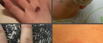
Despite the fact that moles are benign neoplasms, sometimes it is necessary to remove them. They contain a large amount of melanin, so they have a darker color than the underlying skin. They can be large, small, and differ in location. The medical name is nevus. It is concentrated on any area of the skin and even mucous membranes. The more melanocytes accumulate in a mole, the brighter its color becomes. Formations are black, brown, burgundy, red, purple.
What do we know about moles?
What we have always heard about moles: “Does it bother you? Live in peace, don’t touch your mole!” This advice, which was very common in the past, convinced us that moles that were originally given by nature or that appear over the years should remain inviolable. If you touch it, you won’t be able to avoid trouble. This is partly true - indeed, removing moles can lead to the most dire consequences. But in fairness, it should also be noted that not every mole needs to be removed. The most harmless - flat moles
(pink to dark brown).
Usually they behave “quietly”, do not get injured and do not create inconvenience. The only thing you need to remember is to protect such moles (on open areas of the body) from sunlight. Convex moles
, especially if they are often touched by clothing, need to be treated more carefully. If they are on the back and in other places inaccessible to your view, you should entrust someone close to you with monitoring them. And, of course, if any changes occur—the mole becomes larger, turns from flat to convex, or changes its shape—you should immediately contact a specialist.
Classification and characteristics
In total, approximately 60 types of formation have been identified, which are determined by dermatologists or oncologists. They are divided by appearance, size, shape, content and other parameters. Moles can be small, excessively large, up to 15 cm in diameter. If they are localized on the face, this leads to a severe cosmetic defect.
Classification by time of occurrence:
- congenital
- they are formed very rarely, have a small size, which makes them very difficult to see instantly; - acquired
- spread to the upper, deep, middle layers of the skin, between the dermis and epidermis.
The main types of nevi and their exact characteristics are presented in the table.
| Variety | Description |
| Nevus | Has a brown or black tint. Can be located in any area of the body. More common on the face |
| Blue nevus | Smooth, superficial, with a dense consistency. Their diameter is no more than 10 mm. The shade is dark brown or black. Localized singly on the body |
| Angioma | A network of capillaries and blood vessels grows in the epidermis. This leads to the formation of a benign tumor that remains under the skin |
| Lentigo | Presented on the body in the form of a brown or black pigment spot. Singularly located. Rarely found |
It is important to classify the mole before starting treatment so that mechanical impact does not lead to complications.
No independence.
Some people believe that you can remove moles yourself by tying them, for example, with silk thread. This is a deep misconception. Attempts to bandage warts and papillomas with a thin silk thread often only provoke the growth of these formations. Today, there are celandine-based preparations on sale that are intended to eliminate warts. But it is better not to remove moles, papillomas, or warts yourself by any means. Celandine juice is similar in composition to iodine, so prolonged exposure to the drug causes skin burns. Cauterization with iodine is even more dangerous - it is more aggressive than celandine. Trying hard to get rid of papillomas with iodine, you can get scars on the skin. Only a dermatologist can properly remove a mole.
Removal methods.
Removal of moles and warts
- Cost: 2,000 rub.
More details
Mole removal can be carried out using a laser, radio wave coagulation or electrical coagulation. These manipulations are performed on an outpatient basis under local anesthesia. The choice of treatment method is determined by the doctor in each specific case. In my opinion, the most effective and painless method is radio wave coagulation. We use this technique in most cases, although in some cases we use excision with conventional surgical instruments followed by suturing. When using radio wave coagulation, tissue damage during material collection will be minimal, blood loss is reduced, and the rehabilitation period is shortened, while, for example, with the laser method, the healing process is extended over a fairly long period. Repeated formations do not appear after removal, but if precautions are not followed by the patient himself, infection may occur.
brief information
Depending on the thickening of the surface, moles are divided into 2 types
:
- convex;
- flat.
It is the first formations that are most dangerous, since they are often damaged and can therefore become deformed into a malignant composition. This is one of the reasons why a nevus needs to be removed. For this purpose, medications, traditional methods, physiotherapeutic procedures, and surgery are used. The operation is considered gentle, requiring only local application of an anesthetic. Therefore, the rehabilitation period after it lasts a limited time, complications rarely occur. You just need to take proper care of your skin to prevent infection or bruising.
Unattended moles.
Some types of moles can degenerate into one of the most malignant tumors - melanoma (skin cancer). The initial site of formation of most melanomas is congenital pigment spots and warty moles. How does this dangerous development begin?
For example, after damage, a person only notices how the color of the mole begins to change, it enlarges, cracks, turns into an ulcer, and sometimes into a tumor. Therefore, without panicking, pay attention to the above symptoms, especially the progressive enlargement of the mole and change in its color. If all these symptoms occur, immediately rush to the dermatologist. The unfavorable prognosis of melanoma dictates the need for regular monitoring by a doctor. In addition to irritant factors (ultraviolet radiation, trauma), the development of melanoma can also be caused by genetic and endocrine factors. However, in the first place is, of course, excessive exposure to the sun. Ultraviolet radiation in large doses causes irreversible changes in skin cells, greatly increasing the risk of their degeneration. Light-skinned and fair-haired, red-haired people with blue and gray eyes are most susceptible to the mutagenic effects of sunlight. Those at risk also include those who have a lot of freckles, age spots and moles.
Dangerous sun.
For each person, the critical duration of exposure to sunlight is highly individual. It’s not easy to determine this line, so it’s better to just remember that prolonged exposure to the sun is harmful to the body. The skin is forced to protect itself from ultraviolet radiation. Excessive tanning means inevitable burns. If you love beach holidays so much that you simply cannot deny yourself this pleasure, at least listen to the following tips. By following these simple recommendations, you will not only improve your health, but also reduce the risk of a dangerous disease.
- Staying in the sun is safe from morning until 10 o'clock, in the evening after 17 o'clock.
- For people with a large number of moles, it is better to reduce their exposure to the sun.
- Try to avoid redness and sunburn on your skin. If this happens, spend 2-3 days in the shade.
- After swimming in the sea, be sure to rinse your skin with fresh water.
- Even young and healthy people can spend no more than one and a half to two hours a day in the sun.
- Taking certain medications increases the skin's sensitivity to light. Please consult your GP before going on holiday.
- Use sunscreen cosmetics: at the beginning of the holiday, the indicator should be maximum, about 40 units.
- Do not use decorative cosmetics, deodorants and perfumes on the beach - they can cause the appearance of age spots on the skin.
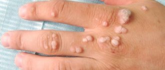
How to get rid of papillomas on the face: professional techniques
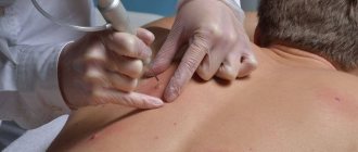
Treatment of all skin tumors begins with a patient interview. It is important for the doctor to find out when the growth appeared, whether it bothers you, and whether its size and shape have changed. This is followed by a visual examination and dermatoscopy - the study of papilloma through the already mentioned dermatoscope. After this, the doctor issues a conclusion about what type of tumor we are dealing with and prescribes the optimal method for its removal.
“To treat papillomas, cytostatics, cryo-laser, diathermocoagulation and electrical destruction are used,” says Anna Torosyan. — Some medicinal formulations that contain, for example, celandine or castor oil, can be effective in the treatment of papillomas, but the use of these drugs without consultation with a specialist is unacceptable, since self-medication can lead to aggravation of the problem. Convex papillomas require immediate removal, especially if they are located on the face. This is due to the high probability of injury to the formation, which can trigger the onset of the inflammatory process and the spread of pathology to nearby areas of the skin.” In some cases, trauma can stimulate the development of cancer.
“Today, the safest and most reliable method of removing papillomas is radio wave,” says Maria Istomina. “Here they use a special device designed specifically for radio wave surgery - a unique non-contact technique for cutting soft tissue using high-frequency radio waves. This occurs due to the heat that tissues emit when high-frequency waves penetrate them. When working with papillomas, this device uses a very thin electrode. A radio wave with a frequency of about 4 MHz is concentrated in it. The papilloma tissue evaporates under its action, the skin does not heat up, and the patient feels practically nothing. The method is universal, since the device regulates the power and wave shape to remove tumors on any area of the skin. The device burns out the papilloma without pain or discomfort for the patient, and completely - only a small crust remains. The radiosurgery technique completely eliminates painful contractions of muscles or nerve endings, so it is one of the most painless techniques in modern medicine.”
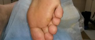
There is also a method of cryodestruction - freezing the tumor with liquid nitrogen. It does not damage the skin, does not cause bleeding, and the procedure itself is quick and painless. Cryodestruction is most often used by specialists to remove papillomas on the face of children.
Histology for removed papilloma is not necessary. The need for this analysis depends on what diagnosis the doctor made when studying the tumor. If during dermatoscopy the doctor has no doubt that he is faced with a benign papilloma, histology can be dispensed with.