Modern plastic surgery has a whole arsenal of tools that allow you to achieve excellent results. In the case of surgery to correct the shape and volume of the mammary glands, this is especially evident. Thousands of patients remain satisfied with the results of mammoplasty. This became possible not only due to the achievement of the desired aesthetic effect, but also due to the minimal risk of complications. At the same time, one should not deny that, although rare, they are possible. One of the complications after mammoplasty is seroma.
Why does it appear after surgery?
During surgery, natural damage to cells and various blood vessels (including capillaries) occurs. As a result, fluid accumulation may begin in the tissues between the dermis and subcutaneous fat. Serous matter is essentially lymph; it contains formed elements of blood, as well as a certain amount of protein fractions. As a rule, such a liquid has a straw color, but occasionally it can be reddish (if there is a significant number of leukocytes in the composition).
The accumulation of serous fluid after surgery does not always pose a threat to human health. However, seroma can cause sagging skin at the site of the intervention and also complicate the healing process.
At-risk groups
The likelihood of seroma occurring is especially high if:
- Sloppy work of the surgeon. If the doctor performs a series of not very careful actions, he can harm the surrounding tissues. Such injuries provoke the release of lymph.
- Inflammatory process. Sometimes it is only a consequence of tissue damage and is non-infectious in nature. But increased formation of serous fluid is characteristic.
- Hypertension. When blood pressure increases, the full distribution of lymph in the body is disrupted, which can lead to its sweating through capillaries damaged during the intervention.
- Excess body weight. Seroma is diagnosed after surgery in approximately 75% of obese patients.
- Diabetes mellitus. This disease is characterized by an increase in blood glucose. Due to its excessive amount, it is difficult for the body to recover after surgery, the process of tissue regeneration slows down (including the restoration of blood vessels).
- Old age. Over the years, metabolism naturally slows down, which is why the rate of regeneration also decreases. The risk of serous fluid formation increases.
- Soft tissue burn. If the surgeon does not use the electrocoagulator carefully enough when cauterizing injured vessels, a small focus of necrosis may appear in the affected area, in which, in turn, serous fluid will accumulate.
A particularly high risk of developing postoperative suture seroma is observed during interventions on the abdominal cavity, as well as on the mammary glands.
Typically, doctors take all possible measures to reduce the likelihood of such a complication.
Due to caesarean section
Seroma of the postoperative suture in women who gave birth to a child through surgery is a fairly common complication. The most common reason is a weakened body caused by a long pregnancy, due to which the regeneration processes slow down by an order of magnitude.
Seroma after cesarean section has a fairly typical appearance - a small, dense ball filled with exudate (lymph) appears on the suture. The appearance of such a disorder is associated with damage to blood vessels as a result of the incision.
Most often, seroma of the postoperative suture, resulting from a cesarean section, does not cause any discomfort to the mother in labor and does not require treatment. But in any case, it is imperative to inform doctors about its appearance.
Symptoms
Suture seroma, which appears during postoperative recovery of the body, is always a local inflammatory process, characterized by the occurrence of concomitant signs of the disease.
They are:
- an increase in body temperature, which reaches 37-39 degrees and directly depends on the level of the human immune system, the presence of infectious agents, and the extent of inflammation;
- a feeling of aching pain that may not stop for several hours, subside for a while, and then resume again;
- swelling of the soft tissues and epithelial surface located around the postoperative suture, which is the first sign of abundant accumulation of seroma between the lipid and dermal layers;
- burning pain syndrome that occurs when the surgical area is inclined and there is an influx of additional blood and lymph;
- redness of the skin around the postoperative suture, the color of which depends on the severity of the inflammatory process and can vary from pale pink to deep purple and bluish.
If the complication occurs without worsening the clinical picture, then upon examination a yellowish-tinged liquid without a foul odor is observed, and its appearance always indicates the addition of a fungal or bacterial infection.
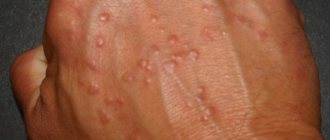
What does the scar area look like?
The most typical sign of seroma formation is swelling and some redness of the suture site. This symptom may be accompanied by aching pain and a feeling of fullness. When you try to palpate the skin near the suture, pain also occurs. Quite rarely, an increase in temperature (up to 37 ° C) is detected, and general malaise is possible.
In severe cases, seroma leads to the formation of a serous fistula. This is an opening through which accumulated liquid is released. The passage may appear in the thinnest areas of tissue. Most often, the hole is located near the suture, and its occurrence warns of the risk of developing blood poisoning. The appearance of a fistula is a serious reason for repeated surgery.
Identifying gray is not difficult at all. When examining a patient, the doctor pays attention to swelling and redness of the skin, and upon palpation he may feel the flow of fluid under the skin.
Seroma formation
Seroma is not related to malignant neoplasms, but requires removal. It increases the risk of infection entering the operated area, so it requires timely treatment.
The formation of seroma is possible from a hematoma. Sometimes it is formed from lymphatic fluid due to capillary leakage. “Leakage” from blood vessels is also possible if there are breaks in them. Another risk factor is dying or damaged cells, which can cause inflammation.
Treatment
The need for treatment for seroma of the postoperative suture is determined individually by the doctor. Minor accumulation of serous fluid requires only observation and goes away in a few days. All this time, the patient must be under the supervision of a doctor and adhere to his recommendations.
But a significant accumulation of fluid in the presence of a threat of infection requires targeted treatment, which can be:
- Surgical.
- Conservative (medication).
Most often, seroma can be dealt with after surgery in just a few days. However, this condition definitely cannot be ignored.
Conservative
If a postoperative suture seroma occurs, the doctor may prescribe the following to the patient:
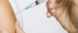
- Antibacterial drugs to prevent infection.
- Nonsteroidal anti-inflammatory drugs for relief of aseptic inflammation.
- Hormonal medications. Such medications are used very rarely, but they can quickly suppress the inflammatory process.
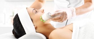
- Carrying out physiotherapeutic procedures.
Conservative treatment will only help with minor accumulation of serous fluid. If the pathology progresses rapidly, an immediate surgical procedure is performed.
Operational
The easiest way to deal with seroma is with a puncture. It does not require any special preparation of the patient or the skills of the doctor. This procedure involves pumping out the accumulated liquid with a syringe. And this is most often enough to get rid of the problem. A positive result can be achieved in 90% of cases. The doctor may also resort to other methods of surgical treatment.
Vacuum aspiration
This method can be used for minor seroma at an early stage of occurrence, which, in the doctor’s opinion, is unlikely to resolve on its own. To carry out the procedure:
- The doctor makes a small incision where the main accumulation of serous fluid is.
- A tube connected to a special vacuum apparatus is placed inside the wound.
- Exudate is removed from the tissues after turning on the suction.
Aspiration helps speed up the wound healing process and avoid complications of seroma. But, unfortunately, such intervention does not protect against recurrence of this condition.
Drainage
This method is very common in surgery, and it really helps to cope with congestion at the site of surgery and prevents its occurrence. To perform this procedure, the doctor:
- An incision or puncture of the skin is made in the area of the surgical field.
- Inserts a special drainage system.
- Attaches a container to collect liquid (not always).
Thanks to drainage, it is possible to achieve a natural outflow of serous exudate. The wound with the system installed is covered with a bandage that needs to be changed daily. Also, the seams are regularly treated with antiseptic solutions.
Treatment must be supplemented by taking antibacterial drugs.
Will folk remedies help?
Doctors strongly warn patients after undergoing surgery not to self-medicate or experiment with their own health. Seroma of the postoperative suture cannot be eliminated by traditional medicine; such attempts can lead to serious consequences.
Diagnosis of pathology
Seroma goes through stages of its development very quickly. In order not to start the disease, it must be detected in a timely manner.
To identify this pathology, the following diagnostics are used:
- Visual inspection. The surgeon's responsibilities include daily inspection of the patient's wound. If undesirable changes in the scar are detected, the doctor may perform palpation. If he feels fluid flowing under his fingers, he will prescribe an additional examination.
- Ultrasound examination of the surgical area. It allows you to confirm or refute the presence of fluid in the postoperative suture area.
It is extremely rare that a puncture is performed if a gray tumor is suspected. It is mainly needed to determine the qualitative composition of serous exudate. Based on these data, treatment tactics are subsequently developed.
Complications
If detected early, seroma can be successfully treated, but in the absence of adequate correction, this condition can lead to:
- The development of infectious processes - inflammation and suppuration of the wound. A cavity filled with serous fluid is an ideal incubator for the activity of pathogenic bacteria. Moreover, they can get into the wound even with complete sterility during and after surgery. The infection easily spreads throughout the body along with lymph and blood, for example, from chronic lesions in the nasopharynx.
- Intensive growth of connective tissue at the site of intervention. As a result, a rough scar may remain on the skin, and if a seroma appears after implantation, the implant subsequently becomes deformed.
- The occurrence of a serous fistula. This condition, as we have already found out, increases the likelihood of secondary infection several times.
- Sepsis (blood poisoning).
Postoperative period
Removal of breast fibroadenoma
Removal of an adenoma is an invasive intervention, which is carried out using several techniques:
- sectoral resection;
- enucleation;
- laser removal, for which puncture is performed.
Compression underwear
The need to remove the formation, including mastopathy, is due to the fact that the disease may very rarely recur in the future. Each technique has its own level of trauma, which requires a certain amount of pain relief. Before the operation, the woman undergoes preparation, the doctor makes sure that there are no contraindications. Regardless of the chosen operation, further tissue restoration depends on the implementation of rehabilitation measures, which include:
- drug treatment;
- general recommendations with limitations;
- diet;
- wearing special underwear.
Kinds
The reasons why a doctor decides to perform a cesarean section can be very different. Depending on the delivery process and complications encountered during the delivery, incisions may be made in different ways, resulting in different types of sutures that require special care.
Vertical seam
If acute fetal hypoxia is diagnosed or the woman in labor begins to experience heavy bleeding, a caesarean section is performed, which is called corporal. The result of this operation is a vertical suture starting from the navel and ending in the pubic area. It is no different in beauty and in the future it will spoil the appearance of the body quite strongly, since the scars are nodular in nature, very noticeable against the background of the abdomen, and are prone to compaction in the future. This type of operation is performed quite rarely, only in emergency cases.
Horizontal seam
If the operation is performed planned, a Pfannenstiel laparotomy is performed. An incision is made transversely, above the pubis. Its advantages are that it is located in a natural fold of skin, the abdominal cavity remains unopened. Therefore, a neat, continuous (special application technique), intradermal (so that there are no external manifestations) cosmetic suture after a cesarean section is invisible on the body.
Internal seams
Internal sutures on the wall of the uterus in both cases vary in the way they are applied. The doctor is guided here by the goal of achieving the best possible conditions for faster wound healing without complications and reducing blood loss. You cannot make a mistake here, since the course of subsequent pregnancies depends on this. During a corporal operation, a longitudinal internal suture is made after a cesarean section; during a Pfannenstiel laparotomy, a transverse suture is made:
- the uterus is stitched with a continuous single-row suture made of synthetic, very durable, self-absorbing material;
- the peritoneum, like the muscles, is sutured with continuous catgut stitches after cesarean section;
- the aponeurosis (muscle connective tissue) is sutured with absorbable synthetic threads.
The speed of healing, features of care, various complications - all these important points directly depend on what kind of incision was made during the caesarean section. After childbirth, doctors advise patients on all issues that cause them doubts, anxieties and fears.
Preparation
Preparation for sectoral resection of the mammary gland includes undergoing a thorough examination of the woman, when tests are taken for:
- prothrombin index, INR, fibrinogen, free heparin level;
- thyroid hormone levels;
- prolactin, testosterone, estradiol;
- blood levels of urea, bilirubin, AST, glucose, ALT;
- microscopy of urine sediment;
- determination of blood group and Rh factor.
If the above tests deviate from the norm, you will need to consult with a physician or a specialist specified by the physician. They will tell you what actions need to be taken to normalize the function of an organ whose indicator has deviated. In this case, rehabilitation after resection of the sector should proceed without complications.
In addition, preoperative preparation includes a blood test for the presence of antibodies to the HIV virus, hepatitis B virus, and RW. If the results of at least one test are positive, the intervention will have to be postponed and appropriate treatment will be carried out by an infectious disease specialist (if hepatitis B or HIV is detected) or a venereologist (in the case of a positive RW test).
Before the operation, it is necessary to undergo laboratory tests:
- fluorography;
- ECG;
- Ultrasound of the thyroid gland;
- mammography – ultrasound (up to 45 years) or x-ray (after 45 years).
If sectoral resection is carried out for cancer, distant metastases are excluded using tomography - computed tomography or magnetic resonance imaging - because this involves a completely different operation. Also, as preparation for the intervention, radiation therapy can be performed.
Actions before surgery
Before surgery, a woman must do the following:
- exclude taking birth control pills (in consultation with the operating surgeon and gynecologist who prescribed the drug);
- stop taking vitamin E 5 days before the intervention;
- After consulting with a therapist or cardiologist, at least 3-4 days before surgery, discontinue Aspirin, Warfarin, Curantil, Pentoxifylline or other blood-thinning drugs. Otherwise, sectoral resection may be complicated by severe bleeding;
- stop drinking alcohol or smoking, as this leads to a decrease in blood supply to tissues. The healing period in this case will be longer.
If an intervention is to be performed under general anesthesia, the last meal should be consumed 6-8 hours before, and water 4 hours before.
This is important, since the introduction of anesthesia can be complicated by vomiting, and it is dangerous on a full stomach
When planning a sectoral resection under local anesthesia, you should stop eating and drinking 4 hours before surgery.
Options and their features
How breast reconstruction will take place after a mastectomy is determined by several circumstances:
The volume of tissue excised during tumor removal. If the breast was only removed from the tumor, dents and scars may become a problem. This is the easiest option for reconstruction.
Sometimes all organ tissue is removed, leaving the skin and fatty tissue intact. And here restoration will not cause much difficulty. It is more difficult when the gland is completely removed; the pectoralis major muscle, lymph nodes and blood vessels, and subcutaneous fat are affected.
- The patient's condition. It is necessary that she can tolerate general anesthesia without problems.
- Features of the shape and size of healthy breasts. Sometimes it also needs to be adjusted for purely aesthetic reasons. This could be a lift or resizing.
Reconstructive surgery is performed at the same time as a mastectomy or some time after it.
Expander
Breast reconstruction can be achieved using a temporary device. This is an expander, which is a cavity with liquid. It is installed under the skin, then gradually, over several visits to the doctor, the patient is filled with saline solution using a syringe. This is necessary so that a pocket for the implant is formed in the problem area, and the preserved breast tissue is sufficiently stretched.
Breast reconstruction with tissue expander
The expander is used for 3 - 6 months. At this time, the breasts look a little unnatural. In addition, the patient will have to regularly visit the doctor, only he will be able to increase the volume of the device. There is no hurry with this.
Instead of an expander, a vacuum device can be used. This is a whole system that is worn 10-12 hours a day. It is a bowl tightly fixed to the iron. A vacuum is formed inside it, which stretches the skin.
This method is good for small breasts. It will allow you to restore it using lipofilling, that is, reconstruction with your own fat. After this procedure, your breasts will look as natural as possible. And the patient will quickly integrate into normal life after the procedure.
To learn about installing an expander, watch this video:
Musculocutaneous flap transplantation
Breast restoration is possible through transplantation of one's own tissue. Use the latissimus dorsi or rectus abdominis muscle. This method will make the breasts look very natural and will eliminate the danger of the implant moving, because in this case it is not needed. But it lasts up to 5 hours under general anesthesia and is highly traumatic, long-term rehabilitation, and a considerable amount of risks.
Breast reconstruction with tram flap
Breast reconstruction with DIEP flap
Combined method
Delayed breast reconstruction may combine both methods. This operation is appropriate for total tissue removal. First, the adjacent areas of the breast are restored using your own graft. This is done using adipose tissue, that is, by the method of lipofilling. Then the implant is installed and the myocutaneous flap is transplanted.
Of all types of intervention, this is the most difficult. But it allows you to restore the beauty of your breasts after a severe and extensive mastectomy.
Nipple and areola restoration
In almost all cases, part of the reconstructive surgery of the mammary gland is the restoration of the nipple-areolar complex. There are options here too:
- you can use tissue from the same area of another breast;
- perform a skin graft on the labia minora;
- collect the nipple from the tissue of the reconstructed breast, and mark the areola using tattooing.
For information on different options for breast reconstruction after mastectomy, watch this video: