Sometimes after removing a mole, an unpleasant aftertaste may remain in your soul. Either the doctor was fussy and unsure, or the administrator was not very polite, or maybe they just noticed dust in the corners.
It often happens, after removal, people find on the Internet an opinion according to which histology is mandatory for all moles. Otherwise, cancer or melanoma may develop. And the doctor before the operation simply looked at the mole and said: “Histology is not needed - it is 100% benign.” A state close to neurosis sets in...
What to do with moles?
If there are no clear signs of a malignant process, then you can go in 2 ways:
The first way is observation. In this case, we take a picture, a photo of the mole, warn the person about the signs in case the size, shape, symmetry changes, and schedule a return visit in 3-6 months, depending on how much we fear for this formation.
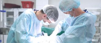
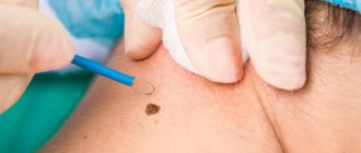
The second way is removal of the formation with histological examination. We even resort to this path more often. There is a common belief that a mole should not be touched. In fact, there is no danger in removal. Moreover, this is even a preferable option, since in this case a suspicious formation can be sent for histological examination. So, we excise the formation, sometimes if it is a suspicious mole, then not with a laser, but with a scalpel, with minimal grip (1-2 mm is enough) and send the material for histological examination.
If we receive the result that it is a benign formation, then we have removed it as benign and no further action is required.
Is histology really necessary?
I am convinced that yes, and that clinical diagnosis alone is not enough. The word “clinical” means the absence of additional studies - histology, dermatoscopy, puncture. Only conversation with the patient, examination and palpation. There are few studies on the accuracy of clinical diagnosis of melanoma, the most malignant skin tumor. According to the most optimistic of them, the clinical method is accurate by no more than 80% [V. V. Anisimov et al. Melanoma of the skin, part 2 - St. Petersburg: Science, -1996. -280 s.]. I don’t know about you, for me 20% percent of errors is a bottomless abyss.
A variety of outcomes are possible after removal of moles - relapses, scars, suppuration. If something goes wrong with healing, it is the histological examination that gives 1) confidence in the benign quality of the mole and 2) restful sleep for the doctor and the patient.
Melanoma and dysplasia
If, according to histology, we receive a result - severe dysplasia or early melanoma, then this can also be regarded as a positive result, since the disease was detected in the initial stage. In this case, we refer the patient to the oncology clinic, where he undergoes further treatment.
Early (thin) melanoma is very treatable, with a good survival prognosis. In contrast to melanomas, which are left untouched, which grow and patients treat them at stages 3-4, when effective treatment is no longer possible.
Who makes mistakes more often?
What I say below is my personal opinion. This is based solely on my experience and is not supported by research.
It seems to me that the probability of a doctor’s error when removing moles is higher, the further he is from skin cancer. A cosmetologist, surgeon or dermatologist is more likely to mistake a melanoma or skin cancer for a regular mole. This probability is much lower for an oncologist. The error for an oncologist specializing in skin tumors (dermato-oncologist) is close, but also not zero.
Dear colleagues, dermatologists, surgeons, cosmetologists! If you are reading these lines, please do not be offended. I know for sure that among you there are those who will give many oncologists (including me) a hundred points ahead in diagnosing melanoma. Unfortunately, my experience in conducting advanced training courses for doctors of these specialties suggests that this is rather the exception than the rule.
Which moles are dangerous?
Every person has moles. These are small spots on the body of various colors: from flesh-colored to almost black. These are nodules or papules consisting of pigment cells. All of them are similar to melanoma, and therefore need to be treated with special attention.
Moles at risk:
- multiple;
- atypical (irregular shape, heterogeneity of coloring).
Who needs to pay special attention to the condition of nevi:
- blondes and red-haired people, people with very fair skin and light eyes;
- people whose relatives have had cases of melanoma or precancerous skin diseases;
- people aged 40–45 years (melanoma is most often diagnosed).
Moles located in places of constant mechanical influence - friction, pressure - are most prone to malignancy (malignant degeneration). These pigment formations are localized on the neck, lower back, folds and folds of the body, in the places where clothing fits most tightly. The most common location in women is the lower leg, in men it is the back. In older people, melanoma most often develops on the skin of the neck and scalp.
In light of the craze for dark tanning and tanning salons, the risk zone includes all moles that are exposed to excessive ultraviolet radiation. People who suffer from sunburn more often are at risk, so they need to use light filter creams.
You should pay attention to the condition of your moles. If you see that they begin to enlarge, change color, bleed, or itch, you should immediately consult an oncologist. The study will rule out melanoma or atypical nevus.
The histology of the mole most accurately differentiates:
- atypical moles;
- intradermal nevi;
- non-pigmented neoplasms.
It helps to prescribe optimal therapy based on the most accurate diagnosis.
What are the risks of melanoma?
Melanoma and mole are very similar in appearance. They consist of identical melanocyte cells. These cells produce melanin. A pigment that protects the skin from high doses of ultraviolet rays. However, when a mole becomes injured or burned, it becomes dangerous. Melanocytes begin to multiply rapidly. They crowd out healthy cells.
A tumor forms. From the surface of the skin it quickly makes its way deeper. At the same time, it affects all layers of the skin. It is important to stop the tumor in time! Otherwise, metastases may appear - cancer cells that separate and affect internal organs (brain, liver, lungs, etc.).
Signs of malignancy
The clinical signs of a malignant mole are very diverse. This is manifested in size, shape, outline, surface character, consistency, color, and dynamics of change.
Common to all forms is a set of characteristics, which is expressed by the initial letters by the abbreviation “AKORD”, developed by dermatologists and cosmetologists:
- Asymmetry (A) - lack of symmetry in the shape and contours of a spot, with the exception of birthmarks present on the child’s body at birth.
- Edges (K) are most often uneven and indistinct (blurry).
- Coloring (O) - uneven; The presence of dots and stripes of various tones of dark brown and black is noted.
- Size (P) - in diameter from 7 mm or more.
- Dynamics (D) of development - an increase in the previous birthmark or a rapid increase in the size of a new pigmented formation.
Is it true that after removing an ingrown toenail, a patient will not be able to walk for several days?
After traditional ingrown toenail surgery, the patient usually requires semi-bed rest for a week. At the very least, there is significant pain that requires the use of analgesics. With marginal laser destruction of an ingrown toenail, the discomfort is minimal and is felt almost only in the first hours, or at most a day, after the operation. Analgesics are usually not required. Of course, there are exceptions, but these are usually advanced cases complicated by large pyogenic granuloma. Active motor mode is recommended after 2-3 days.
You can find out more about the service and its cost here >>
Progress of the laser removal procedure
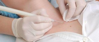
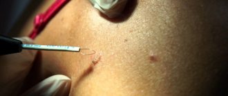
A laser removal tool is a beam of light that destroys tumor cells and evaporates them from the skin. Laser can remove a mole without bleeding. The procedure is performed under local anesthesia.
Removal lasts on average 10–15 minutes. Then a protective crust appears at the site of formation, which will disappear in a few days. There are no scars left at the site of laser treatment. After the procedure, it is necessary to follow medical recommendations: temporarily avoid sunbathing and contact with water, avoid injuring the skin area, treat, if necessary, with products prescribed by a doctor.
Information for foreign patients
Foreign patients wishing to receive qualified oncological care in Israel can count on correspondence consultation with a specialist at the pre-hospital stage. In addition, if necessary, a histological examination of the material can be carried out.
In Israeli clinics, a sentinel lymph node biopsy procedure is used, according to which lymph nodes with possible metastatic lesions are eliminated.
In Russia and the CIS countries, the procedure of sentinel lymph node biopsy has not yet become widespread.
The use of this technique is most relevant if histological examination reveals an increased mitotic index and ulceration in the area of primary melanoma.
The type of melanoma, its classification and determination of the stage of tumor growth are the main characteristics of histological examination.
In specialized cancer centers such as the Melanoma Unit, histological testing of a tissue sample is carried out for more than 20 parameters. In addition to structural features, the pathologist takes into account many additional characteristics, including the properties of cell nuclei and enzyme activity.
Patient Andrey M., 31 years old, diagnosed with melanoma of the right shoulder
The conclusion of Russian pathologists speaks of increased mitotic activity (from 2 to 5 mitoses in the field of view), melanoma is described as a tumor of the 3rd level of invasive growth according to Clark with a thickness of 2.5 mm. Israeli oncologists established level 4 melanoma according to Clark with a tumor thickness of 4.25 mm. The mitotic activity rate was 20/mm2.
Examples of differences in the histological reports of Russian and Israeli pathologists.
The study of tumor material involves determining its following properties:
- Tumor thickness
- Ulceration
- Clark level
- Histological type
- Cell type
- Primary localization
- Signs of regression
- Number of mitoses
- Lymphocytic infiltration
- Vertical growth stage
- Invasion of blood vessels
- Invasion into the lymphocytic zone
- Ploidy
- S phase of the cell cycle
- DR1 gene expression
- DNA Index
- Heat shock protein expression
- Positive staining for HLD-DR
- P53 protein mutation
- Cell adhesion factor expression
- Protease expression
- Migration marker molecule
- Angiogenesis factor
- Oncogene expression
- Presence of an estrogen receptor
- Cytokine, growth factor
Our clinic offers correspondence consultations and remote examination of histological samples. This greatly facilitates diagnosis and increases the efficiency of medical care.
Is histology necessary when removing a mole, fibroma or other neoplasm? Doctor's opinion
Most neoplasms are not life-threatening. They are removed for aesthetic reasons and to prevent injury (for example, convex moles that constantly rub against clothing or get touched when shaving). But we cannot exclude the risk of the neoplasm degenerating into a malignant tumor.
Patients who want to remove a mole, papilloma or other skin tumor often ask: Is it necessary to send the removed tumor for histology?
Natalia Genrievna Yezhova, Deputy Director for the Medical Department of CLINIC21, answers:
“I often come across the desire of patients to simply remove any tumor on the skin without conducting a pathological examination.
Many people think that they just have a harmless growth and there is no danger.
People do not have developed cancer alertness. I will give an example from my experience. Patient X underwent excision of a 'soft fibroma' in the area of the shoulder joint with pathomorphological examination. The study showed that it was nodular basalioma, that is, an oncological formation. The patient was taken under observation, the progress in treatment is positive, because the disease was detected on time.”
Histological analysis allows us to determine the presence of cancer cells in the tumor. This is an important diagnostic procedure that can save lives. Thanks to this study, it is possible to timely determine the malignant nature of the tumor and undergo treatment to prevent the progression of oncology.
Is it true that the cause of calluses and corns is just the wrong shoes?
The cause of the appearance of calluses and “corns” is most often congenital or acquired pathology of the foot (various forms of flat feet, etc.). “Wrong” shoes only aggravate the manifestation of these formations. Very often, viral plantar warts are hidden under the guise of calluses. Unprofessional pedicure very often leads to the appearance of plantar warts and their spread over the skin of the foot.
You can find out more about the service and its cost here >>
Preparing for the removal procedure
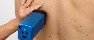
Before removing moles with a laser, it is necessary to examine the neoplasm. The dermatologist conducts a visual examination of the nevus under multiple magnification. A dermatoscope helps to examine the surface of the formation in detail.
The laser itself or in combination with superficial excision can simultaneously effectively remove skin lesions and obtain sufficient tissue for laboratory examination. If malignancy is suspected, the patient is recommended to undergo surgical excision, affecting not only the mole, but also healthy areas.
No special preparation is required for laser removal. The doctor may recommend undergoing general tests to make sure there are no general contraindications and possible restrictions.
Early diagnosis methods
Each person should regularly examine their skin for the presence of suspicious nevi. Paying attention to your body is a simple and effective diagnostic method. However, a person who does not understand medicine cannot always distinguish a healthy mole from a dangerous neoplasm - a sign of oncology.
At the hospital, doctors use the following diagnostic methods.
- History The collection of information by a doctor allows you to determine the root cause of the malignancy. The doctor will ask about sun exposure, injuries to the mole, and any actions taken with the neoplasm.
- Dermatoscopy Examination of the skin with dermatoscopes is an effective diagnostic method. The doctor will examine the color of the nevus, examine the relief and structure of the integument.
- Histological examination Examination of a skin sample under a microscope.