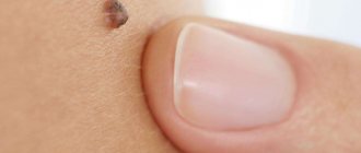
Moles are benign pigmented formations on the skin, which in medicine are usually called nevi. These spots can be present on the body at birth and appear until puberty, and then remain on the body forever. Moles sometimes cause inconvenience and injury, which can ultimately cause malignancy. Or they can simply be located in a visible place and spoil a person’s appearance. In this case, the mole must be removed, one of the methods is the radio wave method.
The essence of the radio wave method
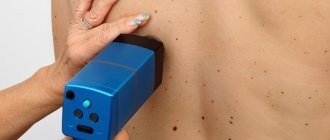
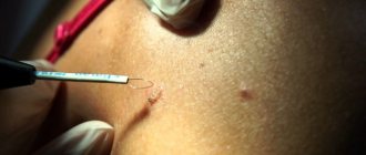
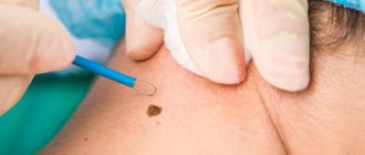
Compared to surgical and other methods of removing moles, the radio wave method is considered very gentle, and at the same time very effective. The high-frequency waves used generate thermal energy, and without contact with the skin, they carefully cut tissue, while evaporating pigmented cells, leaving only a barely noticeable trace. During radio wave removal, no physical pressure is applied to the skin.
Important: Radio wave surgery is the most effective method of removing moles, as it leaves only a small depression. When the operation is over, the wound is treated with an antiseptic, and after a couple of weeks it smoothes out.
Types of skin formations
All benign skin formations are conventionally divided by color, size, shape and surface texture. The generally accepted classification includes only two types of moles - congenital and acquired. Congenital nevi are divided by size into: small (1.5 centimeters in diameter), medium (no more than 10 centimeters), large (more than 10 centimeters), giant.
Giant formations cover the entire anatomical region (for example, the face). Small moles are safe and rarely develop into malignant tumors. Owners of giant moles should be on alert, since in 50% of cases they degenerate into melanoma. To protect yourself, perform regular self-examinations and consult a dermatologist. Acquired moles begin to form in childhood. It is at this time that the restructuring and development of the entire organism occurs.
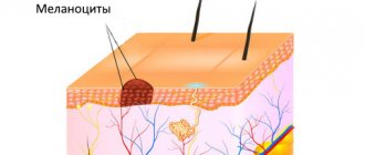
Melanocyte cells rise from the deep layers of the skin to its surface - the epidermis, where they accumulate and provoke the growth of moles. The location, size and number of nevi are determined by genetic factors, lifestyle, and external environment. Acquired moles are classified by location:
- epidermal (accumulation of pigment cells in the upper layers of the skin);
- intradermal (accumulation of melanocytes in the deep layers of the skin or dermis);
- borderline (accumulation of melanocytes at the border of the upper and lower layers of skin).
Benefits of radio wave mole removal
- radio waves do not damage healthy tissues, leaving them completely intact;
- the duration of mole removal using a radio wave knife is only twenty minutes;
- there is no bleeding during the procedure;
- even without anesthesia, the intervention is absolutely painless;
- there are no complications after the operation, and the postoperative period is very short;
- There are no scars left after removal of moles by radio waves;
- the likelihood of inflammation, redness or swelling after the procedure is extremely low;
- During the operation, the tissues are sterilized, thereby reducing the likelihood of complications;
- After surgery, the wound does not need special care;
- radio waves do not destroy tissue during removal, so histological analysis can be carried out;
- Using a radio wave knife, you can remove a mole anywhere.
Causes
The most common causes of ingrown toenails include:
- Wearing tight and narrow shoes;
- Wearing high heels;
- Congenitally large periungual ridges;
- Flat feet;
- Injury to the periungual fold during pedicure.
Quite often there is a combination of various reasons and factors. In some cases, an ingrown toenail occurs during pregnancy and the postpartum period (this may be due to changes in weight, posture, and foot alignment). An integral part of treatment is eliminating the causative factor - wearing comfortable shoes, correcting flat feet with insoles, etc.
Contraindications to radio wave surgery
Removing moles with a radio wave knife has a number of contraindications that need to be taken into account:
- if skin tumors have already degenerated into cancer, then more serious therapy is required;
- It is unacceptable to use a radioknife if there are heart rate sensors in the patient’s body;
- in case of chronic inflammatory skin diseases in the area of mole removal, it cannot be removed with a radio knife;
- The radio knife is prohibited for use during breastfeeding or pregnancy, so as not to accidentally affect the baby;
- In case of a viral infection of the skin, for example, with herpes, using a radio knife will have to wait a little.
Indications
- Benign skin formations. Using a radioknife, birthmarks (nevi), moles, keratomas (skin plaques), angiomas (vascular tumors), lipomas (fat deposits), and hyperplastic (enlarged) sebaceous glands are removed.
- Basaliomas are cancerous skin tumors with a low degree of malignancy.
- Viral lesions. A radioknife is used to remove papillomas, warts, and condylomas (warty growths on the genitals).
- Scars on the skin, including rough keloids. Using a radio knife you can make them softer and more elastic.
- Pathologies of the cervix. Using a radioknife, erosions, ectopion (eversion of the mucous membrane), cicatricial changes, cysts, and polyps are eliminated.
- Leukoplakia and erythroplakia are precancerous conditions of the genital mucosa, accompanied by the formation of dense light or red areas.
- Dysplasia is a precancer of the cervix caused by papillomavirus. Using a radioknife, you can perform conization - an operation to excise a cone-shaped dysplastic fragment of the mucous membrane of the cervix and cervical canal.
- Various benign neoplasms in the genital area and anus.
- Abscesses and cysts of the Bartholin glands - glandular formations located at the entrance to the female genital tract.
- EHF is also used for biopsy - taking samples of skin and mucous membranes to study the composition of cells.
Radioknife is widely used in eye and ENT surgery (rhinoplasty). In this case, the ability of radio waves to make invisible, thin, neat cuts is used.
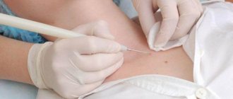
The process of radio wave removal of moles
The radio wave method is effective due to the fact that radio waves act specifically on the area of the mole, eliminating unnecessary and unnecessary damage to healthy tissue. This is done due to steam bubbles, which are formed under the influence of high-frequency radio waves and gently move the tissue apart. As a result, only the cells in the wave zone are destroyed. Thanks to this, there are no scars or scars left on the skin, and the procedure remains painless. To completely eliminate pain, local anesthesia is performed before the procedure. The radio knife, which is used to carry out the procedure for radio wave removal of a mole, performs three functions simultaneously:
- cuts fabrics;
- disinfects the affected area, completely eliminating the possibility of infection;
- stops bleeding in the affected area, eliminating the need for the doctor to apply stitches.
Radio wave surgery is the most effective method for removing moles, as it leaves only a small indentation. When the operation is over, the wound is treated with an antiseptic, and after a couple of weeks it smoothes out.
Differences between Gamma Knife and Cyber Knife
Cyberknife is one of the latest developments in the field of nuclear medical technologies, which is also a safe alternative to aggressive operations involving opening the skull. Based on computer navigation, robotics and radiation surgery. About 300 thousand patients with cancer have been treated with such modernized devices around the world. The non-surgical tactics of “burning out” a tumor using the Gamma Knife principle are applicable not only within the head, but also in other segments of the body. For example, it is often used in oncology of the lungs, breast, prostate, liver, spine, etc.
The Gamma Knife provides 200 angle variations in one procedure. The Cyber Knife has 1400 variations of irradiation angles.
Cyber station is a robotic machine equipped with a compact linear accelerator of radioactive radiation based on X-rays. The accelerator is installed on a flexible movable manipulator - a robot “arm” with 6 degrees of freedom. CyberKnife generates high-energy photon radiation of 6 MeV to target beams and destroy atypical cells. The device instantly responds to the slightest movement of the tumor in the anatomical space (for example, during a person’s breathing), quickly adjusting the direction of the beam flow.
- The fundamental difference between this method and Gamma Knife is that it uses the principle of X-ray radiation. X-rays are characterized by an extranuclear form of origin, while gamma rays are a product of the decay of the atomic nucleus of the radioactive isotope 60Co.
- This tactic is also distinguished by the absolute unnecessaryness of the use of anesthesia and the installation of massive stereotaxic frame structures on the head. The head is fixed using a light mesh mask made of elastic thermoplastic, which does not cause inconvenience or discomfort, which is especially valuable in the practice of treating children.
It is important to note that during the operation of the Gamma Knife, when the head is fixed in a rigid frame, the highest degree of reliability of the correct positioning of the beams is ensured.
So, a high-tech Cyber knife is based on focused high-dose X-ray irradiation of any brain structure that has undergone hyperplasia. The destructive force in the form of numerous rays converging at one point is directed exclusively at the problematic object. Nearby tissues, similarly, are not exposed to the toxic effects of the radiating field, but only if the process is impeccably managed by medical professionals.
Cyberknife surgeries, depending on the histology and size of the lesion, last from 30 to 120 minutes. All this time, the patient lies on the open station table, which provides comfort and does not cause the effect of a closed space (fear, panic, anxiety, etc.). For maximum relaxation, the session takes place to the sound of a pleasant musical composition. Doctors are in the next room, continuously monitoring the treatment process on computer monitors. The patient can return to his normal rhythm of life approximately 1 hour after treatment.
Conclusion: Cyber and Gamma knives are based on the same principle of influencing pathology, with the only difference being the source of active radio emission. The effectiveness, clinical indications and contraindications, and risks of complications are the same for both technologies.
Rehabilitation after the procedure
Since during radio wave removal of moles the electrode does not come into contact with the skin, complications after the procedure are almost completely eliminated, and rehabilitation proceeds smoothly. There are only a number of recommendations that should be followed in order to completely avoid the formation of a scar:
- in the first few weeks after the procedure, do not stay in the bright sun for a long time, and also use sunscreen;
- Do not visit the bathhouse, swimming pool or sauna for several days, and do not wet the site of the removed mole;
- Treat the skin in the affected area with an antiseptic three times a day for several days.
Remember that mole removal is a completely safe operation, and it is neither difficult nor painful, so you should not be afraid of it. If a mole begins to increase in size and bother you, then you should quickly visit a dermatologist to remove a suspicious skin growth. If a mole begins to bother you, then you should quickly visit a dermatologist to remove a suspicious skin growth.
Important: In case of chronic inflammatory skin diseases in the mole removal area, it cannot be removed with a radio knife.
How it works
The effectiveness of radiosurgery is based on the delivery of very high, “radiosurgery” doses of radiation directly to the pathological focus. The radiation dose is so high that it leads to radiation necrosis in one or at most several sessions. By comparison, with conventional radiation therapy, treatment is spread out over several weeks. The possibility of delivering high doses is ensured by their addition at the intersection points of individual beams of ionizing radiation, which are directed into the human body along given trajectories. Modern equipment allows the use of several hundred such beams. Each of them passes through different points of the body, giving only a small dose of radiation (“tolerant dose”) to the tissues lying along its path. This allows you to save vital organs and reduce the radiation impact on them to almost zero.
Efficiency of the procedure
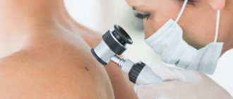
In general, people note that removing moles with radio waves is really quick, simple and without problems, and there are no scars left after this. Before the procedure, many advise first visiting an oncologist to make sure that the mole is not dangerous, and after removing it, take the mole for analysis to completely exclude the possibility of a malignant formation. The only drawback of the radio wave method, which patients call, is its rather high cost compared to the same surgical removal.
Is anesthesia necessary during radioknife treatment?
Since radio waves cauterize nerve endings, very often such operations can be performed without anesthesia. But sometimes, due to the risk of discomfort, it is much easier and simpler to administer local anesthesia, completely removing all discomfort. To do this, when removing moles, warts, papillomas and other pathological formations, a small dose of local anesthetic is used.
When treating cervical pathology, pain relief is often not necessary either. The stage area does not have a large number of nerve endings, and the wave method itself is low-traumatic. However, if the woman experiences discomfort, she will be given local anesthesia.
Where to have surgery in Moscow?
If you want to remove an ingrown toenail in Moscow using a laser or radio wave method, we will be happy to help you. We have all the necessary equipment for treatment, the price of the operation is low.
In our clinic, operations are performed by Dr. Igor Vitalievich Elshansky, surgeon, Ph.D. with more than 20 years of experience. All types of interventions are possible, incl. laser and radio wave method. When you contact, the doctor will carefully listen to your complaints, examine the problem area, and choose the optimal treatment method. If there are indications for surgery, it can be performed on the same day of treatment. The intervention itself takes about 15 minutes, and in most cases eliminates the problem forever.
Recovery and care
Cauterization with Surgitron is not accompanied by a long period of rehabilitation. It will take a short time to recover. In rare cases, complications may develop.
The duration is determined by the doctor individually, taking into account the complexity of the procedure and the reaction. If the patient observes the occurrence of suspicious symptoms, you should contact a specialist.
Standard rules of care for the prevention of infectious lesions.
- After treatment of cervical erosion, it is necessary to avoid physical activity and sports for a month. It is not recommended to walk a lot or lift heavy objects. Sexual activity is prohibited.
- Mole removal is often performed using the Surgitron device. During the rehabilitation period, it is forbidden to take a bath, go to the pool, sauna, or solarium. Swimming in open waters is not permitted. Only showers are allowed. The water should be warm, but not hot.
- When menstruation occurs, it is prohibited to use tampons.
- Massage and cosmetic procedures are not recommended.
- Do not apply gels or scrubs to the affected area of the skin for 1 month. In the first days, the wound is washed with warm water and antiseptics. After a week, you can use baby soap.
- The following creams will help speed up the regeneration of damaged skin: Bepanten, Panthenol.
It is forbidden to use any means, especially medications, on your own. They must be prescribed by a doctor. Otherwise, there is a risk of complications and unwanted reactions.
The occurrence of discomfort and other suspicious signs should be a reason to visit a specialist.
Do lipomas need to be removed?
The wen itself is not dangerous to human health. As a rule, neoplasms are asymptomatic and do not cause any inconvenience. However, those lipomas that reach large sizes can put pressure on adjacent organs and disrupt their functioning. Wen, which are located in places of constant friction with clothing or accessories, can become inflamed and fester. As a result, the skin around the tumor becomes red and swollen. In this case, a person may be bothered by burning, itching and soreness of the skin.
Lipomas are not prone to spontaneous resorption, so they are recommended to be removed. The most modern method of combating wen is the radio wave method.