Drug rash: causes
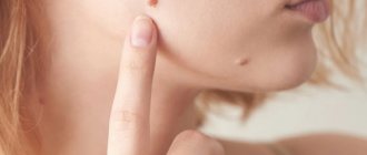
The appearance of red spots on the skin that occurs after taking a certain medication may have the following reasons:
- Allergic reaction. According to statistics, about 10% of the world's population suffers from an allergic reaction to certain groups of drugs. After the initial dose of the drug, sensitization occurs, after the secondary dose, symptoms of urticaria develop.
- Intolerance to the drug or its individual components. A similar reaction develops in individuals with increased immunological reactivity. For example, aspirin intolerance is quite common among asthma patients.
- Side effects. Taking certain medications can lead to adverse reactions, including various skin rashes. Usually, the manufacturer indicates the possibility of such complications in the annotation for the drug.
Also among the prerequisites for the occurrence of drug-induced urticaria, it is worth highlighting the following:
- Treatment using immunostimulants.
- Complex therapy, which includes the simultaneous use of several medications and their incompatibility with each other.
- The patient has endocrine diseases.
- Diseases characterized by immune hyperactivity - for example, asthma.
- Hereditary predisposition.
- Changes in skin microflora caused by taking antibiotics.
- Cross reactions. For example, if a person has previously been diagnosed with a mold allergy, there is a high likelihood of a similar reaction to penicillin and some other antibiotics.
Relevance
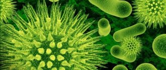
The new coronavirus disease (COVID-19) is a severe respiratory syndrome infection caused by the SARS-CoV-2 virus.
The COVID-19 attack began in Wuhan (China) at the end of 2022 and spread across Europe with the maximum number of infected people showing positive tests: in Spain (about 250 thousand) and Italy (more than 240 thousand people), as of 01.07 .2020. The World Health Organization announced on March 11, 2020 that the spread of coronavirus infection has reached pandemic proportions and as of July 10, 2020, the number of cases worldwide exceeded 12 million people. The main clinical symptoms of COVID-19 are fever, headache, cough and myalgia, as well as sputum production, hemoptysis and loose stools [1]. Lung changes at autopsy include alveolar and interstitial inflammation, epithelial proliferation, and hyaline membrane formation; the infection affects the heart, blood vessels, liver, kidneys and other organs.
Clinical variants of skin lesions with COVID-19
Skin manifestations in patients testing positive for SARS-CoV2 occur in approximately 20.4% of cases [2]. The rash is clinically represented by widespread urticaria, morbiliform rash, erythema, varicella-like symptoms, vesicles, purpura, acrocyanosis, dry gangrene, and petechial rash.
It is known that the expression of endothelial cells in COVID-19 triggers a cytokine storm with the involvement of macrophages and the development of inflammatory reactions similar to vasculitis. Cases of cutaneous vasculitis in patients with pneumonia with COVID-19 [3] in the form of urticarial skin lesions of the lower extremities were similar to lesions that occur in autoimmune diseases. A case of drug-induced vasculitis in a patient with pneumonia of coronavirus origin is described [4]. During the epidemic outbreak in Italy, cases of varicella-like papulovesicular exanthema were described as a rare but specific COVID-19-associated skin lesion. Eight Italian dermatology centers summarized the clinical data of patients with COVID-19 (72.7% of them over 60 years of age) [5]. On average, the skin rash remained on the skin for 8 days, and in 54.5% of patients it was represented by vesicles. In most cases, the rashes were localized on the skin of the torso; the face and mucous membranes were not affected. In 40.9% of patients, moderate itching was noted, in 95.5% of cases - fever, 72.7% - cough, headache (50.0%), weakness (50.0%), runny nose (40.9%) , dyspnea (40.9%), hyposmia (18.2%), hypogestia (18.2%), pharyngodynia (4.5%), diarrhea (4.5%), myalgia (4.5%). Lethal outcome was observed in 13.6% of patients. PCR (polymerase chain reaction) study from skin lesions was not demonstrative.
D. Ortega-Quijano et al. (2020) note that COVID-19 is more common in adults, for whom chickenpox is not common. This may indicate a positive prognostic value for varicella-like lesions compared with other lesions [6]. For differential diagnosis, the Tzanck test and Varicella-Zoster PCR are valuable. D. Fernandez-Nieto et al. (2020) observed monomorphic cavitary rashes with a diameter of up to 3–4 mm localized on the skin of the chest, abdomen, and back in 25% of COVID-19 patients with skin manifestations [7].
It is obvious that skin rashes associated with viral infections can serve as a manifestation of viremia. Most dermatotropic viruses act as inert foreign particles in the skin, causing an inflammatory process as a result of a reaction with circulating antibodies and sensitized lymphocytes. Analyzing cutaneous symptoms of COVID-19 in children, M. Morey-Olivé et al. (2020) pointed out their similarity to exanthemas typical of the acute phase of other common viral infections. However, at the same time, it should be taken into account that these manifestations may reflect inflammatory or microthrombotic phenomena in the phase of the immunological response [8].
In France, a retrospective study was performed involving 77 patients with skin manifestations, including: urticaria (26.9%), vesicles (15%), acral lesions (80%), morbiliform (25.9%), petechial (7.3). %) manifestations, livedo reticularis (4.1%) and other types of rashes (15%). It was noted that some patients experienced several types of rashes at once. Chills were the most common (75%) sign of acral lesions, symptoms of dyshidrosis were detected in 14% of patients.
28% of patients with acral lesions had positive results of PCR tests for SARS-CoV2. Histological studies in areas of chills revealed lichenoid dermatitis with perivascular and eccrine mononuclear infiltrate, as well as microthrombi [9].
Acroischemic foci have been described in individuals with certain characteristics of the course of COVID-19: in patients with severe ischemia in the lower extremities and in young patients with a clinical picture of skin chills. We describe 7 patients with severe coronavirus pneumonia and acroischemia, accompanied by a significant increase in the levels of D-dimer, fibrinogen and abnormal coagulation function (mainly prolonged prothrombin time was observed). This may be due to the cytokine storm caused by the virus, due to unwanted activation of the coagulation cascade and the development of microthrombosis. The skin lesions completely regressed within two weeks. Histological examination showed ischemic necrosis affecting the epidermis and dermis with signs of re-epithelialization [10].
A. Saenz Aguirre et al. (2020) observed in 74 patients with skin manifestations of coronavirus infection (56.8% of them men) skin lesions of the lower extremities (in 95.94% of patients, in 74.3% of cases - on the toes); the skin of the upper extremities was affected in 8.1% of patients (in 100% of cases - on the fingers). Bilateral lesions were observed in 68.91% of patients, symmetrical – in 51.35%. Skin biopsy of the acral lesions showed a lymphocytic perivascular and perieccrine infiltrate, with no vascular occlusion or intravascular thrombus observed. The etiology of these lesions remains unclear. It is described that at the onset of the disease microangiopathies and inflammation develop; later, complement activation occurs, leading to inflammation and blood clot formation [11].
For acral skin manifestations of COVID-19, the term “frostbite” is used [12], however, D. Fernandez-Nieto et al. (2020) note the advisability of using the term “acroischemic lesions” rather than “chiblains”, despite the morphological similarity of skin lesions in COVID-19 with classic frostbite or chills, because Lesions of the acral region with minor or no signs of frostbite have also been described. The authors suggest that acral skin lesions associated with coronavirus infection represent a spectrum of manifestations ranging from erythematous macules to frostbite-like lesions to gangrene or ischemia [13].
R. Rivera-Oyola et al. (2020) described skin manifestations in 60-year-old patients with COVID-19 in the form of erythematous spots, papules and urticarial plaques localized on the skin of the trunk [14]. A family case of urticaria-like rashes with COVID-19 in Monterrey (Mexico) has also been described [15]. Infected members of one family had anosmia, loss of taste, chills and dizziness, and two out of five cases had skin manifestations: a 50-year-old woman and her 20-year-old daughter had a widespread rash on the skin of the upper and lower extremities, erythematous ring-shaped rashes, regressing within 24 hours.
Another clinical variant of the skin manifestations of COVID-19, simulating common allergic dermatosis, is atypical exudative erythema multiforme with palmar localization of rashes [16]. Two patients are reported with a diagnosis of COVID-19 and similar rashes, which can also be regarded as cutaneous manifestations of a viral infection with acral localization. The first case: a 17-year-old teenager with a mild infection, who on the 15th day of illness developed erythematous and maculopapular atypical rashes located exclusively on the palms, painless, accompanied by mild itching. The mucous membranes were not involved. Case 2: A 29-year-old man treated with hydroxychloroquine and azithromycin developed fixed erythematous and urticarial target-like lesions on the palms without subjective sensations on the 16th day from the onset of COVID-19 symptoms and on the 4th day from the start of treatment. The treatment was continued, the rash on the palms gradually regressed. The authors suggest that the pathophysiological mechanism of these manifestations may be a hypersensitivity reaction of lymphocytes, mediated by the production of proinflammatory cytokines directed at Sars-Cov-2 antigens present in the skin.
V. De Giorgi et al. (2020) note that dermatological manifestations may correlate with the severity of COVID-19. Acroischemia (mainly cyanosis of the fingers and toes) has been observed in more severe cases. These manifestations have been found to be associated with coagulation disorders reflected in increased levels of prothrombin, fibrinogen and D-dimer, which are observed in patients in intensive care units [17].
N. Skroza et al. (2020) present a case of histologically confirmed urticarial vasculitis in a 47-year-old patient with COVID-19 and concomitant arterial hypertension and diabetes mellitus as an example of a toxic-allergic reaction to drugs. The patient received an antibiotic, antiviral and anticoagulant agents (ceftriaxone, lopinavir/ritonavir, hydroxychloroquine, enoxaparin) [18].
It was reported that in patients with a subsequently confirmed diagnosis of COVID-19, periorbital erythema was not accompanied by subjective sensations until the onset of systemic symptoms; therefore, this symptom can be regarded as an early sign of the presence of coronavirus infection [2].
U. Wollina et al. (2020) draw attention to the high incidence of androgenetic alopecia (AGA) in men with COVID-19.
In a clinical study in a group of 41 middle-aged men 58 years old with bilateral pneumonia (COVID-19 disease), it was revealed that 29 (71%) had AGA (Hamilton-Norwood scale > 2), while 39% had AGA classified as a severe variant (Hamilton-Norwood scale ≥4). Several pathways have been suggested for androgens to be involved in the spread of COVID-19. The androgen-regulated protease TMPRSS2 is a cellular co-receptor required for SARS-CoV-2. Another link is androgen-driven immune modulation, since androgens serve as an immunosuppressive agent. Indeed, among adult patients with COVID-19, there is a predominance of male patients. Genetic factors may influence the geographic distribution of COVID-19, as there are regions with higher rates of androgen-related diseases. The adrenal-permissive phenotype of the HSD3B1 gene encodes 3β-hydroxysteroid dehydrogenase-1, which is involved in the conversion of dehydroepiandrosterone into active and more potent androgens [19].
In an analysis of a group of 20 pediatric COVID-19 patients, BB Duramaz et al. (2020) suggested that maculopapular rashes are a side effect of hydroxychloroquine [20].
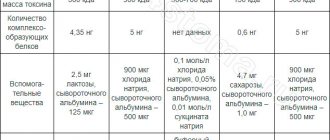
U. Wollina et al. (2020) provide the following list of clinical manifestations [19] (see table).
JL Jia et al. in May 2022, we analyzed the PubMed/MEDLINE database from 12/31/2019 to 05/03/2020. Published data from 997 patients from 9 countries who had cutaneous manifestations associated with COVID-19. The most common skin symptoms were chills (400/40.1% of cases), maculopapular lesions (230/23%), vesicles (101/10.1%), urticarial rashes (87/8.7%), and areas of necrosis (23/2.3%) and other undifferentiated skin lesions (197/19.7%). Pain and burning were reported in 70 cases, and itching in 268. Future studies are planned to include information on race/ethnicity [21].
S. Young, A.P. Fernandez (2020) note that some patients experienced several different rashes at the same time. A case of severe COVID-19 was described in a 68-year-old seriously ill man with morbiliform rashes on the skin of the torso, acral purpura resembling chills and ulcerated plaques on the buttocks [22].
Features of the course of severe chronic dermatoses against the background of coronavirus infection
A potentially vulnerable group during the COVID-19 pandemic are patients receiving immunosuppressive drugs for severe chronic dermatoses; the benefit-risk ratio should be assessed for each of them. In these patients, if COVID-19 is suspected or confirmed, immunosuppressive medications should be immediately suspended (with the possible exception of systemic corticosteroids). For persons with respiratory symptoms, but without confirmation of COVID-19, it is recommended to reduce the dose of the drug or suspend its use for 1-2 weeks.
Lidia Rudnicka et al. (2020) (https://pubmed.ncbi.nlm.nih.gov/32376422/?from_single_result=32376422&expanded_search_query=32376422 - affiliation-1) believe that the risk of treatment with cyclosporine during the COVID-19 pandemic is low, comparable to that of persons receiving placebo. Of the 225 patients in the study who took cyclosporine for 12 months, none experienced reactivation or onset of viral infections, including varicella zoster virus, herpes simplex virus-1, herpes simplex virus-2, Einstein-Barr virus, cytomegalovirus, human immunodeficiency virus or other infectious diseases.
Numerous in vitro data indicate that cyclosporine has a broad spectrum of antiviral activity. It inhibits the replication of viruses such as hepatitis B virus, hepatitis C virus and human immunodeficiency virus. Cyclosporine also inhibits the replication of influenza A virus (FLUAV), West Nile virus, Rift Valley fever virus and Zika virus by blocking the interaction of cellular cyclophilins with viral proteins and inhibition of viral RNA synthesis. It has been established that in vitro cyclosporine reduces the replication of two coronaviruses dangerous to human life - MERS-CoV and SARS-COV [23]. A similar inhibitory effect of cyclosporine was observed with other coronaviruses, including HCoV-229E, TGEV, FCoV, PEDV and MHV. Research results have led to the suggestion that cyclosporine may be a promising treatment option for severe acute respiratory syndrome. There is no reliable data on the effect of cyclosporine on SARS-CoV2, which causes COVID-19, but it is possible that cyclosporine taken by patients with autoimmune skin diseases may also have an antiviral effect: these patients have a lower risk of developing severe COVID-19. 19 [23].
Cases of regression of COVID-19 symptoms in dermatological patients receiving biological agents have been described [24, 25]. We describe a 40-year-old woman who suffered from psoriasis since 2000 and was treated with guselkumab from January 2022 until complete regression of psoriatic rashes. On 03/03/2020, she had contact with relatives infected with SARS-CoV2, and after 6 days she manifested a severe cough, myalgia, increased fatigue and fever up to 39.4°C. The patient did not cancel the guselkumab injection scheduled for March 16, and the day after the injection she reported significant improvement in her respiratory condition, normalization of body temperature, and a gradual decrease in symptoms of myalgia and fatigue. Thus, this finding supports the hypothesis of a potential role for interleukin-23p19 (IL-23p19) inhibitors in counteracting the cytokine storm triggered by SARS-CoV2, potentially influencing symptom severity.
Cytokines key to the pathogenesis of psoriasis, tumor necrosis factor α (TNF-α) and IL-17, enhance the inflammatory response to viral pneumonia, while IL-23 appears to be less important for an effective immune response. Increased inflammatory cytokines are associated with worsening clinical course of SARS-CoV-2. Based on these observations, it was hypothesized that biologic therapies against TNF-α or IL-17 may help alleviate the COVID-19 cytokine storm and acute respiratory distress syndrome. For this reason, the use of ixekizumab and adalimumab is currently being studied in China for the treatment of COVID-19 [24, 25].
V. DiLernia et al. (2020) described a patient who suffered from psoriasis and psoriatic arthritis and was treated with the biological drug secukinumab, against which, for mild COVID-19, hydrochloroquine was prescribed 800 mg daily for 2 days, then 400 mg daily for 5 days. After 28 days, two consecutive PCR tests were negative. It has been suggested that inhibition of IL-17 in severe forms of psoriasis may have a beneficial effect on the course of COVID-19, because it is one of many cytokines released when the human body is attacked by the coronavirus.
Consequently, further research into the use of biological products in patients with SARS-CoV2 will make it possible to use their capabilities to reduce the body’s immune response [26].
M. Shah et al. (2020) analyzed the tactics of managing patients with severe atopic dermatitis (AD) during the COVID-19 epidemic, and noted the need to optimize the skin care regimen [27]. Two cases of continued treatment of severe AD due to COVID-19 with dipilumab have been described (S. Ferrucci et al., 2020). Dupilumab is a fully human monoclonal antibody against the α subunit of the IL-4 receptor that blocks signaling from IL-4 and -13, which are key type 2 cytokines in the pathogenesis of AD. The European AD Working Group recently argued that dupilumab does not increase the risk of viral infections and therefore has advantages over traditional systemic immunosuppressive agents in the COVID-19 pandemic [28].
Thus, most dermatologists negatively assess the cessation of immunosuppressive therapy for severe dermatoses during the COVID-19 epidemic.
Of course, more intensive use of hand washing and disinfection products, as well as prolonged wearing of masks and gloves lead to an exacerbation of dermatitis of the hands and face. Higher stress levels during the pandemic increase the risk of AD flare-ups. Caution should be exercised when prescribing high doses of prednisolone in epidemic conditions, given its pronounced immunosuppressive effect. Other immunosuppressants, such as methotrexate, mycophenolate, azathioprine and cyclosporine, should be reduced to the minimum dose as possible increased susceptibility to viral infections, including SARS-CoV2. Prescribing lower doses of immunosuppressive drugs will help minimize the patient's risks. With regard to dupilumab, an inhibitor of monoclonal antibodies of the IL-4/IL-13 signaling pathway, an opinion has been expressed about the advisability of its use for the treatment of AD during a pandemic; If symptoms of a viral infection are present, the decision to continue treatment should be made individually for each individual patient. C. Patruno et al. (2020) analyzed the risks of exacerbations of blood pressure during quarantine, highlighting the main unfavorable aspects: increased itching due to neuroendocrine modulation of skin inflammation as an adverse psychological effect during quarantine; violation of the hypoallergenic diet and decrease in physical activity during self-isolation, as well as some patients stopping taking immunosuppressive medications on their own initiative due to fear of contracting COVID-19 [29].
Hydroxychloroquine is included in treatment regimens for coronavirus disease. Hydroxychloroquine has been shown to reduce viral load in patients with COVID-19 infection, but a number of reports have noted adverse dermatologic effects of this therapy. M. Sachdeva et al. (2020) indicate the need for a systematic review of previously reported cases of onset, exacerbation, or relapse of psoriasis following treatment with hydroxychloroquine. The authors conducted a comprehensive search for original studies examining the side effects of hydroxychloroquine treatment in psoriasis. It has been revealed that treatment with hydroxychloroquine can lead to induction, exacerbation or relapse of psoriasis. It was concluded that monitoring of side effects is necessary and clinical trials are needed to characterize the safety profile of hydroxychloroquine use in patients with COVID-19 infection [30].
N. Litaiem et al. (2020) describe acute generalized exanthematosus pustulosis (AGEP) as a rare and severe complication of treatment for COVID-19 with hydroxychloroquine, which developed in the form of itchy erythema and pustules in a 39-year-old patient 18 days after the start of treatment for COVID-19 with hydroxychloroquine. daily dose 600 mg. The diagnosis of AGEP was made by a combination of clinical and histopathological studies. Discontinuation of hydroxychloroquine led to significant regression of skin lesions, but the patient died from a massive pulmonary embolism.
AGEP is usually induced by antibiotics and develops within 48 hours of starting treatment. AGEP occurring after treatment with hydroxychloroquine has a longer incubation period (up to 2–3 weeks) and atypical clinical manifestations, including blisters and target-like papules that regress after drug discontinuation. Side effects such as exanthema, stomatitis, itching and hyperpigmentation, as well as Stevens-Johnson syndrome and toxic epidermal necrolysis have also been described. However, AGEP is the most common serious side effect associated with hydroxychloroquine [31].
Dermatological problems of using personal protective equipment during the COVID-19 pandemic
Coronavirus infection spreads through the respiratory route, but also through contact with contaminated surfaces. Thus, hand hygiene is the cornerstone of effective infection control during the COVID-19 outbreak [32].
Frequent hand washing, however, can lead to the development of eczema. Any dermatologist is well aware of the phrase “eczema does not like water.” On the one hand, frequent hand washing is important to stop the cycle of sustained transmission of coronavirus, but on the other hand, frequent use of hygiene products, particularly harsh soaps and other strong detergents, can initiate the development of simple contact dermatitis and associated complications, including methicillin -resistant skin colonization by Staphylococcus aureus, especially in critical work areas such as intensive care units, emergency departments and isolation wards during an ongoing epidemic.
It is recommended to use synthetic detergents instead of soap, because... They have a neutral to slightly acidic pH and relatively high free fatty acid content, are less irritating to the skin and have a moisturizing effect, helping to prevent dry and irritated hands. To clean and disinfect hands, it is advisable to use alcohol-based detergents. Frequent use of hot water can lead to excessive dryness of the skin, so you should use warm water at a temperature of 45–50°C when washing your hands. From a hygienic point of view, paper towels are preferable to electric dryers.
The use of emollient external products during working hours after washing hands and after working at home supports the regeneration of the skin barrier. The use of water-based moisturizers (glycerin, panthenol, urea, gelatin, hyaluronic acid, α-hydroxy acids) should be avoided as this may increase transepidermal water loss. Direct contact with chemicals used to disinfect surfaces is prohibited.
Coronavirus disease has necessitated the use of personal protective equipment (PPE), which has led to a number of problems with skin lesions: 97.0% (n=526/546) of healthcare workers reported skin side effects from the use of PPE, most often on the skin of the bridge of the nose (83 .1%) and cheeks (78.7%). Associated burning, itching, or tingling sensations may predispose health care workers to touch the facial area, which may lead to exposure to the virus. Some additives (for example, formaldehyde textile resins) to fibers in PPE can cause allergic contact dermatitis. It is known that 22.8% of the population have increased sensitivity to formaldehyde, which is also aggravated by friction, sweat, and tight-fitting clothing. Up to 99% of skin reactions associated with PPE are caused by wearing gloves, because their long-term use can lead to allergic contact dermatitis, subsequent maceration and erosion of the skin. 59.6% of healthcare workers who regularly used masks during the epidemic developed acne-like rashes. Prolonged wearing of safety glasses can lead to urticaria, pressure injuries, contact dermatitis and acne vulgaris in healthcare workers. Some face masks are secured by clamping to the ear flaps, resulting in damage due to pressure and/or prolonged exposure [33].
Thus, the use of PPE by healthcare workers during the COVID-19 outbreak has provoked multiple inevitable dermatological problems, because... Strict adherence to PPE guidelines is paramount to reducing the spread of infection.
Exposure to detergents and soap, frequent (more than 20 times a day) hand washing and the use of gloves during an epidemic are all known risk factors for the occurrence of eczematous process and/or its exacerbation, therefore, during the COVID-19 pandemic, intensive prevention of eczema should be carried out : use of moisturizers in combination with protective creams [34].
L. Dong et al. (2020) propose hydrogel patches as a means of preventing facial skin damage caused by mask compression: water, sodium polyacrylate, cellulose gum and sodium hyaluronate are the main components of the hydrogel, which serves as a shock absorber for the soft tissues of the face when compressed by the mask. In addition, glycerin and several plant extracts in the hydrogel can keep the skin hydrated and protect the skin barrier [35].
Changes in the organization of dermatological services during the COVID-19 epidemic
Teledermatology technology has been widely developed to optimize dermatology care during the COVID-19 pandemic. The American state of Ohio, where the epidemic situation was most tense, is a leader in the use of this technology in a crisis. A simple algorithm for counseling dermatological patients in a hospital setting was developed, and ethical issues and restrictions were regulated [36]. The Ohio Dermatological Association has issued guidance to physician offices and academic centers on patient selection and use of personal protective equipment. The Ohio State University has provided a telemedicine platform for outpatient and inpatient clinics. When consulting new patients, an initial assessment of photographs was carried out for referral to telemedicine consultations. If the clinical case was not suitable for telemedicine, the consultation was carried out with the least risks (initial evaluation of photographs, limited number of people in the room). Limitations on the use of telemedicine in hospitals are associated with financial problems and distortion of photographic images. Of course, the experience gained during the COVID-19 epidemic in using telemedicine consultations will be used in the subsequent period [37].
Italian dermatologists G. Nazzaro et al. (2020) in the midst of the COVID-19 pandemic indicate the need for a temporary reorganization of dermatology services: all outpatient consultations were postponed, with the exception of consultations for patients receiving biological agents (to ensure continuity of therapy) and dermatological surgery for patients with skin neoplasms suspected of malignancy. At the same time, dermatosurgical manipulations in patients with facial injuries were postponed as they interfered with the retention of the surgical mask during surgery. In addition, planned hospitalizations and the work of day hospitals were stopped [38].
F. Al-Niaimi, FR Ali (2020) point out the danger of surgical smoke during dermatological operations for patients and staff during a pandemic, given the high contagiousness of coronavirus when transmitted by aerosol. Dermatosurgeons consider bipolar coagulation advisable, in which the concentration of small aerosol particles is reduced. Protection is provided by the use of specialized masks capable of filtering particles smaller than 5 microns. Air filtration in operating rooms is also important [39].
K. Damevska et al. (2020) summarized the experience of the Macedonian dermatology service under stressful conditions during the epidemic: the fourth diagnosed case of COVID-19 in the Republic of Macedonia was a dermatologist working at the University Clinic of Dermatology in Skopje. Before the diagnosis was made, the doctor contacted almost all the clinic staff, and also held a previously planned seminar, which was attended by 95 dermatologists from all over the country. Within hours of his diagnosis, swift action was taken: 128 dermatologists were quarantined; only 9 of them were able to resume practice between 9 and 26 March 2022. A number of socially restrictive measures taken further reduced the availability of dermatological services. In the absence of an official teledermatology platform, conventional telecommunications and social media were used to maintain communication with other specialists and patients. As a result, treatment for most chronic patients occurred without interruption. To monitor dermatological conditions that require immediate attention, the authors of the article conducted a survey in which 77 dermatologists participated: 91% of respondents received requests for consultations from inpatients, 82% of doctors believed that consultations through mobile applications were useful for monitoring patients. However, these methods were useful in less than 30% of initial consultations. The most common reasons for consulting patients were to monitor prescribed therapy. Thus, the COVID-19 pandemic has become a necessary stimulus for the development of teledermatology [40].
As experience accumulated in reorganizing dermatovenereology services during the pandemic, PA Cerro et al. (2020) summarized the experience of several countries (Italy, China, Spain), highlighting the key points: dermatologist visits only for emergency cases, the use of telephone consultations to reduce the flow of patients in the clinic, patients wearing masks, thermometry, use of PPE by medical personnel throughout working day, periodic disinfection, training on personal protective measures, cancellation of most dermatosurgical interventions, widespread use of teledermatology, special consultations for patients with skin lesions suspected of coronavirus infection with a preliminary assessment through teledermatology [41].
T. Y. Loh et al. (2020) note the critical importance of teledermatology in the dermatology education of medical students [42]. While the benefits of teledermatology are evident, it is important to recognize that important elements of dermatology education cannot be achieved virtually, such as the ability to evaluate lesion texture, perform biopsies, or use instruments such as a dermatoscope, Wood's lamp, or fungal scraping.
US Muzumdar et al. (2020) state various aspects of changes in the dermatoesthetics industry during the COVID-19 epidemic. In particular, there was a reduction in financial costs for organizing events. At the same time, virtual communication with patients does not always guarantee the receipt of reliable information and the ability to correctly assess the situation [43]. After the restrictions were lifted, many patients still do not want to leave home and visit medical facilities. Under current conditions, aesthetic dermatological procedures have temporarily faded into the background due to restrictive measures, as well as fear of the risk of infection in the clinic during the procedure. In addition, many patients have been affected by the economic downturn, limiting the availability of aesthetic procedures. In the practice of dermatologists against the backdrop of an ongoing pandemic, it is advisable to find a balance between the use of teledermatology, reducing the share of face-to-face consultations and simplified administration and logistics [44].
Conclusion
Thus, the coronavirus pandemic, which began in 2019, serves as a unique challenge to global dermatology, which has forced us to think about the development of new therapeutic approaches to skin lesions in patients with COVID-19, to optimize the tactics of treating severe dermatoses with immunosuppressive agents; increase the use of teledermatology; evaluate the side effects of using various PPE for medical personnel and patients, and determine methods for their neutralization.
Symptoms of drug rash
The time for the first signs of drug-induced urticaria to appear can vary: it usually ranges from a few minutes to several days. Among its manifestations it is worth highlighting the following:
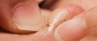
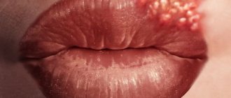
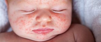
- The appearance on the skin of small pale pink, red or purple blisters - hollow spots of oval or irregular shape, slightly rising above healthy areas of the skin. Gradually, individual elements of the rash merge into large spots. Their localization may vary.
- In addition to blisters, small nodules with a diameter of about 5 mm may appear on the skin, containing purulent contents mixed with blood.
- The rash is accompanied by severe itching, tingling, burning and skin irritation.
- A local increase in temperature is possible.
- Symptoms such as lacrimation, nasal congestion, headache, bronchospasm, swelling of the mucous membranes, etc. may be present.
Diagnostics
If you find that you have a strange-looking rash on your body after taking a particular medication, stop taking the medication temporarily and consult a doctor. After collecting anamnesis and visual examination, the doctor will draw conclusions about the possible cause of urticaria. In some cases, special diagnostic tests may be required to determine the sensitivity of the immune system to a range of medications.
To make an accurate diagnosis, differential diagnosis is important, since drug-induced rashes can have the same symptoms as some infectious and dermatological diseases.
Drug rash: treatment in children and adults
The first thing to do after the appearance of hives is to stop taking the drug that caused it. If therapy involves taking several medications at once (both for internal and external use), it is advisable to temporarily stop treatment as such. After the “culprit” of the rash is identified, the doctor will select a safer analogue of the drug for the patient, which will differ in composition.
Treatment for drug-induced hives usually involves taking antihistamines. They are necessary to reduce the intensity of skin itching and burning.
In some cases, it may be necessary to use local hormonal agents. However, you need to remember that such ointments and creams are characterized by a rather aggressive effect, so they should not be used unless necessary. This is especially true for the treatment of drug rashes in infants and older children.
An important role in the treatment of rashes is given to the intake of enterosorbents, which absorb harmful substances and remove them from the body.
During treatment, you must follow a diet and drink enough fluids. It is advisable to exclude spicy, fried, smoked and salty foods, simple carbohydrates, and sweets from the diet.
What is Omicron?
Omicron
(B.1.1.529) is a SARS-CoV-2 variant of concern (WHO).
The new variant of coronavirus is characterized by a less aggressive course, but higher contagiousness (infectiousness).
According to the latest data, the omicron carrier infects up to 12 people (with delta, this number hardly reached 6). After entering the body, the virus multiplies in the nasopharynx and oropharynx, main bronchi 70 times faster than the delta variant, that is, it reaches a number sufficient for clinical manifestations and further transmission of infection much earlier. At the same time, Omicron penetrates more easily into the upper respiratory tract and affects the lungs much less often.
According to one version, omicron developed in the body of a person with a sharply weakened immune system; according to another, it was the result of mass vaccination. One way or another, despite the increased contagiousness, the milder course of the infection allows epidemiologists to consider the appearance of Omicron as a turning point in the pandemic, a consequence of the fact that the vaccine gave it a controlled nature.
Treatment of medicinal urticaria using folk methods
Traditional medicine methods can act as an additional measure in the treatment of drug rash. However, we must remember that using exclusively “grandmother’s” recipes is unacceptable, as it can further aggravate the situation. Before using traditional medicine, consult your doctor.
What treatment options for medicinal urticaria does traditional medicine offer?
- If hives that appear after taking medications are accompanied by severe itching, you can reduce it with the help of grated raw potato gruel. The potato tuber should be washed and grated. The resulting paste is applied to the rash for 10-15 minutes, after which it is carefully removed. There is no need to wipe off the remaining potato juice.
- Baths with herbal infusions help relieve irritation and itching caused by medical urticaria. To prepare them, 1 tablespoon of dry herbs (chamomile, string, calendula or mint) is poured into 0.5 liters of boiling water and allowed to brew for half an hour. The resulting infusion is added to a bath with warm water.
- In the fight against skin itching caused by medical urticaria, propolis tincture helps well. It is recommended to wipe the affected areas of the skin with a cotton pad soaked in a 10% tincture.
- You can make lotions from nettle decoction. 1 tbsp. l. dry herbs pour a glass of water, bring to a boil and remove from heat. The resulting broth is cooled to room temperature, moistened with a clean bandage and applied to the rash.
Omicron and vaccination
Does Satellite V protect against Omicron?
The ability of antibodies produced after full vaccination with Sputnik V to neutralize omicron is somewhat lower than to neutralize other variants. However, complete antibody evasion does not occur, suggesting that Sputnik V vaccination provides immunity of varying degrees of effectiveness against all currently known coronavirus variants. It should be remembered that when speaking about the effectiveness of vaccines, we do not mean the absence of clinical manifestations of the disease, but a decrease in the severity of the disease and a reduction in the number of the most dangerous complications and manifestations of post-Covid syndrome.
A number of scientists believe that the best immunity against omicron is formed in vaccinated individuals and patients who have recently recovered from a coronavirus infection caused by other variants. Vaccination forms antibodies to the surface of the virus, but in the case of a previous illness, the immune system involves extensive cellular units that are active against viral particles and infected cells. An important role is played by how soon after recovery the omicron infection occurs; depending on this, the strength and form of the immune response also differ.
Is Sputnik Light effective?
According to the latest data, if Sputnik Light is used as a booster vaccine, it provides 100% of the appearance of antibodies effective against Omicron (after two doses of Sputnik V, the appearance of such antibodies is 75%).
What is important to remember about vaccines?
First of all, the vaccine does not make people invulnerable and does not eliminate the need to use personal protective equipment.
Hope for vaccination, but don’t take off your mask!
For clarity, you can turn to arithmetic. Let's imagine a conscientious citizen who has been vaccinated with Sputnik V. He has n antibodies in his body, among which 0.7n are effective against omicron.
If, out of naivety, he walks without a mask and encounters a carrier of the infection, who for some reason is also without a mask, the citizen can receive 2n viral particles and get clear symptoms of coronavirus despite being vaccinated.
If our citizen is lucky and the sick person he encounters is wearing a mask, he will receive n viral particles and after a couple of days he will feel slightly unwell, ignore it, go to work as if nothing had happened, infect 12 people along the way and continue enjoy life.
And only if both people are wearing masks, the citizen will receive 0.5n viral particles that his antibodies can handle. This is the only way he will protect his family, loved ones and random passers-by.
What to do to prevent the occurrence of drug-induced urticaria?
To protect yourself from developing a rash after taking medications, it is important to increase the body's defenses and not take medications uncontrollably. Among the preventive measures are the following:
- You cannot self-prescribe certain medications (especially antibiotics).
- You must take prescribed medications strictly according to the regimen recommended by your doctor.
- If you have had problems with a rash after taking medications in the past, be sure to tell your doctor.
- In the autumn-winter period, it is advisable to increase the body's defenses in every possible way, since it is the imperfection of the immune system that can lead to the development of a rash. To do this, it is recommended to take multivitamin complexes, eat fresh vegetables and fruits, drink natural juices, eat right and follow a drinking regime, play sports and spend time in the fresh air.
Pinpoint rash
Characteristic for: scarlet fever, staphylococcal infection, pseudotuberculosis, toxicoderma, drug rash. Scarlet fever is characterized by the appearance of a rash on the first day of illness. Most often, it is a monomorphic rash against the background of hyperemic skin (concentration in natural folds) with a pale nasolabial triangle. Brief fever, sore throat, crimson tongue, burning pharynx. After 5-7 days, peeling begins at the site of the rash. The more profuse the rash, the stronger the peeling (can be pityriasis or lamellar). First appears on the neck and torso. By the end of the second week, peeling begins around the nail folds, the thick epidermis of the palms and soles peels off in layers. Staphylococcal infection Presence of staphylococcal disease: osteomyelitis, arthritis, other purulent diseases.
Unlike scarlet fever, the rash usually occurs on the 3rd-4th day of illness. Having disappeared, it can reappear. The appearance of a rash is preceded by purulent foci: boil, panaritium, abscess, etc. Regional lymphadenitis is unusual. Pseudotuberculosis (scarlet fever) Rash (polymorphic) appears on the 3-4th day of illness or later. General hyperemia, a symptom of “socks”, “gloves”, “hood”. The combination of these symptoms is possible only with pseudotuberculosis. Characterized by the absence of sore throat. Catarrhal phenomena and a crimson tongue are possible, but such pronounced manifestations as with scarlet fever are not observed. It may be accompanied by damage to other organs and systems, primarily the gastrointestinal tract with the development of terminal ileitis, enlargement of the liver and spleen. Prolonged fever for 7-10 days.
La-Cri products and their help in fighting rashes
In addition to medications prescribed by your doctor to treat drug rashes, you can use La Cree Cream for Sensitive Skin. Its use helps reduce the intensity of rashes, redness and skin itching. Due to the fact that the product contains such active ingredients as string, licorice, violet and walnut extracts, as well as panthenol and bisabolol, the cream has a pronounced anti-inflammatory, regenerating, antipruritic effect. Since La-Cree Cream for sensitive skin does not contain hormones, it can be used for a long time.
Clinical researches
The effectiveness, safety and tolerability of La-Cri products have been clinically proven. The products are recommended by the Union of Pediatricians of Russia. During clinical studies, specialists were able to record excellent results.
It has been proven that La-Cri diaper cream:
- creates a protective barrier on the skin;
- relieves redness and irritation;
- provides gentle skin care.
Sources:
- Molochkova Yulia Vladimirovna, Dermatology. Brief reference book, GEOTAR-Media, 2022.
- Baumann Leslie, Cosmetic Dermatology. Principles and practice, MEDpress-inform, 2016.
- Ratner Desiri, Avram M.R., Avram M.M., Procedures in Dermatology. Clinical cosmetology, GEOTAR-Media, 2022.