Clinical manifestations of rosacea
The disease begins after 25-30 years. First, there is a tendency to frequent redness of the face, less often the neck and décolleté. “Burst blood vessels” (telangiectasias) appear. Then the redness in some areas becomes persistent. Subsequently, inflammation, peeling, and rashes (outwardly resembling pimples) may develop at the site of persistent redness. Without treatment, the number of such elements increases. Dryness and redness of the eyes often develop. In a small proportion of patients, with age, the so-called rhinophyma develops, a thickening and growth of the skin of the nose (“bumpy nose”).
Types of rosacea: A - Erythematotelangiectatic rosacea, B - papulopustular rosacea, C - rhinophyma, D - ocular rosacea. Photo: Dermatology Reports / Open-i (CC BY-NC 3.0)
The cause of rosacea is impaired vascular tone of the face and inflammation in the vascular wall. They can be caused by various factors: hormonal disorders, characteristics of psycho-emotional status, diseases of the cardiovascular or digestive system, microbial factors. A doctor must understand the possible causes of the development of rosacea in a particular person and analyze the degree of influence of a particular health feature. The main task is to minimize external and internal factors that aggravate rosacea, and to directly affect the blood vessels of the face. Treatment regimens are chosen by the doctor; they are strictly individual in nature.
Photo: Dermatology and Therapy / Open-i
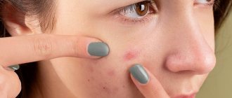
Red spots under the eyes (around the eyes)
Our skin is one of the first to react to any negative changes in the body. The more sensitive the skin, the faster and brighter the signs of any disease appear on it. The thinnest layer of skin is located in the area around the eyes. As a rule, the skin in this area is uniform in color, one tone lighter or darker than the base color of the facial skin. But sometimes red spots appear under or around the eyes, which can signal a number of diseases of the visual organs or the entire body as a whole.
Causes of red spots under the eyes
Various factors can cause the appearance of red spots around the eyes, including:
- Stress, lack of sleep and nervous tension. When the daily routine is normalized and the emotional state is restored, the redness disappears
- Dermatological problems. For example, with eczema and dermatitis, the skin around the eyes not only turns red, but also flakes and may also become crusty.
- Kidney diseases - stones, kidney failure and other pathologies make themselves felt by the appearance of bags, swelling, as well as redness or darkening under the eyes
- Inflammatory processes - abscesses and phlegmon (purulent inflammation). In such cases, redness is accompanied by increased body temperature.
- An allergic reaction in which in addition to redness there is also itching
- Vitamin deficiency and unhealthy lifestyle: lack of exercise, lack of air, poor diet and bad habits often contribute to the deterioration of skin condition
- Congenital skin abnormalities.
Do not forget that only a specialized specialist can establish the true cause of the disease and help get rid of unpleasant symptoms.
What happens in the skin?
Redness, no matter what causes it, represents increased blood flow to the skin. The vessels dilate greatly and contract with difficulty. Some of the fluid from the overcrowded vessels can seep into the surrounding tissues - swelling occurs.
Proteins and blood cells may also leak out. The condition of connective tissue fibers and muscles that help blood vessels contract and drain blood and lymph from the skin deteriorates. Stagnation and low-grade inflammation occurs in and around the vascular wall. Opportunistic and pathogenic microorganisms can join this inflammation, which weakens the skin’s defenses.
Rosacea treatment
Treatment of rosacea using intense pulsed light (IPL) technology.
Photo: BioMed research international / Open-i (CC BY 3.0) Therapy for this disease depends on the totality of manifestations and characteristics of a particular person. There is a classic treatment regimen for rosacea, accepted in dermatology. These are antibiotics, metronidazole, retinoids, antihistamines, local therapy.
Creams, gels, ointments containing azelaic acid, metronidazole, retinoids, pimecrolimus, and antibiotics are used topically. Treatment lasts several months. As prescribed by a doctor, various combinations of medications and their alternation can be used. Historically, ointments and pastes with ichthyol, tar, sulfur, and salicylic acid were widely used. Often they are still used today due to their low price, but all these products irritate the skin.
The main treatment should be aimed at eliminating provoking factors and restoring the normal state of blood vessels. Triggers vary from person to person, but the clear leader is excessive emotional reactions.
Physiotherapy has all these qualities.
The first stage of therapy, the longest, is to reduce swelling and inflammation in and around the vascular wall. Two methods are suitable for this. Laser-magnetic therapy is a combination of red and infrared laser radiation with a constant magnetic field. A powerful anti-edematous and anti-inflammatory effect on the vascular wall promotes the outflow of excess blood and lymph from the face.
Mens therapy, better known as microcurrent . A mild anti-inflammatory effect, restoration of normal tone of the neuromuscular system that regulates vascular contraction, normalizes vascular reactions to external influences.
Both procedures are prescribed 2 times a week, a total of at least 12-20 procedures are required. Depending on the prevalence of specific symptoms, you can start with microcurrent (redness and hot flashes predominate) or laser-magnetic therapy (swelling and inflammation predominate). For optimal results, it is advisable to use both methods. The procedures are inexpensive and painless, but require regular visits to the doctor (2 times a week for 30-40 minutes). The earlier treatment is started, the more effective and shorter it will last. After the procedures, maintenance therapy is prescribed.
In addition to physical therapy, medications may be prescribed that are selected individually. Efficacy is assessed as treatment progresses; drugs may be changed or combined.
Second phase. After stabilization of the situation, persistently dilated vessels that cannot be eliminated by therapeutic methods are removed.
One of the most effective and gentle methods for removing “vessels” is the use of a neodymium laser with a short powerful pulse. Delivery of such a pulse with an absorption spectrum in oxyhemoglobin (a blood component) promotes the removal of blood vessels without damaging the surrounding tissue.
Why does my cheek swell?
Periostitis of the jaw
It is the most common dental cause of the symptom. Develops against the background of dental diseases: periodontitis, pulpitis, alveolitis, periodontitis, suppurating jaw cyst. It is provoked by open fractures of the jaws, infected facial wounds, operations, and tooth extraction. In some cases, it becomes a consequence of hematogenous or lymphogenous spread of infection from distant foci. Depending on the form, it is accompanied by the following manifestations:
- Acute serous periostitis.
Moderate swelling of the soft tissues of the cheek, redness of the mucous membrane, and enlargement of regional lymph nodes are detected. The general condition suffers slightly, sometimes there is an increase in body temperature to subfebrile levels. - Acute purulent periostitis (flux
)
.
The contours of the face are sharply changed, the predominant localization of edema is determined by the location of the inflammatory focus in the periosteum. Patients complain of sharp pain radiating to the temple, eye, ear, and neck. General hyperthermia, chills, weakness, headache, and regional lymphadenitis are observed. When examining the oral cavity, an area of swelling with a fluctuation in the center is detected - a subperiosteal abscess. - Chronic periostitis.
Slight swelling of the cheek, thickening of the jaw, and enlargement of the submandibular lymph nodes are typical. The patient complains of periodic moderate pain. The mucous membrane of the affected area is swollen, hyperemic, with a bluish tint.
Other dental diseases
Other possible dental causes of cheek swelling include the following:
- Perimaxillary abscess.
It occurs as a result of infection of the soft tissues of the perimaxillary area due to boils, tonsillitis, wounds, abrasions, and some dental diseases. Swelling is preceded by toothache, which gets worse when biting. Then dense swelling appears, the temperature rises, and appetite disappears. After spontaneous opening of the abscess, the condition improves, but subsequently the pathology may recur. - Vincent's stomatitis.
Develops when immunity decreases due to diseases, injuries, and stressful situations. The leading symptom is the formation of multiple ulcers on the mucous membrane. Swelling of the cheek is detected in severe cases of the pathology. - Noma.
An area of ulcerative-necrotic lesions of the lip or oral mucosa appears. Necrosis covers the gums, tongue, cheek tissue, and facial bones. The tissues around the area of necrosis are swollen; in severe cases, swelling from the cheek spreads to the chin and infraorbital area. - Salivary gland adenoma.
The parotid gland is most often affected. Local swelling with clear boundaries forms on the outer surface of the cheek, in the parotid zone, and spreads to the angle of the jaw, the area under the earlobe. Characterized by slow growth, painlessness at the initial stage, progression of discomfort and dry mouth as the formation increases. - Tumors of the salivary glands.
Along with adenomas, benign connective tissue neoplasia, intermediate and malignant neoplasms can form in the area of the salivary glands. The area of swelling is located in the same place as with adenomas. The swelling slowly increases in benign tumors and spreads quickly in malignant ones. - Purulent parotitis.
Swelling occurs in the parotid area, which spreads to the adjacent part of the cheek. There is severe pain, difficulty when trying to open the mouth, severe intoxication, and severe hyperthermia.
Cheek swelling
Traumatic injuries
All facial injuries are accompanied by rapidly increasing soft tissue swelling, spreading to adjacent anatomical structures. Swelling of the cheek of traumatic origin can occur with the following injuries:
- Injury.
Swelling without clear boundaries. Along with edema, pain, hyperemia, and sometimes hemorrhage are detected. The pain intensifies when opening the mouth and active facial movements. Speech and the ability to eat were preserved. - Hematoma.
Formed against the background of a bruise. A compaction appears in the area of diffuse swelling, which, as a rule, resolves on its own within 1-2 weeks. - Fracture of the upper jaw.
The most pronounced swelling is observed in Le Fort type 1 fractures, combined with neurological symptoms, hemorrhages in the conjunctiva and periorbital area. Type 2 is manifested by edema, hemorrhages in the periorbital zone, and changes in facial parameters. In type 1 fractures, swelling is more noticeable in the area of the upper lip and medial cheek. - Fracture of the lower jaw.
Injuries to the ramus, lateral and angular fractures of the bone body are characterized by swelling of the lower or outer edge of the cheek. Facial asymmetry, hematomas, bruises, articulation disorders, and stepped dentition are observed. - Fracture of the zygomatic bone.
Swelling appears in the cheekbone area, quickly spreads down the cheek, up the infraorbital region. Bruises and hemorrhages form in the conjunctiva. Along with pain, victims are sometimes bothered by nosebleeds and double vision.
Allergic reactions
Swelling of both cheeks is observed with angioedema, combined with swelling of the eyelids and lips, and breathing problems. The condition develops acutely, within a few minutes, less often – hours. It is provoked by contact with an allergen, insect bites. Along with the listed symptoms, angioedema in children may be accompanied by abdominal syndrome, and sometimes by neurological symptoms.
The cause of swelling of the cheek from the oral cavity may be an allergy to prosthetic materials. The pathological condition occurs several months or years after the installation of prostheses and is characterized by a burning sensation in the area where the prosthesis is fixed, tongue, cheeks, soft palate, changes in taste sensitivity, thirst, and dry mouth. When using metal products, a metallic taste may appear.
Ophthalmic diseases
In patients with acute dacryocystitis, swelling of the lacrimal sac area is complemented by swelling of the cheek, eyelid, and dorsum of the nose. In the chronic form of the pathology, swelling is noticeable along the upper inner edge of the cheek and covers the inner edge of the lower eyelid. Cellulitis of the lacrimal sac is characterized by sharp pain and swelling along the inner edge of the eyelid, combined with fever, weakness, weakness, headache, swelling of the cheek and paranasal area.
Neurological pathologies
Local symptoms of cavernous sinus thrombosis are exophthalmos, blurred vision, swelling and pain in the eyeball, swelling of the temple, part of the forehead, cheek, upper lip, and mastoid process. The clinical picture also includes headache, nausea, vomiting, and in case of infectious genesis of the pathology - hyperthermia, intoxication syndrome.
Swelling of the cheeks can be detected with one of the types of angioneurosis - rosacea. There is constant redness of the cheeks, nose, forehead, chin, and the formation of spider veins. The cause of swelling is persistent dilation of blood vessels, which, with a long course of pathology, leads to skin changes.
Skin lesions
Minor swelling of the cheek may result from simple contact dermatitis. The severity of the symptom increases against the background of prolonged contact with the irritant and secondary infection. With allergic dermatitis, the swelling is more noticeable and is combined with itching of the skin. Atopic dermatitis is characterized by mild swelling combined with the formation of vesicles.
Due to the abundant blood supply to the face and the structural features of the soft tissues, a boil on the cheek is accompanied by significant swelling. In the center of the swelling area there is a limited round or cone-shaped formation with a black rod in the center. After the boil has matured, yellowish pus appears around the shaft. Increasing twitching pains are noted.
In young children, swelling of the cheeks is often provoked by superficial pyoderma and occurs against the background of pustular rashes. A severe form of facial skin lesions in adults and children is erysipelas. The disease manifests itself with itching, bloating, and a burning sensation. Subsequently, the cheek swells, and a focus of clearly defined hyperemia with uneven edges, reminiscent of a geographical map, forms on it. Fever and intoxication syndrome are observed.
ENT diseases
Slight swelling of the cheeks is possible with the development of acute sinusitis or exacerbation of chronic inflammation of the maxillary sinuses. Other symptoms include pain, impaired nasal breathing, nasal discharge, weakness, fever, and signs of intoxication. Patients with large odontogenic cysts of the paranasal sinuses are bothered by a feeling of tension and heaviness. Objectively, diffuse swelling of the cheek on the affected side and protrusion of the bottom of the nasal cavity are detected.
Other reasons
Swelling of one or two cheeks is observed with the following pathologies:
- Myxedema.
The swelling is bilateral, uniform, covering the forehead and chin, making the face look puffy. In severe cases, swelling spreads to the entire body. Symptoms of hypothyroidism are observed. - Parotitis.
Due to inflammation of the salivary glands, the perimaxillary area, the outer part of the cheeks, swells. The deformation is bilateral, often uneven. The disease manifests itself acutely, accompanied by fever, chills, and signs of general intoxication. - Melkersson-Rosenthal syndrome.
The leading manifestation is periodic swelling of the lips. Possible swelling of the tongue, cheeks, eyelids. Neuritis of the facial nerve is often detected. - Phlebolith.
Stones in the veins of the cheek are often asymptomatic, but can manifest as pain and swelling, and sometimes inflammation of the affected area.
Differences between rosacea and rosacea
Cuperosis.
Photo: evgeniasheidt / freepik.com Cuperosis is the appearance of “bursted” blood vessels (telangiectasia) on the face and body. This may be the initial manifestation of rosacea or an independent disease. The difference from rosacea is that there is no progression in the form of inflammation and the appearance of rash elements. The concern is not so much the persistent diffuse redness as the presence of “vessels”. If rosacea is mild, manifested by single vessels, then treatment is limited to their removal. If the presence of “vessels” is accompanied by “hot flashes” to the face, heaviness and swelling in the legs, etc., you should consult a doctor so that he can conduct a consultation and find out whether there are signs of rosacea or, if the “vessels” are located on the legs, varicose veins.
The editors thank the specialists of the BioMI Vita clinic for their assistance in preparing the material.
Diagnostics
Dentists and maxillofacial surgeons are involved in determining the cause of swollen cheeks. According to indications, the patient is referred to other specialists: otolaryngologists, ophthalmologists, dermatologists. To clarify the diagnosis, the following procedures can be performed:
- Questioning, general examination
. The doctor finds out when and under what circumstances the cheek began to swell, and determines the presence and dynamics of the development of other symptoms. Detects pain on palpation, changes in skin color and temperature. Evaluates the prevalence of edema and tissue density. - Dental examination.
The specialist assesses the condition of the teeth, gums, oral mucosa, and bone structures. Detects signs of inflammation and suppuration, the causative tooth (if present). - Ophthalmological examination.
Indicated for damage to the lacrimal sac and suspected cavernous sinus thrombosis. It is possible to perform ophthalmoscopy, nasolacrimal test, probing of the lacrimal ducts, and other diagnostic procedures. - Otolaryngological examination
. Necessary for sinusitis, paranasal sinus cyst. Can be supplemented with echosinusoscopy and diagnostic puncture. - Radiography.
An x-ray examination of the tooth is prescribed to clarify the condition of the teeth, an x-ray examination of the jaw or zygomatic bone is prescribed to detect fractures due to injuries of the maxillofacial area, an x-ray examination of the accessory sinuses is prescribed to confirm sinusitis, maxillary sinus cysts. - Sonography
. Ultrasound of the salivary glands is recommended for tumors, adenomas, and other pathologies. Allows you to assess the size and structure of organs, identify stones, space-occupying formations, and signs of inflammation. For symptoms of ENT pathology, an ultrasound of the paranasal sinuses is performed to visualize fluid or tumor. - Lab tests
. Most often, a general blood test is used to confirm the inflammatory process, and a microbiological blood test to determine the pathogen. If a tumor process is suspected, a cytological or histological analysis of the biopsy specimen is performed.
Dental examination