The penis is the male external genital organ, consisting of a head, a body and a root. If a healthy representative of the stronger sex does not have sexually transmitted or other diseases of the genital area, including allergic ones, then there are no spots or formations on any part of the phallus.
If large or small red dots appear on the penis, this is a serious reason to consult a doctor - a dermatovenereologist or urologist. Because soon other symptoms of the pathology may appear, and if left untreated, the man will not only experience discomfort in the groin, but also reproductive dysfunction, including infertility.
Causes of fungus on the testicles
Excessive sweating allows the fungus on the skin to actively develop if hygiene is ignored
The disease may not manifest itself for a long time, while living on the testicles, absorbing dead epithelial cells. Fungal activity can begin at any time; it is enough just for the infection to wait for the desired provoking factor:
- humid environment - in the scrotum it is created due to increased sweating and non-compliance with personal hygiene rules,
- high air temperature - this disease is especially important in the hot season, including in summer,
- excess body weight,
- impaired metabolism,
- the presence of untreated fungal infections,
- reduced immunity,
- frequent stress,
- presence of cardiovascular diseases,
- excessive sweating,
- predisposition,
- taking certain medications and sudden changes in diet or these medications,
- work in chemical production,
- frequent exposure to ionizing radiation (radiologists or men undergoing radiation therapy),
An important nuance for a fungal infection is the presence of concomitant diseases, such as tuberculosis, diabetes, oncology, hormonal imbalances and an immunodeficiency state. The risk of infection through contact with an infected animal or person cannot be excluded. Just a single contact is enough, after which the infection will have its hands free.
Infection with such an unpleasant disease occurs in the following ways:
- contact,
- household
Types of fungus that affects the scrotum
Varieties of the disease are based on the bacteria that provokes the disease, thus there are:
- The yeast type is the previous stage of candidiasis or the so-called thrush. The disease affects the area of the scrotum, penis and even the genitourinary canal.
- Epidermophytons are a provocateur of dermatomycosis or, more simply, lichen. Most often, this disease affects men who lead an active lifestyle. There are often cases when athletes face exactly this problem, since they sweat significantly in the groin area, thereby providing two fundamental factors for the disease: a humid environment and high body temperature.
- Pityrosporum is a pathogenic fungus that manifests itself in the form of pityriasis versicolor. The main cause of this problem is a change in hormonal levels and is much less likely due to the amount of progesterone in the body.
Read also: Flucostat for thrush: instructions for use
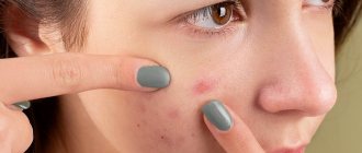
Reddish spots that itch and cause discomfort
By type of skin manifestations:
- Pityriasis versicolor - middle-aged people encounter it, while children under 7 years of age and elderly people are practically not susceptible to it. The risk of encountering this disease increases during periods of hormonal imbalance. This type of disease is especially dangerous during puberty. The provocateur of the testicular fungus of this group gathers in colonies, thus, smooth cream-colored bulges are observed in the groin.
- Lichen versicolor is a dermatological disease of an infectious nature and infection occurs through contact with a patient or his personal belongings (touching a sick animal is also true). At the same time, this group of fungus is characterized by low contagiousness, thereby posing an immediate danger to the skin of the scrotum.
Why do red dots and spots appear on the penis?
This sign signals the obvious presence of a pathological process in the body, both infectious and non-infectious.
The first group includes diseases that are often transmitted during unprotected coitus. The causative agents are viruses, bacteria or fungi. Red spots and dots may appear during the development of:
- candidiasis - in addition to isolated formations, a man is bothered by severe itching of the phallus, a whitish coating on the mucous membrane and white curdled discharge from the urethra;
- genital herpes - in this case, blisters with watery contents appear on the head, accompanied by severe unbearable itching;
- chlamydia - with this pathology, red spots on the head of the penis do not itch or hurt, but pus is released from the urethra, and the patient complains of pain in the scrotum, perineum and lower back;
- HPV - the main sign of the disease is a profuse rash; after some time, the rash increases in size, but there is no pain or itching;
- trichomoniasis - in this case, the spots are the result of irritation of the mucous membrane with pus released from the urethra; in addition to these symptoms, the man complains of swelling of the head and pain when urinating;
- balanitis and balanoposthitis - with these diseases, red spots on the penis itch, a burning sensation when urinating is disturbed, a white coating will be noticeable on the mucous membrane;
- syphilis - in the case of this pathology, one pimple turns into an ulcer, the formation does not hurt or itch, disappears after a while, and the patient may think that he has recovered without treatment (but this is a mistaken opinion).
There are other infectious diseases that can cause scarlet rashes to form on the penis. For example, scabies. Its causative agent is the scabies mite. When it penetrates the skin, a small rash occurs, accompanied by severe itching. The patient scratches the problem area, resulting in ulcers. Itchy pimples on the phallus are also characteristic of a viral infection such as molluscum contagiosum.
It's also worth noting that small red spots on the penis that turn into watery, itchy blisters can occur with chickenpox. In this case, the patient’s body temperature will rise, weakness and headache will occur. The rash associated with this disease usually appears on the skin all over the body.
Signs of mycosis
Despite the fact that cutaneous mycosis is quite problematic in treatment, its manifestations are not long in coming. The problem is that the disease is prone to relapse after incorrect treatment. The disease can manifest itself in a variety of ways, but in the early stages the following occurs:
- small cracks appear on the skin in the scrotum area (on the inside of the thigh) (painful sensations depend directly on the characteristics of the body and the individuality of the case),
- the formation of bubbles is possible, due to which the skin becomes rougher and denser,
- over time, the pain may quickly go away, the skin will soften, return to its normal color and begin to peel off,
- under the condition of bacterial infection, in any case, the blisters will transform into ulcers.
All of the above signs are extremely general, but the following factors cannot be excluded: fungi on the testicles cause a feeling of itching, along with peeling off areas of the skin. Next, diaper rash appears, reddish spots appear, actively growing in size. Periodically, the spots begin to itch, causing discomfort, and all standard ointments for inflammation or disorder do not help. They only partially relieve inflammation and stress on the affected area, while the disease only temporarily stops progressing.
Read also: Nogtimycin: instructions for use, features of manicure and pedicure, analogues of the drug
If left untreated, the fungus spreads to neighboring areas
Lichen planus
Lichen planus (Lichen ruber planus) is a chronic, inflammatory, common skin disease. Lichen planus for 0.78 to 2.4% of all skin diseases and 7 to 10% of diseases of the oral mucosa.
According to reports from domestic authors, in recent years the frequency of recurrence of this disease has increased, and there has been an increase in the number of patients suffering from atrophic, verrucous, erosive and ulcerative forms of lichen planus . Malignant degeneration of lichen planus . This was the reason for classifying it as a precancrosis disease with a malignancy rate of 10-12%.
The term “ lichen planus ” was introduced by F. Gebra in 1860. The English dermatologist E. Wilson in 1869 first gave a clinical description of this disease. The first report about it in Russian literature was made by V. M. Bekhterev and A. G. Polotebnov in 1881.
Despite the fact that the history of the study of this disease goes back more than 100 years, a single, universally accepted hypothesis of the etiology and pathogenesis of lichen planus does not yet exist. Currently, lichen planus is generally considered to be a multifactorial disease in which endogenous and exogenous factors, along with genetic defects, can play a role in the formation and nature of the pathological process. In the development of dermatosis, a large role is given to infectious factors (viruses), neurogenic disorders, toxic-allergic effects (including drugs), as well as immune disorders.
With lichen planus, the skin most often suffers, although in 3-26.5% of cases isolated damage to the mucous membranes of the oral cavity occurs. Lesions of the vulva, bladder and urethra, rectum, and digestive tract may occur. In 1-13%, isolated damage to the nail plates is observed.
Characteristic signs of skin lesions in lichen planus are uneven granulosis and band-like lymphocytic infiltration of the papillary dermis.
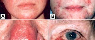
The typical form of lichen planus is characterized by a monomorphic rash in the form of small, flat, shiny (especially in lateral lighting), polygonal papules that are not prone to peripheral growth. The elements are reddish-pink in color with a characteristic lilac or purple tint. In the center of the papules, as a rule, there is a small umbilical depression. On the surface of the formed relatively large (0.5 mm in diameter) nodules, one can find the Wickham grid, pathognomonic for the disease, characterized by opalescent white or grayish dots and stripes. The mesh becomes more noticeable if the surface of the papules is moistened with water or oil; its formation is explained by uneven granulosis. The rashes can be grouped with the formation of small plaques covered with scales, along the periphery of which new, isolated small papules appear, which is explained by the “push-like” nature of the appearance of rashes in this dermatosis. In the progressive stage of the disease, a positive Koebner phenomenon is observed (the appearance of rashes in the area of even minor skin trauma), patients are bothered by intense itching. When the process regresses, secondary hyperpigmentation usually remains in place of the papule.
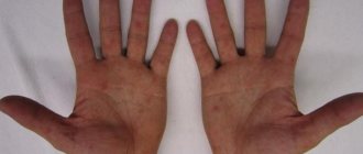
As a rule, lichen planus rashes are localized on the flexor surfaces of the wrist joints and forearms, the anterior surfaces of the legs, in the sacrum, and in men - on the penis. The rash can be very common, even erythroderma.
The mucous membranes are most often affected in the oral cavity (the inner surface of the cheeks, tongue, gums, palate, tonsils), less often - the genitals. Initially, small (miliary) papules of a grayish-white color appear, clearly standing out against the pink background of the mucous membrane; papules then form plaques. The plaques acquire a whitish or grayish-white color, not waxy, as a result of constant maceration in the oral cavity. Papules located on the mucous membranes do not have a characteristic shine, the infiltrate is slightly expressed, and the elements almost do not rise above the surface of the mucosa.
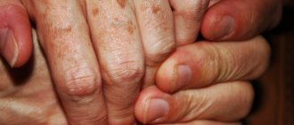
Changes in the nail plates with lichen planus are characterized by the formation of furrows, depressions, and areas of opacification; nails may become thinner or even partially or completely destroyed, and therefore there are two types of nail changes with this dermatosis - onychorrhexis and onycholysis.
Atypical forms of the disease include: ring-shaped, erythematous, warty, pemphigoid, atrophic, erosive-ulcerative lichen planus .
The ring-shaped (circinar) form of lichen planus is characterized by the presence of ring-shaped rashes. This type of dermatosis is more common on the genital organs (glans penis, scrotum, etc.).
The erythematous form of lichen planus is characterized by the sudden appearance on a large part of the skin (mainly on the trunk and extremities), diffuse crimson redness, swelling and peeling. Nodules with this form are soft. Elements typical of dermatosis can be detected in small quantities after the general erythema has decreased.
The warty (verrucous) form of lichen planus is diagnosed by the formation (usually on the lower extremities) of flattened, warty, as if pricked with a pin, spongy, with a honeycomb surface, pinkish-red (in some cases with a purple tint) papules and plaques, covered with a small amount scales. The lesions are round or oval, with clear boundaries. Individual small papules are often visible along their periphery. The distinctive features of this form of the disease are painful itching, very long-lasting rashes and their extraordinary resistance to therapy.
The pemphigoid form of lichen planus is characterized by the appearance of blisters on papules and plaques of lichen planus , as well as on erythematous areas and unchanged skin. With this form of the disease, the general condition of the patient is often disturbed. When the rash resolves, atrophy and hyperpigmentation may remain. When the elements are localized on the scalp, cicatricial alopecia may develop. In some cases, this form is a manifestation of toxicoderma or paraneoplasia.
The atrophic form of lichen planus can be primary or secondary. The primary atrophic form is considered as a type of superficial scleroderma or as an independent disease. The clinical picture of the disease is characterized by the appearance of white spots the size of lentils, ivory or gray, with a pearlescent sheen. The spots are localized on the neck, upper chest, shoulders, genitals, and less commonly on the back, abdomen, and thighs. The skin pattern within them is smoothed, they sink somewhat relative to the skin unaffected by the disease. In some cases, a purple corolla is visible along the periphery of the spots. In the secondary atrophic form, atrophy remains after regression of the rashes typical of the disease.
Rare forms include erosive-ulcerative lichen planus , which is characterized by the formation of erosions or small ulcers of irregular or round shape with a pinkish-red velvety bottom on the mucous membrane of the mouth (cheek, gums, red border of the lips) or on the skin of the legs. At the base and along the periphery of the lesions, a sharply demarcated plaque infiltrate of bizarre shapes or rashes characteristic of typical lichen, giving a lace pattern on the mucous membranes, can persist for quite a long time. Erosive-ulcerative lesions are usually combined with typical rashes on nearby and distant areas of the skin and mucous membranes. Rashes on the mucous membranes are characterized by severe pain. This form of dermatosis may be one of the components of Potekaev-Grinshpan syndrome (a combination of erosive and ulcerative lichen planus with diabetes mellitus and arterial hypertension).
Very often, when the process subsides, skin changes do not regress everywhere, remaining more often on the legs and genitals. The warty, erosive-ulcerative and ring-shaped forms are particularly “persistent”. In some patients, the process may begin acutely. In the acute course, fever, rapid generalization of rashes are noted, skin swelling and erythema are possible, and erythroderma with fine-lamellar peeling (as opposed to psoriatic) may develop. Acute lichen planus can regress relatively quickly, but more often it becomes chronic. Localized forms of dermatosis usually have a long-term chronic course from the very beginning with periodic exacerbations.
The diagnosis is made on the basis of clinical and histological data. Differential diagnosis is carried out with psoriasis, toxicoderma, neurodermatitis, flat warts, papular syphilis.
Treatment is prescribed depending on the severity of the process. For severe itching, sleeping pills, sedatives and antihistamines are indicated.
When foci of chronic infection are detected in patients, antibiotic therapy (penicillin, erythromycin in medium therapeutic doses) is used.
In a common process, quinoline drugs are also used - delagil, plaquenil. The drug is prescribed one tablet twice a day for, as a rule, three weeks.
In acute and widespread cases, systemic corticosteroid therapy is carried out with prednisolone tablets or the long-acting corticosteroid diprospan, intramuscularly, 1-2 ml once a week for two to three weeks.
Photochemotherapy - PUVA therapy - has a high therapeutic effect for common forms.
Vitamins of groups A, B, D, E, nicotinic acid, and immunomodulators (decaris, neovir) have a certain positive effect.
In the treatment of lichen planus, acupuncture, spa treatment, and electrosleep are used.
For all forms of lichen planus , local therapy is used, with the main emphasis being on the use of a variety of corticosteroid ointments (celestoderm, advantan, elocom, etc.). Also shown are ointments with a reducing (absorbable) effect, which contain ichthyol, naphthalan, sulfur, tar, and salicylic acid.
As a preventive measure, it is recommended to normalize the work and rest regime.
K. M. Lomonosov, Doctor of Medical Sciences, Professor of MMA named after. I. M. Sechenova, Moscow
Diagnostics
At first, it may seem that there is no particular point in diagnosis. “The first signs have appeared, which means it’s a fungus,” this is what most patients think, coming with advanced inguinal fungus and numerous consequences of self-medication, such as skin burns and others.
First, everyone must undergo a full diagnosis, during which the doctor will be able to determine the stage of development of the disease along with the optimal method of treatment. The doctor first conducts an examination (dermatologist), examining the skin infected with the fungus. After this, the specialist directs the patient to scrape the lesion, examine the damaged area using a Wood's lamp and the Balzer test.
During the penultimate manipulation, a special device is used to highlight hidden problem spots of the epithelium. It is important to understand that the outcome of treatment, as well as the expenses incurred by the patient, depend on a complete diagnosis.
The Balzer test is a procedure for applying an alcohol solution of iodine to a problem area. Instead of this solution, aniline dyes are often used, which also highlight problem areas. After applying a special composition, the affected skin begins to peel off, which makes it possible to determine the stage and stage of development of the disease. Pityriasis versicolor, when illuminated with a special lamp, stands out as brown spots, and pityriasis versicolor shows up as mesh spots.
Iodine test is a simple and accessible diagnostic method
Tinea versicolor
Lichen versicolor is often confused by patients during self-diagnosis with other diseases with similar symptoms. At the moment there are three stumbling blocks, namely:
- leukoderma syphilitica - the spots are not brightly colored, have a network structure,
- roseola syphilitica - there is simply no peeling, the spots are pink in color and you just need to press on one spot and it disappears without the formation of frames, depressions, bubbles, pimples,
- imbricate mycosis - this disease is relevant in tropical climates, its spots are ring-shaped, the disease develops very quickly, affecting the entire area of the scrotum.
Treatment of scrotal fungus
The treatment period for a disease such as inguinal fungus is two months, but it is possible to completely safely speed up this process. Recommendations:
- purchase and wear loose underwear, changing it daily,
- use medicinal ointments only after taking a bath and washing the genitals with baby soap,
- treat infected and near-infected areas of skin with ointments,
- to avoid genital friction, use special talcum powder,
- shave your groin hair,
- avoid sexual intercourse during therapy (this is not only to protect yourself, but also your sexual partner).
Read also: The danger of toenail fungus for the whole body
Usually effective therapy is prescribed by doctors. It includes medication and the use of special ointments. The ointment is selected solely based on the type of disease and stage of development (volume of affected tissue).
Some people decide to use folk recipes to combat groin fungus, which, in principle, only gives the desired result in 20% of cases, especially if you have not consulted a doctor.
Traditional medicine involves the use of alcohol tincture of iodine for two weeks. You need to lubricate the affected area once a day, preferably before bed after a shower. The product has proven itself effectively in the fight against fungus; the main thing in the process is not to overdo it with the dosage.
Lichen planus (LP) is a chronic inflammatory non-infectious skin disease that affects 0.5-1% of the world's population. Symptoms of LP typically include polygonal, flat, red-violet papules and plaques on the skin, as well as itching. At the same time, there is a wide variety of clinical forms of this disease, such as LLP of the oral mucosa, nail plates, linear, annular, atrophic, hypertrophic, inverse, eruptive, bullous, ulcerative, pigmented, palmoplantar, genital, bullous forms, as well as actinic LCP.
Involvement of the genital organs in LP is considered a relatively rare occurrence. However, in recent years, dermatosis of this localization has been increasingly observed, especially in Caucasian women in the perimenopausal period. Often this form of the disease is combined with damage to the oral mucosa. Thus, about 1% of the world's population suffers from LP of the oral cavity, while 20-25% of women and 3.7% of men with this localization of rashes have manifestations of the disease on the genitals [1, 2].
The nature of LP rashes on the genitals is characterized by a wide morphological diversity: papular rashes, erosions and ulcers, often accompanied by itching and pain; resolve with the formation of deforming scars.
It should be noted that the incidence of the genital form of LP is probably underestimated. Patients' anogenital area is not always examined, and LLP lesions on the genitals may be scanty and asymptomatic. The latter determines the absence of complaints from patients.
The variability of clinical manifestations of genital LP often causes misdiagnosis. The same patient may have different morphological elements, which significantly complicates diagnosis.
According to the classical ideas about LP, thin whitish lacy spots and papules appear on the mucous membranes of the genital organs, sometimes in the form of fern leaves, which can form both on an erythematous and unchanged background. This is the so-called reticular form of the disease. Such rashes are usually not accompanied by itching and are localized in women on the mucous membrane of the vulva and vagina, in men - on the glans penis, in the coronary groove, on the inner layer of the foreskin.
In the erosive-ulcerative form of LLP on the genital organs, the rashes are often localized in fairly large areas of the mucous membrane (in women) and are accompanied by erosions and small ulcers, as well as whitish plaques and papules on a pronounced erythematous background. This form of the disease is usually accompanied by intense pain and burning.
The plaque form of LP localized on the genitals manifests itself in the form of edematous papules covered with a whitish coating.
The atrophic form of LP on the genital mucosa is characterized by the appearance of slightly sunken whitish spots.
Rashes with LP on the genitals can be located not only on the mucous membranes, but also on the smooth skin of the penis, the scalp of the scrotum, the labia majora, in the perineum, on the pubis, and also spread to the inner surface of the upper third of the thighs. In this case, the manifestations of the disease have signs of the classical form of LP and largely depend on the location and severity of the dermatosis. Typical symptoms of LP on the skin are red-purple polygonal papules and plaques with a dry, shiny surface on which a thread-like mesh is visualized, known as Wickham's mesh, a pathognomonic symptom of the classical form of the disease. This reticulation is caused by uneven granulosis. On the surface of the skin elements, whitish-opaline dots are clearly outlined, which intersect in the grid line.
The erosive form of LP is most common on the moist mucous membrane of the vulva and vagina. On the head of the penis, this form of the disease is much less common.
A characteristic feature of LP, including genital localization, is a chronic course with periods of exacerbations and remissions. During exacerbation, patients complain of irritation in the external genital area, itching, burning, soreness, and dyspareunia. If the vaginal mucosa is affected, these symptoms may be accompanied by discharge, often purulent. Upon examination, areas of inflammation, hyperemia, and erosion are found on the mucous membrane of the vulva and vagina, which, upon epithelialization, become covered with a grayish-white coating.
Rash with LP on the vulva, especially with the erosive form, is usually associated with vaginitis. A whitish coating on the vaginal walls is rarely observed with LP. Upon examination, the vaginal walls are erythematized, sometimes with single erosions, which leads to the development of bleeding during sexual intercourse or gynecological examination. Microscopy of a vaginal smear visualizes immature parabasal and basal epithelial cells, small, flattened, cuboidal, with a relatively large nucleus, as well as accumulations of leukocytes. Lactobacilli are usually absent, and the vaginal pH is relatively high, at least 5-6.
The development of severe vulvovaginitis in patients with the genital form of LP can lead to the occurrence of cicatricial changes, synechiae, which disable the woman. In addition, the development of erosive LP on the external genitalia in individuals of both sexes is currently regarded as a risk factor for squamous cell carcinoma of the corresponding localization [3, 4].
The vulvovaginal form of LP is often associated with lesions in the oral cavity. However, in patients with combined lesions of the mucous membranes of the genital organs and the oral cavity, atypical manifestations are often observed, such as diffuse erythema and superficial erosion of the gums, desquamative gingivitis. The triad of desquamative gingivitis and erosive vulvovaginitis is often referred to in the literature as “vulvo-vaginal-gingival syndrome” [5].
Differential diagnosis of LP of genital localization often causes certain difficulties due to the fact that this disease is sometimes almost indistinguishable from other pathologies of the skin and mucous membranes of the genital organs. LLP papules may resemble genital warts when they occur on the glans penis; for psoriatic papules and plaques, intraepithelial neoplasia (eg, Bowen's disease, squamous cell carcinoma in situ
, as well as candidiasis).
Whitish lesions of LP are also often mistaken for lichen sclerosus, especially when the erythema is little noticeable, for example in patients with severe pigmentation. However, unlike LP, lichen sclerosus never affects the vaginal mucosa. However, differential diagnosis between these two dermatoses may be clinically infeasible and histologically difficult. In addition, lichen sclerosus and LP can occur in combination, and in recent years such an association has been observed more and more often.
Rashes with LP localized on the mucous membrane of the genital organs may be similar to similar manifestations of cicatricial pemphigoid and pemphigus vulgaris. Making a correct diagnosis can be helped by extragenital detection of blisters and erosions in patients with blistering dermatoses, as well as microscopy of fingerprint smears.
Erythema multiforme exudative (EME) can also be similar in clinical picture to the genital manifestations of LP. A thorough history taking, assessment of the patient’s general condition and extragenital rashes during MEE can help in making the correct diagnosis.
Thus, almost any lesion of the skin and mucous membranes of the genitals must be differentiated from LP and a thorough additional examination of the oral mucosa, as well as the skin, must be carried out in order to detect morphological elements typical of LP. In difficult cases, a diagnostic biopsy is performed to make a diagnosis, which is very informative.
Taking into account the physiological characteristics of the anogenital area against the background of damage to the integrity of the skin (especially when erosive and ulcerative elements occur), therapy for LP localized on the genital organs is a difficult task. Erosive and ulcerative rashes of LLP in the anogenital area predispose to the addition of a secondary infection, both bacterial and mycotic, which creates additional difficulties in diagnosing the disease, aggravates its course, and complicates treatment.
Before starting therapy, patients should be informed that erosive LP is a disease that is difficult to treat. Adequate treatment is often determined by trial and error, can be lengthy, and the main goal is control of the disease.
An important aspect of therapy is also emotional support for the patient, since even papular LP in the genital area, which is not accompanied by itching and pain, can cause anxiety, a feeling of embarrassment or shame in the patient. Erosive LP often occurs with severe pain, which makes it difficult not only to sexual activity, physical exercise, but also to perform everyday activities and the ability to wear ordinary clothes.
Nonspecific methods of therapy for genital LP are aimed primarily at reducing discomfort in the genital area. In case of severe pain, short-term relief can be brought by cool compresses with clean water or non-alcoholic antiseptics. Using topical anesthetics and covering erosions with petroleum jelly or zinc paste before urinating or washing can reduce burning and pain.
To prevent vaginal synechiae for women who are not sexually active, daily insertion of dilators into the vagina is recommended. Men with uncircumcised foreskin should be warned about the possibility of developing phimosis, so it is extremely important for them to ensure daily and regular retraction of the foreskin with full exposure of the glans penis.
The first-line drugs for the treatment of the genital form of LP are topical glucocorticosteroids (GCS). In the absence of erosions, but in the presence of papules and itching, applications of 0.1% betamethasone valerate or triamcinolone once a day are recommended. Erosive forms of the disease require more powerful topical corticosteroids (0.05% fluocinonide or 0.05% clobetasol propionate). Applications of these medications are also carried out once a day.
Currently, there are no topical corticosteroids that are officially recommended for insertion into the vagina. Therefore, for the treatment of erosive lesions of the vaginal walls in patients with LP, suppositories with hydrocortisone acetate, approved for administration per rectum, or topical corticosteroids in the form of ointments are recommended.
The use of GCS in the genital area, as well as the anatomy and physiology of this area against the background of erosive changes in the mucosa, creates conditions for the addition of a secondary infection. Most often, the course of genital LP is complicated by the development of urogenital candidiasis. A bacterial infection is also possible. In such cases, it is advisable to use systemic antifungal or antibacterial drugs. Nevertheless, the use of topical forms of drugs remains a priority. Local azole antifungals in some cases have an irritating effect on the mucous membrane, and their use can aggravate local discomfort in patients.
In connection with the above, the use of combined topical corticosteroids is most optimal for the treatment of genital LP. One such medicine is Tetraderm
is a unique drug that is a combination of mometasone, econazole, gentamicin and dexpanthenol. Mometasone belongs to the class of strong topical corticosteroids with a high safety profile (according to the European classification Miller and Munro). Econazole is a modern, highly effective antifungal drug with a bactericidal effect, the basis of which is the suppression of the synthesis of ergosterol, which regulates the permeability of the cell wall of microorganisms. Gentamicin is a broad-spectrum antibiotic. Dexpanthenol, which is provitamin B5, has an anti-inflammatory and wound-healing effect by stimulating the expression of IL-6, IL-8 and antioxidant genes, stimulating the proliferation and migration of fibroblasts to the site of dermal damage, as well as collagen formation. In addition, dexpanthenol helps reduce transcutaneous water loss, which reduces inflammation and moisturizes the skin. These properties of dexpanthenol justify its use as a means to accelerate epithelization. Dexpanthenol is used in gynecology, in particular for the treatment of cervical erosion, as well as for the treatment of inflammatory diseases of the oral cavity, nose, larynx, respiratory tract (including after tonsillectomy), gastric mucosa, and urinary system organs.
Thus, Tetraderm
is a combined topical drug that is optimal for local treatment of the genital form of LP.
In addition to topical corticosteroids, cyclosporine for external use is used for the treatment of LP. The effectiveness of this drug has been demonstrated in the treatment of lesions of the oral mucosa in patients with LP [6]. One study demonstrated a significant reduction in erosions after 1 and 3 months of topical cyclosporine therapy [7]. This immunosuppressive drug is prescribed in cases where other drugs for external therapy are ineffective. For erosive vulvovaginitis in patients with LP, 1 ml of cyclosporine injection solution (contains 100 mg) is applied to the vulva or injected into the vagina 4 times a day daily until the rash resolves. However, vaginal administration of cyclosporine is limited due to its local irritant effect [7].
A remedy for topical therapy of LP with localization on the mucous membranes of both the genital organs and the oral cavity is tocrolimus ointment, which is used for lesions of the vulva 2 times a day, but its use, as well as in treatment with cyclosporine, is limited by the development of local irritation [8 ]. Topical use of tacrolimus for vaginal lesions is not recommended due to the high degree of penetration of the drug into the systemic circulation through the mucous membrane.
Systemic therapy for LP is indicated only after failure of local therapy. The only group of drugs with systemic action that are effective in the treatment of erosive forms of LP are GCS for oral administration. In some cases (for example, in severe cases of LP), systemic administration of GCS is used in initial therapy. Prednisolone in a daily dose of 40-60 mg is used for 2-4 weeks until the symptoms of the disease resolve. After the patient's condition improves, topical corticosteroids are used for further treatment.
As an alternative method of systemic therapy for torpid genital LP, hydroxychloroquine is used at a dose of 200 mg 2 times a day [9]. Also for LP, systemic retinoids are used, for example, isotretinoin at a dose of 1 mg/kg or acetretin at a dose of 25 mg once or twice a day, as well as oral cyclosporine, azathioprine, methotrexate at a dose of 10-20 mg once a week and dapsone [10-14].
In addition, to eliminate deforming scars and synechiae that develop in severe genital LP in the absence of adequate drug therapy, surgical treatment methods are used to restore sexual function.
Thus, LP with damage to the genital organs, especially in its erosive form, is painful, accompanied by cicatricial deformation, and, consequently, a sexually disabling disease. The prognosis for LP is favorable, but unpredictable. Nevertheless, adequate therapy with the use of topical corticosteroids as first-line drugs in the vast majority of cases can improve the course of the disease. Combined topical corticosteroids are the optimal drugs for the treatment and prevention of complications of the genital form of LP.
The author declares no conflict of interest.
The author declare no conflict of interest.
Information about authors
Dvoryankova E.V. — https://orcid.org/0000-0002-2458-419X
HOW TO QUOTE:
Dvoryankova E.V. Anogenital forms of lichen planus. Clinical dermatology and venereology
. 2019;18(6):762-766. https://doi.org/10.17116/klinderma201918061762
Corresponding author:
Dvoryankova E.V. — email
Corresponding author:
Svechnikova E.V. — email
Prevention of inguinal mycosis
As with every disease, prevention of this disease involves eliminating all underlying factors. Thus, the patient is obliged:
- compliance with personal hygiene rules (including the fight against sweating),
- restore metabolism (consultation with a specialist is required),
- taking a shower at the end of the working day upon returning home and after physical activity,
- change of underwear daily,
- using powder to combat excessive sweating,
- use of strictly individual personal hygiene items (do not trust or lend your own personal hygiene products),
If you have been in contact with an infected person or things they use, you should wash your hands well with soap and preferably take a shower. This measure will significantly reduce the risk of disease.