What is atheroma
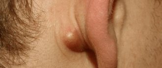
Atheroma is a benign growth of tissue on which increased sweating from the sebaceous gland is observed. A capsule is formed around the fat content. The formation is also called a “skin cyst” because it develops in the dermis, the last layer of skin.
They are most often noticeable on the face, on the back and behind the ears, although they can appear anywhere. Cysts are elastic to the touch and can be of very different sizes - from 5 mm. Only inflamed formations are painful - in other cases they do not cause discomfort, which is why the patient is not interested in the cost of removing atheroma. Once inflamed, the cyst increases in size, turns red, looks like a pimple and causes pain.
Reasons for the appearance of wen
The cause of lipoma formation is unknown. The risk of developing a tumor increases with a family history. Other risk factors include:
- morbid obesity (adirositas dolorosa) is a rare disease characterized by multiple lipomas);
- Cowden's syndrome;
- Gardner's syndrome;
- Madelung syndrome.
Atheromas are formed when the outflow of sebum is obstructed. Provoking factors are considered:
- compaction/thickening of the epidermis;
- hyperhidrosis – increased sweating;
- hormonal imbalances;
- use of antiperspirant deodorants;
- incorrect personal hygiene.
What about lipoma?
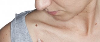
This benign formation consists of adipocytes (fat cells) and is located under the skin. Sometimes under its mask hides a much more insidious pathology of a malignant nature - liposarcoma. Lipomas are also painless and round in shape, but they are firmer to the touch. They appear, as a rule, on the back and neck, on the arms and shoulders, and less often on internal organs.
Suddenly, a person discovers a flat lump under his skin with a soft or spongy texture that moves easily without causing pain or discomfort. Few people in such cases are in a hurry to find out the prices for lipoma removal, unless its size or location causes concern. After injury or tissue damage, the cyst can grow, and it occurs mainly in older age groups.
Hygroma treatment
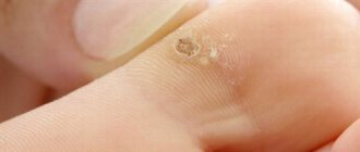
Hygroma is a benign cystic formation of the synovial bursa associated with the joint.
Clinically, this is a tumor of a round shape, dense consistency, covered with normal skin, with a diameter of 0.5 to 3 cm, inactive, as it is fixed at the base.
There is no clear view of the cause. There is a connection with injuries and excessive physical exertion, but in some cases, hygroma appears for no apparent reason. In this case, a slight bulge of the skin forms, as if there is a pea or cherry inside.
The favorite localization of the formation is the area of the wrist joint, and it is in this area that it often causes discomfort. Although it happens that it appears in other places.
Since the hygroma is associated with the joint, it happens that fluid flows into its cavity. Then for some time it may seem that the formation has disappeared, but, as a rule, after some time it appears again.
Hygroma can exist for a long time without causing any discomfort. Many people live with this education all their lives and do not pay any attention to it. Surgery should be considered in cases where the hygroma creates an unaesthetic appearance, causes inconvenience, causes pain during movement, or in case of its active growth.
Treatment of hygroma
Experience in treating hygroma indicates that conservative treatment methods are ineffective and in the vast majority of cases produce relapses.
An absolutely terrible and painful method is crushing the hygroma. In this case, the liquid is forced into the joint cavity, or the membrane of the hygroma ruptures and the contents pour into the tissue. Over time, at best, everything returns to normal. At worst, an inflammatory reaction may develop in the area of the injured hygroma, leading to suppuration. After crushing, sooner or later the membrane heals, restores its tightness, and the hygroma appears again.
They try to puncture the hygromas - suck out the contents with a syringe and inject various substances into it. In this case, the cavity collapses for a while, but the shell itself does not disappear anywhere, and the liquid sooner or later accumulates again.
The most effective surgical treatment of hygroma involves complete excision of the ganglion.
Radical surgery is the main guarantee of the absence of relapse.
The operation is performed under local anesthesia, lasts 20-30 minutes, the sutures are removed 7-10 days after the operation. For large sizes and complex localization, the operation is performed under anesthesia.
What happens if unqualified specialists undertake the operation?
Only the upper, most accessible part of the formation is isolated, which is removed after evacuation of the fluid that filled the hygroma. Most of the shell remains deep in the tissue. The first 2-3 months after the operation create the appearance of a favorable outcome of the treatment, but as the remaining part scars, the tightness of the hygroma is restored, and fluid begins to accumulate in it again.
In fact, instead of a good, beautiful operation, only the appearance of intervention is created.
Therefore, beware if you are told that the operation is “nonsense, for 5 minutes.” It takes much more time to thoroughly isolate the hygroma. The operation is very delicate and painstaking. If the hygroma is not completely isolated from the surrounding tissues to the very point where its pedicle communicates with the joint, the risk of relapse increases many times over.
Guaranteed treatment effectiveness is achieved with modern equipment and more than 15 years of experience of a highly qualified surgeon.
Complications
A complication of a lipoma should be considered torsion of the leg and disruption of the blood supply to the neoplasm, if we are talking about a lipoma on the leg. In this case, the node may undergo necrosis or ulcerate, which is accompanied by severe intoxication syndrome and can be life-threatening, so the tumor in this case must be immediately removed.
Another complication of lipoma should be considered its malignancy. Therefore, when active growth of the tumor begins, it is necessary to remove the tumor as quickly as possible and perform a tissue biopsy.
Preparation for the procedure
In case of subcutaneous lipoma removal, no special preparation is required. The mini-surgery is performed on an outpatient basis, meaning it does not require hospitalization. The surgeon performs all necessary manipulations under local anesthesia. Thus, the procedure is painless for the patient.
Giant subcutaneous lipomas, as well as neoplasms of the intestines, internal organs, and peritoneum require more serious and thorough preparation. Operations of this type are carried out with hospitalization of the patient. Before the intervention, samples are taken and, if necessary, additional studies are done. Operations performed under general anesthesia require restriction of water and food on the eve of the operation.
Services and prices
Removal of lipoma more than 3 cm
7000 ₽
Sign up
Removal of lipoma up to 3 cm
4000 ₽
Sign up
Lipoma is the most common soft tissue tumor and consists of fat cells surrounded by a thin fibrous capsule. Popularly, such a neoplasm is called a wen.
Often, a person who is attentive to his body may stumble upon such a subcutaneous formation. Such a finding can cause, at a minimum, caution, and sometimes fear of the oncological process. The unknown is always scary. If you have any concerns, you should consult your doctor. In most cases, subcutaneous neoplasms in soft tissues are benign and do not pose a threat to the owner of the wen. However, lipomas still require the supervision of a specialist who must recognize a malignant process, if one occurs.
Peculiar red flags, signs that make you wary, are rapid tumor growth, pain, and the presence of two or more similar formations on the body.
By external signs, lipoma is little distinguishable from liposarcoma, hygroma, subcutaneous cyst, hematoma, parasitic invasion, inflammation or consequences of injury. Therefore, it is important that any neoplasms be examined by a doctor.
First of all, the question of the malignancy of the tumor is decided at the appointment. Liposarcoma occurs more often in middle-aged and elderly people. This is an aggressive and fast-growing tumor that can put pressure on surrounding organs and tissues and cause pain. Situations when an existing lipoma degenerates into a malignant tumor rarely occur. However, the oncological process requires a radically different approach to diagnosis and treatment, so it is important to diagnose it as early as possible.
Lipomas may have a hereditary predisposition. This fact, combined with the spread of wen to other parts of the body, makes the doctor suspect lipomatosis. Lipomatosis accompanies a number of hereditary syndromes such as Madelung's disease and Dercum's syndrome. Diseases with a family history require a special approach to therapy.
Symptoms and classification
Lipomas are distinguished by anatomical location into lipomas of the head, face and neck, lipomas of the trunk, extremities, chest (mediastinum), mammary gland, gastrointestinal tract, internal organs, retroperitoneal tissue, spermatic cord. There are also rare localizations in the myocardium, lungs, and meninges.
Another classification of adipose tissue tumors involves a clinical division:
- Lipoma surrounding nerve structures is called perineural . Due to compression of the nerves it can cause severe pain. Removal of perineural lipomas differs from subcutaneous lipomas and requires a highly qualified surgeon;
- A tumor growing in the spinal canal (usually in the lumbar region) is called lumbosacral lipoma; Mostly occurs in children and is combined with underdevelopment of spinal structures;
- Lipoma of the joint and its structures (synovium, vagina, tendons);
- Intermuscular lipomas are formed from areas of adipose tissue between muscle fibers;
- Angiomyolipoma is a tumor of fatty and muscle tissue, which in most cases grows in the kidneys and pancreas. Middle-aged and mature men are more predisposed to its formation;
- Subcutaneous lipoma is a formation of varying sizes in the subcutaneous fat tissue. In everyday life it is usually called wen.
Lipomas usually occur alone. However, some patients discover several tumors at once. Such cases are most often associated with hereditary diseases and require careful study by specialists. The most common places for lipomas to form are the neck, back and limbs.
A subcutaneous lipoma is a mobile, elastic seal in the form of a lump or ball, which does not cause pain when pressed. A neoplasm can cause pain if it grows beyond its capsule into healthy tissue or due to compression of adjacent nerves.
For example, a wen located on the head can cause headaches, and the same formation on the neck can cause hoarseness and difficulty swallowing.
Gastrointestinal lipomas differ from their subcutaneous counterparts. Small formations in the intestine do not cause symptoms and are often discovered incidentally during an instrumental examination of the gastrointestinal tract. However, if it increases in size, this happens when the tumor reaches 2 or more centimeters in diameter, the lipoma can block part of the intestinal lumen and cause intestinal obstruction, intussusception, stool problems, abdominal pain and even bleeding.
Mesenchymoma
Mesenchymoma
– a malignant formation related to sarcoma. Angiosarcoma and liposarcoma can be found in its composition.
The exact causes of the occurrence have not been clarified. Perhaps this is the effect of carcinogens on the fetus. Risk factors are:
- Vibration, hypothermia and ionizing radiation;
- Chemical exposure – toxic substances, drugs;
- Bacterial and viral infections.
The location is predominantly the chest and abdominal cavity, mediastinum and retroperitoneal space. The formation is manifested by pain, cough, heartburn, shortness of breath and a feeling of fullness.
Diagnosis is carried out using radiography, ultrasound, MRI, biopsy and endoscopic examination. Treatment is carried out surgically. Auxiliary methods are radiation therapy and chemotherapy.
Osteochondroma
Osteochondroma
- a benign formation over the bone in the form of a smooth protrusion of cartilage tissue up to 12 cm in size with bone marrow contents. Osteochondromas can be multiple or single.
The reasons for the appearance of such formations are not fully understood. The risk of developing osteochondromas is believed to increase:
- Intrauterine anomalies of skeletal formation;
- Hereditary factors;
- Irradiation.
Osteochondroma of the bone occurs:
- In the area of the joints, eventually spreading to the middle of the bone;
- Localization occurs on the ribs, femur, vertebrae, tibia, foot and scapula;
- Less commonly, the hands, heel bone and humerus are affected.
The bones of the skull are not affected. There is no pain or mobility. The tumor has clear boundaries. If the tumor is small, it does not bother you; pain may occur when the blood vessels are compressed.
How is the diagnosis done?
- After an external examination, the patient is sent for an x-ray, which will show the formation and its connection to the bone.
- MRI or CT is used if the situation is unclear.
- Morphological analysis may be performed.
Treatment
The only method used is surgery - removal of the formation with its base and stem. The removal process is necessary if:
- Education is increasing;
- Has large sizes;
- Accompanied by pain;
- Causes skeletal deformation.
In other cases, the patient is observed and monitored using x-rays.
Forecast
The disease has a favorable prognosis - its growth stops with the end of the formation of the skeleton. Degeneration into a malignant state ranges from 1 to 10% (more often with multiple formations).
Recovery after surgery is quick. Patients are prescribed physical therapy and physiotherapeutic procedures.
Where can you remove atheroma and lipoma?
Even the simplest surgery should only be performed by an experienced physician and performed in a facility that is responsible for the end result and has a genuine concern for the health and safety of the patient. The Femina women's health center in Simferopol fully meets the necessary requirements, whose clients can expect:
- doctors of the highest and first medical categories who know exactly how a lipoma differs from an atheroma and are able to quickly diagnose these neoplasms;
- best-practice laboratory and surgical conditions;
- Convenient work hours and competitive prices for the full range of services.
Ask the administrator of the Femina clinic any questions you have and make an appointment by phone, online chat, or email right now!