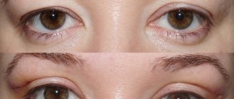
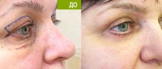
Increasing the incision of the eye along the line of the upper and lower eyelids is called canthoplasty, from the Greek cantos - angle. Blepharoplasty also works on the eyelids, but the purpose of the intervention is different - to hide signs of age-related changes, tighten overhanging skin folds, and get rid of fatty hernias. The main task here is to make the look visually more open and the angle of the neckline graceful. As a result, the impression of a youthful open gaze is created, an emphasis is created on it, the depth and color of the iris is emphasized. Canthoplasty is often combined with blepharoplasty to obtain a complex effect: rejuvenation and aesthetic perfection.
How to enlarge the eye section
In the literal sense of the word, it is physiologically impossible to make it larger - the size is determined genetically. The doctor can:
- excise part of the skin of the upper eyelid;
- remove the fold above the upper eyelid (more common in Asian faces);
- do a lift along the eyelash edge;
- change the shape and diameter of the palpebral fissure by working with the eyelids.
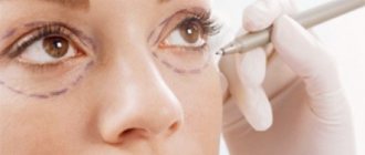
In each case, the surgeon’s manipulations are variable. Schematically, the procedure looks like this.
- Anesthesia is performed (local anesthesia or general anesthesia).
- An incision is made.
- The necessary tissues (skin, fat, muscle) are excised.
- Cosmetic stitches are applied.
Upper eyelid blepharoplasty with a short incision is a sham!
Capital specialists offer many proprietary techniques, as they say, “not all of them are equally useful”! Type in the search bar: blepharoplasty Moscow! You'll probably see ads about short seams.
Blepharoplasty in Moscow can be performed using a technology where the shortened incision does not extend beyond the fold of the upper eyelid. Such an operation always leads to a “rounding” of the eye - these are the laws of geometry that simply cannot be violated without resulting in a “gathering” on the upper eyelid and a round, rather than almond-shaped, incision.
But we women want everything at once! We need eyes like Cleopatra's and no scars around them! I want to please you - this is possible, but not due to a shortened incision (it does not make it possible to completely remove excess skin and restore the shape of young eyes) but due to fine surgical technique and the use of an invisible suture method.
I will tell you in more detail next time how blepharoplasty of the upper and lower eyelids uses these surgical technologies.
Nuances of the procedure
The period for performing a simple intervention is half an hour; with combined techniques (when several tasks are performed simultaneously), the time increases to 2 hours.
When the patient can leave the clinic largely depends on the type of anesthesia. With local anesthesia - after 3 hours, with general anesthesia after 5-6 hours or the next day.
How much surgery to enlarge the eye shape costs depends on the chosen technique and the scope of the intervention. The price is negotiated before manipulation and remains unchanged.
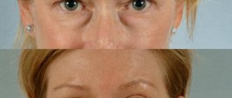
Fat-sparing blepharoplasty with redistribution of fat bags
Fat-saving (fat-preserving) blepharoplasty is very popular among patients who want to completely maintain their usual appearance, only slightly adjusting their appearance. The innovative technique is not aimed at removing excess fat deposits, but at partially redistributing them.
Blepharoplasty with redistribution of fat bags prevents the possibility of the “sunken eyes” effect. High-quality separation of the eyelids and cheeks, elimination of tear grooves harmoniously transforms the appearance, making the appearance young, bright, and contrasting.
The essence of the fat-sparing blepharoplasty technique
The classic approach recommends that the plastic surgeon completely remove the maximum possible amount of excess fat and excess skin that spoils the appearance. Today, such operations are performed using the transconjunctival method, which does not involve external incisions on the lower eyelids.
Transconjunctival lower blepharoplasty does not violate the integrity of the anatomy (the orbital septum and orbicularis oculi muscle are not intersected), as with external percutaneous blepharoplasty.
- With transconjunctival blepharoplasty, the frequency of fat movement and rounding of the outer corner of the eye is significantly less than with external blepharoplasty.
- Understanding the anatomy of the lower eyelid and proper incision placement (4 to 5 mm below the eyelid cartilage) is critical to achieving successful results.
- Fat can be removed or retained (transferred) depending on the patient's wishes.
- Fat repositioning (moving) is an advanced technique performed with a detailed knowledge of the anatomy of the eyelids.
- To obtain excellent results, it is necessary to know and practice various surgical nuances.
- Adding various procedures (canthoplasty, canthopexy, fat transfer, skin removal, etc.) as needed can improve the final results.
- The patient receives excellent results when the surgical technique is mastered and refined.
Lower eyelid rejuvenation with fat redistribution is a complex aesthetic procedure. One of the key aspects of lower fat-sparing blepharoplasty is the surgical approach through which orbital fat hernias are exposed. There are two standard approaches to the fat pads of the lower eyelid: percutaneous (through the skin and muscles) and transconjunctivally (through the inside of the eyelid).
Percutaneous lower blepharoplasty is a more traditional method in which the skin, orbicularis oculi muscle and orbital septum are divided. This approach carries a higher complication rate, including lower eyelid retraction, ectropion, and lower eyelid rounding. This is primarily due to lack of eyelid support, excessive skin removal, and disruption of the orbital septum (with subsequent scarring) and orbicularis oculi muscle (with subsequent muscle weakness) when accessing fatty hernias.
With transconjunctival access to the orbital fat, an incision is made on the inner (conjunctival) surface of the eyelid, thereby maintaining the integrity of the orbital septum and the orbicularis oculi muscle. This is a very important step in preventing malposition of the lower eyelids, since there is no distortion of the anatomy of the eyelid and postoperative gross scarring.
For this reason, transconjunctival lower blepharoplasty with fat redistribution has become widespread in aesthetic surgery.
Transconjunctival blepharoplasty does not remove excess skin. This requires additional procedures. However, if necessary, excess skin can be removed using pinch blepharoplasty.
Adding these additional procedures to transconjunctival blepharoplasty does not result in the same rate of eyelid malposition complications seen with percutaneous lower blepharoplasty.
The advent of volumetric fat preservation through fat redistribution or grafting has become an important adjunct to lower eyelid blepharoplasty and can be used to improve cosmetic results.
Anatomy of the lower eyelid
The incision for transconjunctival lower blepharoplasty is made from inside the eyelid. This can make anatomical orientation and surgical maneuvering difficult because the operative field is small and narrow compared to an open percutaneous approach. For this reason, performing this operation requires the surgeon to have a thorough knowledge of the anatomy of the lower eyelid.
The lower eyelid is divided into three layers or plates (lamellae).
- AL - anterior lamella (skin/muscle)
- PL - posterior lamella (cartilage/conjunctiva and lower eyelid retractors)
- ML - middle lamella (orbital septum) and fatty hernias.
- FP point of fusion of the lower eyelid retractors and the orbital septum,
- FP is critical for incision placement during surgery
Cartilage forms the skeleton of the eyelid, giving it both support and flexibility. Its dimensions are 4-5 mm in height, 30 mm in length and 1 mm in thickness. It is connected to the conjunctiva on its inner surface and to the orbicularis oculi muscle and skin on the outer surface.
Below the cartilage, the conjunctiva continues to the lower fornix. The lower eyelid retractors, also called capsulopalpebral fascia, are located just anterior to and closely connected to the conjunctiva. This sympathetically controlled muscle (similar to the Müller muscle of the upper eyelid) originates from the tendon of the inferior rectus muscle and continues to fuse with the inferior border of the cartilage. There is fatty tissue just in front of the retractors.
In front of the adipose tissue is the orbital septum, which limits them anteriorly. This is a layer of connective tissue originating from the periosteum on the orbital rim (arcus marginalis) and inserting into or connecting with the capsulopalpebral fascia 2-3 mm below the lower edge of the eyelid cartilage.
A conjunctival incision just below the cartilage will separate the conjunctiva and lower eyelid retractors from the cartilage.
This plane is best suited for surgical intervention needed in the fascial plane between the orbicularis oculi muscle and the orbital septum, such as accessing the orbital floor to repair an orbital fracture. Because the dissection in this plane occurs anterior to the septum, fat does not protrude into the surgical field.
A transconjunctival incision below the junction of the orbital septum and lower eyelid retractors (4 to 5 mm below the cartilage) is the preferred approach for transconjunctival blepharoplasty as it is a direct route to fatty hernias.
There are three fatty hernias on the lower eyelid: nasal, central and lateral. Nasal and central fat hernias are separated by the inferior oblique muscle
Care must be taken not to damage this muscle during surgery.
The central fatty hernia is separated from the lateral inferior arcuate ligament.
The separation of the central and temporal (lateral) fatty hernias by the arcuate ligament can often be seen clinically by asking the patient to look upward. But at the same time, the separation between the nasal and central fat pads is not pronounced, since hernias tend to overlap the inferior oblique muscle and dull this separation (this is visible during surgery).
Fat-sparing blepharoplasty of the lower eyelids is based on the rational correction of fatty hernias. They are not removed, but evenly distributed. This allows you to maintain the required volume, getting rid of unsightly “bags under gases”.
Comparative analysis of traditional and fat-saving blepharoplasty
Long gone are the methods in which plastic surgeons used simple skin tension to eliminate aging changes. This technology made the face lifeless, depriving it of facial expressions. Today, the face is perceived as a 3D relief object that requires more subtle surface treatment. It is necessary not only to get rid of the furrows and wrinkles that appear with aging, but also to preserve the usual relief characteristic of a young person. Redistribution blepharoplasty corrects volume, making the eyes look youthful.
With the old traditional approach, the eyelids were completely devoid of fat deposits. The consequence was a tight fit of the tissue to the orbit. Outwardly, the eye looked like it had sunk inward, which revealed the work of a surgeon. It is enough to look at photos of famous artists, athletes or politicians from past years to notice which of them used traditional blepharoplasty. Today, only specialists can determine the work of a plastic surgeon.
The main disadvantage of the traditional method and significant advantages of the new technique
The work of a plastic surgeon may be completely satisfactory for the patient, but after the operation the eyes will look the same as those of other people. Individuality is completely lost, zest is lost. Today, such work is considered unsatisfactory, sometimes requiring rework.
Volume adds charm to the look, making the look unique, beautiful, and bright. This is possible due to the high quality of work of a plastic specialist who uses new methods.
Fat-preserving blepharoplasty makes the eyelid voluminous, preserving the natural beauty of the eyes, its individual attractiveness, completely eliminating age-related skin changes. The periorbital area gains a second youth, without looking like it has been operated on, which is important for public people. The maximum, long-term effect is achieved to maintain beauty, youth, and health.
Surgical technique of fat-sparing blepharoplasty
The eyelid is infiltrated transconjunctivally with 2 ml of 1% xylocaine with adrenaline 1: 100: 000. In order to perform fat transfer, the tear trough (the area into which the fat will be transferred) is marked before the operation.
1-2 ml of the same local anesthetic is injected into the area of the tear trough. Usually the nasal and central hernias move.
After the time required for hemostasis and anesthesia, the lower eyelid is moved with the index finger of the non-dominant hand, while the second finger of the same hand moves the eyeball slightly back. This maneuver elevates and exposes (inflates forward) the incision site on the transconjunctival surface of the eyelid.
The free dominant hand is used to make an incision 5 mm below the cartilage to directly enter the fat compartment of the lower eyelid.
An electric knife (Valleylab) with a Colorado tip or surgical scissors can be used for this incision.
The orbital septum is attached to the capsulopalpebral fascia 2–3 mm below the cartilage. An incision below this insertion point enters the retroseptal space, provides direct access to fatty hernias and preserves the integrity of the orbital septum. This is a critically important point, since it allows you to leave the orbital septum intact and avoid severe scarring.
The conjunctiva is then sutured with a 4-0 suture, which is pulled upward and protects the eyeball during surgery.
Next, all three packages of fatty hernias are highlighted
To eliminate the tear trough, the nasal and median hernia are moved to its projection, after placing separate sutures on them, which are brought out to the skin and a knot is tied on the roller.
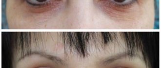
Postoperative care and rehabilitation
After surgery, patients are given the following recommendations. Necessary
- Apply ice compresses ten minutes per hour while awake for 2 days.
- Instill eye drops containing an antibiotic and a steroid (Tobradex) three times daily for the first week after surgery
- Sleep with your head elevated (on two pillows) for the first week.
- Avoid strenuous activities for the first 10 days after surgery.
In the postoperative period, bruising and swelling usually resolve quickly. Watery eyes usually disappear within the first week. The stitches are removed on the fourth day.
By giving priority to the new technique, the patient receives an excellent result that meets international standards.
- After the operation, the patient returns to his usual lifestyle in 3-6 days.
- You can fully evaluate the result of a plastic surgeon’s work in 1-2 months.
- Strict adherence to the specialist’s recommendations during the rehabilitation period will speed up the recovery process.
- The shape and appearance of the periorbital area will remain for 5-7 years.
Complications
Most surgical complications associated with fat removal or transfer are similar. These typically include excessive bruising and swelling, chemosis, and subconjunctival hemorrhage.
- Edema. To reduce swelling, in the absence of medical contraindications, a higher dose of prednisone may be prescribed. In cases of prolonged or refractory edema, when the patient violates the diet and consumes excessive salt, a change in diet is required.
- Chemosis is subconjunctival swelling that can occur when tissue is excessively cauterized during surgery. There are different treatments for chemosis depending on the severity.
- Subconjunctival hemorrhage - bleeding under the conjunctiva, can be very distressing for the patient, which usually reaches its maximum extent a few days after surgery and resolves within 1-2 weeks.
- Excessive fat removal. Dangerous and may cause sunken eyes.
- Incorrect position of the eyelids. Not typical for transconjunctival blepharoplasty.
- Entropion. May occur with a transconjunctival incision. Usually it is spastic, cicatricial. Correction depends on the etiology. Postoperative ectropion occurs rarely in cases of undiagnosed preoperative decrease in eyelid tone.
- Finally, in rare cases, postoperative trichasis may develop. Most likely, it is associated with impaired blood supply to the eyelids and/or post-operative scarring.
When fat transfer is performed, complications unique to this procedure may occur. These include diplopia (in the postoperative period), fatty granulomas, prolonged swelling, unevenness in the projection of the tear trough, pigmentation in the area of the suture of the roller.
- Diplopia is usually temporary and is associated with anesthetic injection, swelling, or injury to the inferior oblique muscle. Treated with higher doses of oral steroids, which should be tapered over 10 days.
- Prolonged swelling is treated similarly to diplopia with higher doses of steroids.
- Irregularities in the projection of the tear trough are usually caused by granulomas and are resolved with steroid injections (Kenalog).
- Temporary hyperpigmentation may occur at the site where the skin prolene suture exits.
New plastic surgery techniques make it possible to quickly and radically change one’s appearance, eliminating congenital or acquired defects. The operation takes little time, but the results will last for years. To learn more about the work and make an appointment, call 89673108595 or fill out an application on the website.
Indications
- The patient's desire to look different.
- Injuries with changes in appearance.
- Asymmetrical eye shape.
- Correction of complications after inflammatory diseases of the eyelids, operations.
- Genetic pathologies (for example, incomplete cleft eyelid).
- Disproportionately narrow palpebral fissures in relation to facial proportions.
You can choose a canthoplasty technology that suits your individual needs, find out how much surgery to enlarge the eye shape costs, and ask any questions you may have at an initial consultation with Dr. Guzal Melisovna Isamutdinova. It is free, you can sign up using the feedback form on the website.