If you find a slight swelling or edema on a child’s body, you can think about the following conditions: inflammation, injury, cyst. When inflamed, the swollen area is pink or red, feels warmer to the touch than the surrounding tissue, and is painful when touched. Swelling after an injury may be a normal color warmer or cooler than the surrounding tissue, and a bruise may appear after some time. Sometimes, with increased sensitivity to any type of food, cold or other factors, edematous areas with clear boundaries quickly develop on the skin - such edema is called angioedema. They are often accompanied by hives (see Allergic reactions), which is another indication of the allergic nature of the condition. Swelling of the skin may be associated with enlarged lymph nodes (see Enlarged Lymph Nodes). General swelling of a limb or torso may indicate an allergic reaction or kidney disease.
THE DOCTOR'S CONSULTATION
Contact your doctor if the swelling:
- does not go away, grows or arose for no apparent reason
- hot to the touch, painful
ATTENTION!
If swelling occurs for no apparent reason, consult a doctor. In any case, it's better to be safe than sorry.
| ASK YOURSELF A QUESTION | POSSIBLE REASON | WHAT TO DO |
| Do you have one or more painful, raised lesions on your skin with a white center? | Boils (inflammation of hair follicles) | Do not try to open a boil by squeezing or piercing it, as this may spread the infection. Wash the area around the boil frequently with soap and water (not the boil itself!). When the boil opens, apply a bactericidal patch. If there is no improvement or new boils form, contact your pediatrician (or dermatologist ): you may need local treatment and antibiotics. |
| Does your child have several painless raised lesions with a rough surface on their skin? | Warts | Warts are caused by viruses and often go away without treatment. Warts that are a cosmetic defect, as well as those that are often injured, can be removed. Talk to your child's doctor (or dermatologist ) |
| Does your child have a soft, painless lump in the groin? Does it disappear with light pressure? | Inguinal hernia | Contact your pediatrician. If necessary, he will refer you to a pediatric surgeon |
| Does the boy have a soft, painless swelling of the scrotum on one side? Does one testicle look significantly larger than the other? Does the swelling decrease when the child lies down? | Hernia; hydrocele | Make an appointment with your pediatrician . He will determine whether there is dropsy, a congenital condition in which fluid accumulates in the scrotum. If your child has a hernia, your pediatrician will refer you to a pediatric surgeon if necessary. |
| Does your child have a protruding formation on the skin of the sole? Is it mildly painful when walking? | Plantar wart | Seek advice from your pediatrician . If necessary, he will recommend treatment |
| Does your child have many red swellings in the shin area? Is he denying the injury? Has he recently had an infection or is he taking medication? | Erythema nodosum | Go see your pediatrician . If necessary, he will prescribe examination and treatment |
| Does your child complain of pain and swelling in the bone area? | Infection; another disease requiring diagnosis and treatment | Contact your pediatrician immediately . Rest the affected limb. |
FOR INFORMATION
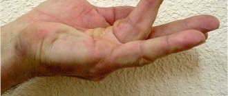
Swelling in the wrist area
Sometimes in children, cysts containing fluid appear under the skin of the wrist on top. This happens like this: synovial fluid - the “lubricant” found inside the joint - leaks through the damaged joint capsule and accumulates in the tendon sheath. Most often this happens in the area of the wrist joint. They occur when there is increased load on the joint, for example, when a child holds a pen incorrectly or writes a lot. As a rule, such cysts are painless and not dangerous. They gradually disappear without treatment. However, if your child's cyst is large, painful, or interferes with normal hand function, treatment may be required. In the latter case, a small operation is performed - the cyst is removed and the “leak” is closed. Do not self-medicate.
A hard, smooth, raised lump, usually about the size of a peanut, just under the skin. Most often it forms on the dorsum of the wrist joint, but can also occur on the ankle or finger.
Skin formations. What could it be?
Various types of formations often appear on human skin: pits, swellings.
Including nodules, the variety of which is also rich. They differ in color, shape, and also in their involvement in the disease, as a result of which they appear on the human skin. How to determine which disease a particular nodule belongs to? They come in different sizes. There is also a division of nodes into subcutaneous ones and those that grow directly on the human skin itself. So, let's look at the most common variations of formations.
The initial form of formation on human skin is called tubercles. They look dense. The color of the tubercles varies from flesh-colored to red, sometimes even black. The color depends on the person’s disease.
Since the tubercles are practically the initial stage of the formation of nodes, they practically do not cause discomfort. Basically, they are painless, but in some cases, with a serious form, they are painful and cause pain on palpation.
Papules usually come in several types. There are subcutaneous and tubercles directly on the skin itself. Subcutaneous nodes may be painless, but they often cause pain and discomfort when pressed.
Benign growths:
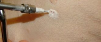
Warts
The formations are caused by the human papillomavirus as a result of immune diseases or metabolic disorders. Most of them are harmless; only warts larger than 0.5 cm, flat or without clear contours and ovoid are dangerous.
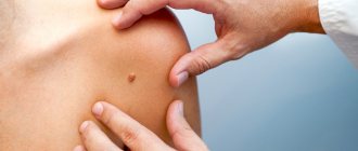
Moles
Formations that arise due to excess in the skin of the coloring substance - melanin. Some of them can transform into malignant neoplasms – melanomas. To do this, the mole must be knot-like, dry, hairless, or bleeding.
Fibroids
Formations that look like a nodule that has grown deep into the skin and protrudes from it. The surface of fibromas is smooth and even, sometimes they develop into a malignant tumor - fibrosarcoma.
Zhiroviki
Tumors of the fatty layer that do not change skin color. They are carried painlessly and can be easily moved to the side with a finger. They form in areas of the body that are poor in adipose tissue: hips, shoulder blades, and upper back.
Atheromas
Formations resulting from blockage of the sebaceous gland duct. Painful to the touch, inflamed atheromas secrete purulent contents. Occurs in areas of increased sweating.
Hygromas
Cysts appearing in the joints and tendons. Hygromas do not change skin color and protrude greatly above the level of the body. They are usually harmless and painless.
Hemangiomas
Formations from cells of capillary or venous vessels are red and blue-black.
Examples of different types of knots and knots
Subcutaneous
Whiteheads are also a type of nodule on the skin. Also, formations may be evidence of a disease such as basal cell carcinoma or basal cell skin cancer
.
Let us consider in a little more detail the diseases, evidence of which may be the symptom in question. Whiteheads. A fairly common pathology of the dermis. Acne, or milia, is a collection of sebaceous secretions under the skin. Often they do not cause discomfort to a person. Unless it concerns the aesthetic side of the issue.
Whiteheads may also indicate the presence of gastrointestinal diseases. To treat milia, a special diet is used, as well as the use of special-purpose medications.
With skin diseases, the bumps can be not only white, but also red. This may be evidence of acne.
Acne
They begin with a small red tubercle, at the top of which a white purulent head forms over time.
After a short period of time, the pus comes out and a crust forms on top. When the crust heals, a keloid forms in place of the pimple.
Lipomas, hygromas, and atheromas are also distinguished. Let's look at each type in more detail.
Lipoma
Lipoma or wen is a subcutaneous formation, soft to the touch, mobile. Wen can only form in that part of the body where there is adipose tissue.
Hygroma
Unlike lipoma, hygroma is a sedentary formation similar to a tumor. Hygroma is filled with serous fluid. It is formed as a result of herbs received by a person during a fall or blow. The tumor is removed surgically.
Atheroma
Atheroma is partially similar to lipoma; it also forms in the sebaceous duct. Atheroma is a cyst formed as a result of blockage of the sebaceous duct. The size of the formation can be either the size of a pea or the size of a chicken egg.
As mentioned above, nodes can be a manifestation of a disease such as basal cell epithelioma. This is a type of skin cancer. If basal cell tumors appear on the skin, this indicates that the disease is progressing from a mild to a more severe form.
Although not in all cases, nodules on the skin indicate a serious illness, a person in any case should contact people specially trained for this. If something bothers you, let alone causes pain, you need to immediately contact a specialist.
You can find such specialists at the Doctor Nearby clinic. All applicants will be given attention and given a clear and correct diagnosis. Naturally, the treatment will be of the highest level. You can make an appointment by phone without leaving your home.
Skin vasculitis
Skin vasculitis is a group of diseases of a multifactorial nature, in which the leading symptom is inflammation of the blood vessels of the dermis and subcutaneous tissue.
The difficulty in covering this topic is that until now there is no generally accepted classification or even agreed upon terminology of vasculitis. Currently, about 50 different nosological forms have been described, and understanding this diversity is not easy. The diversity of clinical manifestations and insufficiently studied pathogenetic mechanisms have led to the fact that under different names only a variant of the main type of skin lesion may be hidden. Also, in addition to primary vasculitis, which is based on inflammatory damage to the blood vessels of the skin, there are also secondary vasculitis (specific and nonspecific), developing against the background of a certain infectious (syphilis, tuberculosis, etc.), toxic, paraneoplastic or autoimmune (systemic lupus erythematosus, dermatomyositis etc.) process. It is possible to transform skin vasculitis into a systemic process with damage to internal organs and the development of severe, sometimes life-threatening complications.
Skin vasculitis is a polyetiological disease. The most common connection is with a focal infection (streptococci, staphylococci, mycobacterium tuberculosis, yeast, viruses, etc.). Hypersensitivity to a number of drugs, in particular to antibiotics and sulfonamide drugs, is of particular importance. Often, despite a carefully collected anamnesis and examination, the etiological factor remains unclear. Among the risk factors for vasculitis, one should take into account: age (children and the elderly are most vulnerable), hypothermia, excessive insolation, severe physical and mental stress, trauma, surgery, liver disease, diabetes, hypertension. The pathogenetic mechanism for the development of skin vasculitis is currently considered to be the formation of circulating immune complexes with their subsequent fixation in the endothelium, although this has not been definitively proven for all diseases of this group.
Skin vasculitis is a heterogeneous group of diseases, and their clinical manifestations are extremely diverse. However, there are a number of common features that unite these dermatoses:
1) inflammatory nature of skin changes; 2) symmetry of rashes; 3) tendency to edema, hemorrhage and necrosis; 4) primary localization on the lower extremities; 5) evolutionary polymorphism; 6) connection with previous infectious diseases, medication, hypothermia, allergic or autoimmune diseases, and impaired venous outflow; 7) acute or worsening course.
Skin lesions with vasculitis are diverse. These may be spots, purpura, nodules, nodes, necrosis, crusts, erosions, ulcers, etc., but the main clinical differential sign is palpable purpura (hemorrhagic rash that rises above the surface of the skin and is felt on palpation).
There is no generally accepted classification of vasculitis. Vasculitis is systematized according to different principles: etiology and pathogenesis, histological picture, severity of the process, features of clinical manifestations. Most clinicians use predominantly morphological classifications of cutaneous vasculitis, which are usually based on clinical changes in the skin, as well as the depth of location (and, accordingly, the caliber) of the affected vessels. There are superficial (damage to the vessels of the dermis) and deep (damage to the vessels at the border of the skin and subcutaneous tissue) vasculitis. Superficial ones include: hemorrhagic vasculitis (Henoch-Schönlein disease), allergic arteriolitis (polymorphic dermal angiitis), leukoclastic hemorrhagic Miescher-Storck microbiota, as well as chronic capillaritis (hemosiderosis): Majocchi's annular telangiectatic purpura and Schamberg's disease. To the deep: cutaneous form of periarteritis nodosa, acute and chronic erythema nodosum.
Hemorrhagic vasculitis is a systemic disease that affects small vessels of the dermis and manifests itself as palpable purpura, arthralgias, gastrointestinal (GIT) lesions and glomerulonephritis. It occurs at any age, but boys aged 4 to 8 years are at greatest risk. Develops after an infectious disease, after 10–20 days. The acute onset of the disease, with fever and symptoms of intoxication, is most often observed in childhood. The following forms of hemorrhagic vasculitis are distinguished: cutaneous, cutaneous-articular, cutaneous-renal, abdominal-cutaneous and mixed. The current can be lightning fast, sharp and protracted. The duration of the disease varies - from several weeks to several years.
The process begins symmetrically on the lower limbs and buttocks. The rashes are papular-hemorrhagic in nature, often with urticarial elements, and do not disappear with pressure. Their color changes depending on the time of appearance. The rashes occur in waves (once every 6–8 days); the first waves of the rash are the most violent. Articular syndrome appears either simultaneously with skin lesions or after a few hours. Large joints (knees and ankles) are most often affected.
One of the variants of the disease is the so-called necrotic purpura, observed during the rapid course of the process, in which necrotic skin lesions, ulcerations, and hemorrhagic crusts appear.
The greatest difficulties are caused by the diagnosis of the abdominal form of hemorrhagic vasculitis, since skin rashes do not always precede gastrointestinal phenomena (vomiting, cramping pain in the abdomen, tension and pain on palpation, blood in the stool).
The renal form is manifested by impaired renal activity of varying degrees of severity, from short-term unstable hematuria and albuminuria to a pronounced picture of acute glomerulonephritis. This is a late symptom and never occurs before the skin is affected.
The fulminant form of hemorrhagic vasculitis is characterized by an extremely severe course, high fever, widespread rashes on the skin and mucous membranes, viscerapathies, and can result in the death of the patient.
Diagnosis of the disease is based on typical clinical manifestations; in atypical cases, a biopsy is performed. In the abdominal form, surgical supervision is necessary. Observation by a nephrologist is recommended for three months after resolution of purpura.
The term “allergic arteriolitis” Ruiter (1948) proposed to name several related forms of vasculitis, differing in clinical manifestations, but having a number of common etiological, pathogenetic and morphological features.
The pathogenetic factors of the disease are considered to be colds and focal infections. The rashes are usually located symmetrically and are polymorphic in nature (spots, papules, vesicles, pustules, necrosis, ulcerations, telangiectasia, blisters). Depending on the predominant elements, three forms of the disease are distinguished: hemorrhagic type, polymorphic-nodular (corresponds to three-symptomatic Gougerot-Duperre disease) and nodular-necrotic dermatitis (corresponds to Werther-Dümling nodular-necrotic dermatitis). When the rash regresses, cicatricial atrophies and scars may remain. The disease is prone to relapse. Often before the rash, patients complain of malaise, fatigue, headache, and at the height of the disease - pain in the joints (which sometimes swell) and in the abdomen. Diagnosis of all types of the disease is difficult due to the lack of typical, characteristic symptoms. Histological examination reveals fibrinoid lesions of small-caliber vessels with the formation of infiltrative accumulations of neutrophils, eosinophils, lymphocytes, plasma cells and histiocytes.
The clinical course of hemorrhagic leukoclastic microbid Miescher-Storck A sign that makes it possible to distinguish this disease as an independent one is the presence of a phenomenon - leukoclasia (disintegration of the nuclei of granular leukocytes, leading to the formation of nuclear dust) during histological examination. Thus, hemorrhagic leukoclastic microbide can be interpreted as a dermatosis caused by chronic focal infection (intradermal tests with streptococcal antigen are positive), occurring with severe leukoclasia.
Chronic capillaritis (hemosiderosis), in contrast to acute purpura, is characterized by a benign course and is exclusively a skin disease.
Schamberg's disease is a lymphocytic capillaritis characterized by the presence of petechiae and brown purple spots, occurring most often on the lower extremities. Patients are concerned solely as a cosmetic defect.
Majocchi purpura is characterized by the appearance on the lower extremities of pink and liquid-red spots (without previous hyperemia, infiltration), slowly growing to form ring-shaped figures. In the central part of the spot, slight atrophy and achromia develop, and vellus hair falls out. There are no subjective sensations.
Periarteritis nodosa is characterized by necrotizing inflammation of small and medium-sized arteries of the muscular type, followed by the formation of vascular aneurysms and damage to organs and systems. Most common in middle-aged men. Of the etiological factors, the most important are drug intolerance (antibiotics, sulfonamides), vaccination and persistence of HbsAg in the blood serum. The disease begins acutely or gradually with general symptoms - increased body temperature, rapidly increasing weight loss, pain in the joints, muscles, abdomen, skin rashes, signs of damage to the gastrointestinal tract, heart, peripheral nervous system. Over time, polyvisceral symptoms develop. Particularly characteristic of periarteritis nodosa is kidney damage with the development of hypertension, which sometimes becomes malignant with the occurrence of renal failure. There are classic and cutaneous forms of the disease. Skin rashes are represented by nodules - single or in groups, dense, mobile, painful. The formation of nodes along the arteries is typical, sometimes they form strands. Localization on the extensor surfaces of the legs and forearms, on the hands, face (eyebrows, forehead, corners of the jaw) and neck. They are often not visible to the eye and can only be determined by palpation. Necrosis may develop in the center with the formation of long-term non-healing ulcers. Periodically, the ulcers may bleed for several hours (a symptom of a “bleeding subcutaneous node”).
Sometimes the only manifestation of the disease may be reticular or branched livedo (persistent violet-red spots), localized on the distal parts of the extremities, mainly on the extensor surfaces or lower back. It is typical to detect nodules along the course of livedo.
Diagnosis of the disease is based on a combination of damage to a number of organs and systems with signs of significant inflammation, fever, changes primarily in the kidneys, heart, and the presence of polyneuritis. There are no laboratory parameters specific for this disease. Dynamic clinical observation of the patient is crucial for diagnosis.
Acute erythema nodosum is a panniculitis that is characterized by the presence of painful pink nodules on the extensor surface of the lower extremities. Accompanied by fever, malaise, diarrhea, headache, conjunctivitis and cough. Among adults, erythema nodosum is 5–6 times more common in women, with a peak age of 20–30 years. The disease is based on hypersensitivity to various antigens (bacteria, viruses, fungi, neoplasms and connective tissue diseases). Half of the cases are idiopathic. Diagnosis is based on history and physical examination. A complete blood count, chest X-ray (detects bilateral adenopathy in the hilar region), a throat swab, or a rapid streptococcal test should be performed.
Chronic erythema nodosum is a group of different types of dermohypodermatitis nodosum. Women aged 30–40 years are most often affected. Nodes of various sizes appear on the legs with reddened skin over them, without a tendency to necrosis and ulceration. Inflammatory phenomena in the area of the rash and subjective sensations (arthralgia, myalgia) are mild. Clinical variants of chronic erythema nodosum have their own characteristics, for example, the tendency of the nodes to migrate (Beferstedt's erythema migrans) or the asymmetry of the process (Vilanova-Pinol hypodermitis).
Tactics for managing a patient with skin vasculitis
- Classify the disease (characteristic clinical picture, anamnesis, histological examination).
- Search for an etiological factor, but in 30% of cases it cannot be established (search for foci of chronic infection, microbiological, immunological, allergological and other studies).
- Assessment of the general condition and determination of the degree of disease activity: general blood and urine analysis, biochemical blood test, coagulogram, immunogram. Degree of vasculitis activity: I. Rashes are not abundant, body temperature is not higher than 37.5, general symptoms are insignificant, ESR is not higher than 25, C-reactive protein is not more than ++, complement is more than 30 units. II. The rash is abundant (extends beyond the lower leg), body temperature is above 37.5, general symptoms are headache, weakness, symptoms of intoxication, arthralgia; ESR is higher than 25, C-reactive protein is more than ++, complement is less than 30 units, proteinuria.
- Assessment of signs of systemicity (research according to indications).
- Determination of the type and regimen of treatment depending on the degree of activity: Art. I. — treatment on an outpatient basis is possible; II Art. - in the hospital. In all cases of exacerbations of skin vasculitis, bed rest is necessary, since such patients usually have pronounced orthostasis, which should be observed until the transition to the regressive stage. A diet excluding irritating foods (alcoholic drinks, spicy, smoked, salty and fried foods, canned food, chocolate, strong tea and coffee, citrus fruits) is recommended.
- Etiological treatment. If it is possible to eliminate the causative agent (drug, chemicals, infection), then resolution of the skin lesions follows quickly and no other treatment is required. But we must remember that when sanitizing foci of infection, an increase in the vascular process may be observed.
- Pathogenetic treatment.
- Preventive measures: medical examination, prevention of provoking factors (infections, hypothermia, insolation, stress, etc.), rational use of medicines, employment, physical therapy, sanatorium treatment.
Treatment of hemorrhagic vasculitis
- Glucocorticosteroids (prednisolone up to 1.5 mg/kg) alleviate the manifestation of skin-articular syndrome, but do not shorten the disease or prevent kidney damage. Prescribed in severe cases and under the cover of heparin, because they increase blood clotting.
- Nonsteroidal anti-inflammatory drugs (NSAIDs) in usual therapeutic dosages. The choice of a specific drug is not of fundamental importance (indomethacin, diclofenac, acetylsalicylic acid).
- Anticoagulants and antiplatelet agents. Heparin for a common process: 300–400 units/kg/day. The duration of the course should be at least 3–5 weeks. Under coagulogram control.
- Therapeutic plasmapheresis, when the manifestations of the disease are not eliminated by the listed means.
- Nicotinic acid in tolerable doses intravenously.
- You should not use: antihistamines (possibly only at the very beginning of the disease), calcium supplements, all vitamins.
Treatment of skin vasculitis
1) NSAIDs (naproxen, diclofenac, Reopirin, indomethacin, etc.); 2) salicylates; 3) Ca preparations; 4) vitamins P, C, antioxidant complex; 5) vasodilators (xanthinol nicotinate, pentoxifylline); 6) 2% solution of potassium iodide, 1 tbsp. l. 3 times a day (erythema nodosum); 7) anticoagulants and antiplatelet agents; detoxification methods IV drip; 9) glucocorticosteroids (GCS) 30–35 mg/day for 8–10 days; 10) cytostatics; 11) ultra-high frequency therapy, diathermy, inductothermy, ultrasound with hydrocortisone, ultraviolet irradiation.
External treatment. For erosive and ulcerative rashes
1) 1–2% solutions of aniline dyes; 2) epithelializing ointments (solcoseryl); 3) ointments containing glucocorticoids, etc.; 4) lotions or ointments with proteolytic enzymes (Chymopsin, Iruksol); 5) Dimexide applications;
For knots - dry heat.
Treatment should not end with the disappearance of clinical manifestations of the disease. It continues until laboratory parameters are completely normalized, and in the next six months to a year, patients are given maintenance treatment
Literature
- Adaskevich V.P., Kozin V.M. Skin and venereal diseases. M.: Med. lit., 2006, p. 237–245.
- Kulaga V.V., Romanenko I.M., Afonin S.L. Allergic diseases of the blood vessels of the skin. Lugansk: “Etalon-2”, 2006. 168 p.
- Berenbein B. A., Studnitsin A. A. et al. Differential diagnosis of skin diseases. M. Medicine, 1989. 672 p.
I. B. Mertsalova, Candidate of Medical Sciences RMAPO, Moscow
Contact information about the author for correspondence
Vulgar warts
They form on the face and hands in both children and adults.
They are also called common or simple warts.
Morphological elements have the appearance of papules.
They can have different sizes.
Sometimes these are small warts on the face, the diameter of which does not exceed 1-2 mm.
There are also larger sizes – up to 1 cm.
In immunodeficiency states, they can grow to impressive sizes.
Upon inspection, it is noticeable that the surface layer of the element is covered with cracks.
Hyperkeratosis is observed on it.
That is, a layer of dead cells.
As a result, simple warts have a hard surface.
As a rule, they do not differ in color from the surrounding skin.
Although in some cases these may be red warts on the face.
At their location, the skin pattern is lost.
However, when the formation disappears, the relief of the epidermis is restored.
If a doctor examines a formation on the face under a magnifying glass, the warts are covered with black or brown dots.
They are capillaries (small blood vessels) that are blocked by blood clots.
This symptom is pathognomonic.
That is, it occurs only in warts.
Detection of this symptom makes it possible to differentiate them from other skin formations.
For example, from keratomas or skin tumors.
As a rule, small warts on the face are round in shape.
If they reach large sizes, they can acquire a polycyclic form.
More often, warts are single.
Less often they are located in groups of several pieces.
At the same time, no systematicity is observed.
The arrangement of the morphological elements of the skin is chaotic and asymmetrical.
If a wart on the face is removed poorly, new formations may appear in the same area.
They are located around the therapeutic area.
Malignant growths:
Lymphomas
Tumors formed due to diseases of the lymphatic tissue. Characterized by enlarged lymph nodes. On the skin they appear in the form of mycosis fungoides, Sezary syndrome, primary anaplastic form, lymphomatoid papulosis.
Myelomas
Formations arising from blood plasma cells. Initially formed in the ribs, spine and bones of turtles. Secondary myeloma may appear on the skin.
Carcinomas
Cancers of epithelial tissue. Carcinomas grow rapidly, metastasizing to cells and organs. A type of carcinoma develops on the skin - basilioma . The tumor appears on the neck or face, forming ulcerating nodules. Basilioma most often does not metastasize, but during development it destroys surrounding tissue. Another type, squamous cell carcinoma , forms nodules, cracks, ulcers, plaques on the skin and quickly grows, metastasizing. A common cause of carcinoma is excess ultraviolet radiation.