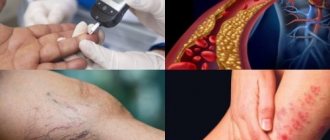
Nowadays, it’s rare for anyone to boast of healthy blood vessels in their legs. At the end of the working day, the legs begin to ache tiredly, buzz and ache, veins treacherously bulge on the skin and more and more spider veins appear.
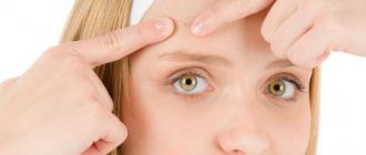
Spider veins (telangiectasia) are dilated intradermal capillaries that form a “star” on the skin. The appearance of spider veins is a signal that you need to strengthen your blood vessels. Unfortunately, this is not just a cosmetic defect, but a manifestation of the initial stage of varicose veins and a serious reason to consult a phlebologist and treat blood vessels in the legs.
Types of spider veins
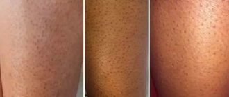
Telangiectasia can be different in color (red and blue), shape (vascular formations in the form of a “tree”, “mesh”, “star”, “spider”), localization (formed both on the face and body).
Red linear telangiectasias most often form on the face (on the nose and cheeks).
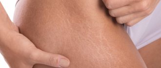
On the legs there are red or blue linear and tree-shaped spider veins, which are formed due to the bursting of blood vessels in the legs. Spider telangiectasias are usually red in color and occur on the back or abdomen
In women with spider veins on the legs, vascular formations are most often located in groups (on the inner side of the thigh they are located parallel to each other and have a linear appearance, and on the outer side they are circumferential and have a “tree-like” shape).
Causes of the disease
Spider veins appear due to the fact that intradermal capillaries lose their tone. There are many reasons for this:
- Pregnancy, postpartum period (often “stars” appear due to hormonal imbalance and compression of the pelvic vessels);
- hormone therapy;
- varicose veins;
- hereditary predisposition;
- hypertension (high blood pressure dilates the capillaries);
- chronic liver diseases;
- excessive physical stress;
- abuse of the sun, solarium, sauna, bath;
- alcohol abuse;
- prolonged load on the legs in a vertical position, etc.
Why do bruises appear on the legs?
A blue-violet spot on the skin of the lower extremities is the result of hemorrhage in the tissue or pathological expansion of the lumen of the venous vessels with stagnation of blood. Hemorrhage usually develops after bruising the tissues of the lower extremities due to a direct blow from an object or a person falling. Bruises on the legs for no reason appear due to the development of various pathological processes, which include:
- Varicose veins, which may be the result of a hereditary predisposition, blood stagnation, or a decrease in the strength of the walls of blood vessels due to a violation of their nutrition, an inflammatory reaction.
- A pronounced decrease in the strength of the walls of microcirculatory structures with insufficient intake of vitamin C, as well as some protein compounds from food.
- Violation of the functional activity of the blood coagulation system (hemostasis), in which the process of thrombus formation worsens to prevent the development of bleeding. This is possible with prolonged fasting of a person, liver failure (liver cells, hepatocytes, synthesize factors that are directly involved in blood clotting, as well as the formation of a blood clot), pathology of the blood system, and red bone marrow.
- Bruises on the legs of women are often a consequence of changes in the body's hormonal levels.
Normally, women may experience bruises on their legs after pregnancy and childbirth, which is caused by changes in the levels of various hormones, as well as congestion in the lower extremities associated with compression of the main vessels of the pelvic cavity by the enlarged uterus.
Dark spots in women and men
The causes of dark rashes on the legs may differ between men and women. Dark, almost black spots with clearly defined boundaries may indicate the development of chloasma.
This is a pathology that is accompanied by excessive accumulation of melanin in one area of the skin, which leads to hyperpigmentation.
Chloasma occurs only in women - and spots can appear not only on the legs, but also on the chest around the nipples, stomach, and face.
Skin pigmentation in men can be caused by Becker's melanosis. Most often, such spots appear at a young age and look like ordinary moles. Hair growth accelerates in darkened areas. The exact cause of the development of Becker's melanosis has not been established.
At the initial stage of development, the rashes have a light beige, pinkish tint. As they progress, they acquire a rich brown, almost black tint. Blackening of the skin indicates the onset of tissue necrosis. In such cases, emergency surgical intervention is required, otherwise gangrene may develop.
Treatment
Modern treatment for the appearance of blue spots and formations of the lower extremities is complex. It involves the use of conservative therapy or surgical intervention, the choice of which is made after the causes of the pathological process have been diagnosed. Conservative therapy includes the use of various medications that improve the functional state of hemostasis, as well as the strength of the walls of venous vessels and microcirculatory structures. Surgical treatment is carried out to remove vessels with irreversible changes in their walls.
If blue spots appear systematically for no apparent reason, you should consult a phlebologist (a medical specialist who deals with the problems of vein diseases), who will prescribe additional research and appropriate treatment.
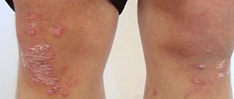
Dark spots due to dermatological diseases
Dark rashes on the shins and other parts of the legs can be caused by various dermatological diseases. These include:
- eczema;
- psoriasis;
- lichen;
- neurodermatitis;
- dermatitis.
Eczema develops against the background of disturbances in the functioning of the endocrine and nervous systems, allergies or slow metabolism. Rashes on the legs are associated with varicose eczema, which is accompanied by circulatory disorders and blood stagnation.
Most often, spots appear on the lower legs, accompanied by redness, peeling and itching.
The cause of the rash can be pityriasis versicolor - with other types of the disease the rash is red. In most cases, it appears on the upper half of the body, but sometimes it can occur on the legs.
No less often, brown spots on the legs appear with psoriasis, dermatitis, neurodermatitis or allergies. Treatment is selected depending on the disease that caused the rash. In the case of neurofibromatosis, rashes on the legs have a different color, location and area of damage. Only a dermatologist can make an accurate diagnosis after an examination.
Types of pigment spots and methods for their removal
Pigment spots can be small, large, single or multiple. They can have different shapes, usually close to oval, and different colors from light red to dark brown. The most common age spots are freckles, birthmarks and age-related pigmentation.
Freckles. This type of pigmentation is characteristic of fair-skinned people. Freckles usually appear on exposed areas of the body - the face, arms, ears, upper back and chest. Their color changes depending on the intensity of the sun. It is believed that freckles are caused by an uneven distribution of pigment in the skin, so over time, when the body gets stronger and adapts, they fade or disappear completely. For many girls, especially red-haired ones, freckles suit them - they add individuality, but this is not always the case. Too contrasting and extensive pigmentation is a reason for complexes, so they are “taken out”.
There are different ways to remove freckles. Traditional medicine, for example, recommends using parsley juice to whiten the skin. Of course, this method will not work immediately - it will take several months. Cosmetology offers much more effective methods. You can quickly remove freckles using chemical peeling with a base of fruit or lactic acids or laser skin resurfacing.
Birthmarks. Moles (nevi) can form on any part of the body. They have an even shape and a rather dark color. Unlike freckles, the melanin that forms the pigment spot is located in several layers of the skin, so removing a mole is more difficult than freckles. Moles have one peculiarity - they can degenerate into cancer, so large and suspicious formations are recommended to be removed. Birthmarks located in places of contact with the seams of clothing, belts and in areas subject to shaving are also removed. An injured mole is a gateway to infection: it can become inflamed, bleed and become wet.
A direct indication for contacting the clinic is a change in the shape, volume and color of the birthmark. Black and asymmetrical moles, bleeding or flaky spots with cracks are especially dangerous.
Moles without signs of cancer are removed with liquid nitrogen, diathermocoagulation or laser. Laser removal of birthmarks is most preferable, since it does not injure surrounding tissues and does not leave scars. And removing moles with a laser does not hurt at all, which is why many patients choose this particular technique.
Age spots. Unfortunately, these signs of impending old age cannot be prevented. Lentigo, as this type of pigmentation is called, appears in people after 40 years of age. Such age spots are especially noticeable during menopause, when pigmentation is spurred by hormonal changes. Lentigines, like freckles, are localized in places where the skin has been exposed to constant sunlight - on the face, arms, chest and back. Women, noticing such pigmentation, are upset, because they cannot hide the brown spots and they clearly show their age.
Age-related pigmentation can only be removed using cosmetic methods - traditional medicine will not help here. You can try medium or deep chemical peeling or low-impact laser removal. The advantage of the latter method is undeniable - after the procedure, only slight redness remains at the site of the spots, which quickly passes. With deep chemical peeling, the cosmetologist completely removes the top layer of skin, so recovery will take at least a month.
Large pigment spots. Melasma is a significant cosmetic problem. Unlike moles, such age spots have an uneven shape and look very unattractive. Their color intensifies in the sun - in spring and summer, and in winter the pigmentation decreases. Large pigment spots are a sign of hormonal changes, so they often occur during pregnancy, taking hormones, and the onset of menopause.
Large age spots can go away on their own, but women do not want to wait for this moment, so many resort to the help of a cosmetologist. Light peeling, special whitening masks, laser skin resurfacing, etc. can reduce the color of such spots.
Diagnosis of acute ischemia during examination by a vascular surgeon
The classic picture of acute ischemia is determined by six symptoms:
- Sudden leg pain
- Pale skin
- Absence or deficit of movement in the affected limb
- Absence of pulse in the affected limb
- Decreased skin sensitivity
- Decreased skin temperature
The pain may be constant or with passive movement of the affected limb. With an embolic blockage, the pain is usually sudden and very intense. With thrombosis, the pain intensity is much less, and sometimes there is a progressive increase in intermittent claudication.
Changes in skin color due to hyperpigmentation
In most cases, darkening of the skin of the legs is associated with hyperpigmentation. They occur in men and women over 45-55 years of age and are called age-related. Such spots can have different color saturation, size and shape. Most often, age-related pigmentation intensifies under the influence of ultraviolet radiation.
Age-related pigmentation of the skin of the legs does not require any treatment and does not cause pain or discomfort; the only concern that age spots can cause is the unsightly appearance of the legs. Age-related hyperpigmentation must be distinguished from carcinoma. This is a malignant disease that affects the tissues of the skin, mucous membranes, and various internal organs. The greatest danger is caused by spots located in the lower parts of the lower leg and rapidly increasing in size. With carcinoma, the darkened areas of the body become rough, and compactions gradually form on their surface. If such neoplasms are detected, you should immediately contact an oncologist to clarify the diagnosis.
Diagnosis of thrombosis or embolism
In addition to the clinical picture, it is necessary to use special research methods for diagnosis.
Ultrasound diagnostics makes it possible to clarify the nature of occlusion and identify atherosclerotic plaques during thrombosis. Thrombosis differs from embolism in the initial damage to the arteries; in embolism, the arteries are most often not affected.
Angiography is performed on the operating table to clarify the receiving vascular bed and allows you to determine the nature of the surgical intervention
Multislice computed tomography is performed when there is time for a detailed diagnosis and allows you to very accurately identify the nature of the lesions and determine treatment tactics.
Ultrasound duplex scanning
Ultrasound duplex scanning allows you to determine the patency of the arteries, localize the site of blockage of the vessel and the state of blood flow below the site of occlusion. Often, in acute ischemia, this diagnosis is enough to determine treatment tactics and send the patient to the operating table. With an embolism or rupture, the arteries below the blockage are usually empty or thrombosed, and blood flow in them is not detectable. Blood flow in the veins is sharply slowed down. With thrombosis, blood flow can be detected below the site of blockage, but its speed is sharply reduced; most often, blood flow through the main vessels cannot be detected, but blood flow can be seen through collaterals. As a rule, this is due to
Angiography
To resolve the issue, surgical tactics require information about the patency of the arteries of the affected limb. The choice of technique for restoring blood circulation depends on the condition of the inflow pathways to the affected limb and the vascular bed below the site of blockage. In addition, angiography can distinguish embolism from thrombosis against the background of atherosclerotic narrowing. During an angiographic examination, endovascular treatment can be undertaken in the form of thrombectomy and angioplasty of the affected segments or local thrombolytic therapy can be performed.
Arterial embolism is an acute blockage of a vessel by a thrombus or other object brought from other parts of the vascular bed. Most often, vascular surgeons deal with thromboembolism from the heart cavity during a heart attack or atrial fibrillation, from the cavity of the aneurysm of the vessel overlying the blocked artery.
Consequences of acute cessation of blood flow
The sudden cessation of blood flow leads to the phenomenon of acute ischemia. The organs and tissues that are supplied by this artery lack nutrition and oxygen, so they begin to slowly die. The most specialized tissues are affected first—nervous, muscle, and finally skin. The body tries to restore blood circulation by opening additional bypass pathways for blood flow, so sometimes tissue death is stopped. But the likelihood of such an outcome is low. In any case, acute ischemia leads either to gangrene or to the development of chronic circulatory deficiency - critical ischemia.
For liver diseases
The appearance of dark spots on the legs can be caused by hepatitis or cirrhosis of the liver. Also, rashes on the legs are characteristic of cholangitis, cholecystitis, cholelithiasis, liver failure, malignant tumors or helminthic lesions of the liver.
The liver plays the role of a kind of filter in the body, removing toxins and toxic substances. When its functioning is disrupted, toxins begin to accumulate, which leads to the formation of dark spots.
Rashes on the legs due to liver diseases can be identified by a characteristic brownish-yellowish or pinkish tint.
Most often they are round or slightly oblong, flat, arranged in a chaotic manner. Such spots are accompanied by severe itching and burning, causing serious discomfort. Liver diseases have other, additional symptoms - redness of the skin of the palms, yellowing of the sclera, the appearance of a pink rash in the abdomen and white plaque on the tongue, small vascular networks on the face, upper or lower extremities.
Stages of acute ischemia
1. Sudden pain in the leg, coldness, reduction in walking distance. If the collateral vessels are good, ischemia may stop at this stage with the development of intermittent claudication or critical ischemia. Most often, this stage is observed with thrombosis of altered arteries, if their lumen was previously narrowed and collateral circulation developed. At this stage, the operation is carried out after the necessary additional examination and preparation. The color of the leg may be pale or take on a bluish tint (cyanosis). The result of surgical treatment is excellent. Recovery of leg function is most often complete.
2. The symptoms described above are accompanied by weakness in the leg, which gradually worsens to the point of paralysis. However, passive movements in the fingers and other joints are possible. Such phenomena are associated with the death of nerve endings and blockade of nerve impulse transmission along the nerves. This stage of ischemia is an absolute indication for emergency surgery, since independent restoration of blood flow is impossible, and delay in intervention will lead to gangrene. Timely surgery restores blood flow with minimal loss of limb function. Numbness of the foot and fingers remains, and swelling of the leg persists for a long time.
3. Muscle death begins, first there are foci of muscle necrosis, muscle pain, and dense swelling of the lower leg. Then comes numbness of the fingers, ankle joint, and knee joint (muscle contracture). The muscles die completely. If the muscles are partially lost after restoration of blood flow and a long postoperative period, the leg may be preserved, but walking will be difficult. In the case of muscle contracture, amputation is necessary, since restoration of blood flow leads to the death of a person from poisoning with decay products.