Human age negatively affects the structure of the periorbital muscles and ligaments. As a result, people after 30 years of age develop wrinkles at the outer corners of the eyes, which are popularly called “crow’s feet.” The face takes on a mournful appearance, which causes negative emotions, especially in women. Disturbances in the area of the eye corners can also be a manifestation of various diseases. When performing plastic surgery, negative changes and aesthetic defects of the face are partially eliminated.
What are canthopexy and canthoplasty operations?
Canthopexy involves lifting the outer part of the canthal ligament to a higher position. This provides additional support for the tissues of the lower eyelid. The operation is usually included as part of a complex surgical procedure - blepharoplasty. When performed, the operation can eliminate asymmetrical facial characteristics and allow you to forget about a dull expression. If it is necessary to correct the shape of the eyes, then patients are prescribed canthoplasty, i.e. complete intersection of the canthal tendon, moving and fixing it in a new position. Canthoplasty is also performed after injuries when the canthal ligament is destroyed due to external influences. This is the fundamental difference between these two operations.
Canthoplasty (canthopexy) - correction of eye shape
Plastic surgery does not stand still, so every day people all over the world have a chance to become more perfect.
Recently, the procedure of canthopexy and canthoplasty has become very popular. Canthopexy and canthoplasty allow you to change the shape of the eyes, tighten the corner of the eyelids, thereby achieving an aesthetic appearance of the entire face. The procedure received its name due to the fact that any operation performed in one way or another affects the canthal tendon in the eyelid area, which is responsible for support.
This procedure is most popular among people who have severe ptosis (sagging eyelids) and too narrow/wide eye shape. Canthoplasty is also suitable for those who have drooping eyelids due to age-related changes. Canthopexy helps to “rejuvenate” the look, since the procedure tightens the lower eyelid and also removes ectropion (a case where the eyelid is slightly inverted).
Features of the intervention of a plastic surgeon
Canthopexy involves the use of local or conduction anesthesia. If necessary, general anesthesia is prescribed. The surgeon makes an incision: on the lower part of the eyelid below the eyelash line, and the length of this incision is only 1-1.5 cm. If the operation is carried out simultaneously with blepharoplasty, then the doctor additionally peels off the skin on the lower eyelid, distributes or, if necessary, removes hernial formations. Fixation of the canthal ligament is carried out to the periosteum of the superior orbital edge. For this, a special non-absorbable thread is used. With its help, a U-shaped suture is applied, holding the eyelid in the desired position. Excess skin is excised.
Canthoplasty differs from the previous operation in that the patient undergoes transection of the lateral lateral tendon. The end of the tendon, simultaneously with the area of the orbicularis muscle, is fixed in the area of the periosteum of the superior orbital edge. The duration of the operation is less than one hour. After surgery, the patient is not recommended to stay in highly lit places for a long time, read or watch TV, work with computer equipment, or visit saunas or steam baths. People who wear contact lenses should stop using such optical accessories for a while. The rehabilitation period after surgery is about 10-14 days.
Nuances of the procedure
The period for performing a simple intervention is half an hour; with combined techniques (when several tasks are performed simultaneously), the time increases to 2 hours.
When the patient can leave the clinic largely depends on the type of anesthesia. With local anesthesia - after 3 hours, with general anesthesia after 5-6 hours or the next day.
How much surgery to enlarge the eye shape costs depends on the chosen technique and the scope of the intervention. The price is negotiated before manipulation and remains unchanged.
Main indications for canthopexy and canthoplasty
Canthopexy is recommended in the following situations:
- the patient’s desire to correct the shape of the lower eyelid;
- formation of fatty deposits or hernias;
- age-related changes on the skin of the face;
- post-traumatic cases that have led to weakening of ptosis or canthus of the lower eyelids;
- bulging eyes caused by myopia or thyrotoxicosis.
The operation helps to get rid of a narrowed eye opening, partial reduction of the eyelids or a shortened palpebral fissure. The manipulations of cosmetologists allow you to forget about trachomatous entropion of the eyelid, which arose as a result of infectious conjunctivitis. A trip to a medical facility is inevitable in case of unsuccessful plastic surgeries related to eye correction. Patients who have suffered chronic inflammation, burns or injuries will need medical care.
Canthoplasty: types and results
Canthoplasty is a surgical correction of the shape of the eye, the purpose of which is to change the shape and cut (raise the outer corner higher than the inner) or (in combination with classic blepharoplasty) eliminate age-related changes in the upper part of the face.
With the help of surgery, the outer corner of the eye is raised higher than the outer corner, which changes the shape and cut. In addition to classic blepharoplasty, the operation will help completely remove age-related imperfections in this area of the face.
Figure 1. The diagram shows that the lateral canthal ligament consists of an upper and lower part. Below the lateral canthus there is a thin ligament called the inferior retinaculum (retinaculum inferior).
With the help of surgery, the outer corner of the eye is raised higher than the outer corner, which changes the shape and cut. In addition to classic blepharoplasty, the operation will help completely remove age-related imperfections in this area of the face. The canthal ligaments are responsible for the location of the corner of the eye. The height of their fixation on the periosteum of the orbital edge will determine how this area will look. There are two such connections:
- medial – holds the angle at the bridge of the nose;
- lateral – secures the outer corner of the eye.
The last type of ligament consists of 2 parts and acts as a suture with a tendon insertion between the outer lobes of the orbicularis muscle, which is located under the eyelids. There are also collagen fibers from the ends of the cartilage, which make it stronger. To determine the shape of the eyes, an axis drawn from the inner corner to the outer corner is used. Based on this feature, all cuts are divided into 3 types.
1. Classic. The most common, more common in youth. Both corners of the eye are located at the same level, the axis is drawn through the bottom of the pupil.
Figure 2. Classic eye shape.
2. Mongoloid or eastern. The outer corner is raised, the connecting axis runs in the middle of the pupil or slightly higher.
Figure 3. Eastern (Mongoloid) section of the eyes.
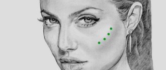
3. European or falling. The outer corner is lowered, the axis goes below the pupil. The form can be congenital or age-related. With such eyes, even a smiling face looks sad. With age, the canthal ligaments stretch and become flabby, which leads to drooping of the angle.
Figure 4. European (anti-Mongoloid, falling) eye shape.
With the help of canthoplasty, you can make your eyes slightly slanted, give them almond-shaped outlines and envelop your appearance in the flair of the mysterious East. As a result, the face is completely transformed. The surgeon cuts the canthal ligament, which supports the angle. Next, the tendon is fixed to the periosteum above the starting point.
There is another similar operation, canthopexy. It differs in that the ligaments are strengthened without incision. This method does not always achieve the desired results. And by separating and freely moving the ligaments, you can radically change the shape of the eyes.
Features of preparation for plastic surgery
Any surgical intervention should begin with a thorough examination of the patient. In addition to an ophthalmologist and a plastic surgeon, you should consult a therapist who can determine the presence of contraindications to the operation by assessing the person’s general health. Before visiting the clinic, you must take a biochemical blood test, undergo fluorography and have a cardiogram. Surgeons at the Beauty Doctor clinic advise: to avoid long-term recovery of the body’s health after surgery, you need to stop taking alcohol and nicotine products, as well as medications that contain aspirin or other blood thinners. The healing process depends on the characteristics of the human body, but lasts no more than a month. The surgical effect lasts for 10 years.
You can learn more about the operation that allows you to correct the shape of your eyes in the “Blepharoplasty” section.
How is canthopexy performed?
The method of performing the operation is very individual.
During a face-to-face examination, the doctor discusses the desired result with the patient, conducts an examination and assesses the amount of work. During the operation, an incision of no more than one centimeter horizontally is most often made above the upper eyelid. This incision helps hide the postoperative cosmetic stitch in the natural crease of the eyelid.
By means of this suture, the surgeon has access to the canthal tendon, which the surgeon subsequently attracts to the ocular periosteum. Again, in some cases, excess skin may need to be excised.
The cost of canthoplasty (canthopexy) is determined after an in-person or correspondence consultation. Approximately $500 for upper OR lower eyelids.
After canthoplasty (canthopexy)
After the operation, there is slight swelling of the eyes, and possible accumulation of blood in the form of hematomas. However, the recovery period is very short: usually within a week, patients can return to their previous work. The pain is minimal and can be relieved with painkillers.
The postoperative suture may subsequently disappear almost completely. In any case, it is absolutely invisible in the natural crease of the eyelid.
The result can be observed immediately after the swelling subsides. The effect of canthoplasty can last more than 10 years.
Our clinics are equipped with the latest equipment, which plastic surgeons have mastered to perfection. That is why the recovery period is minimal, and surgical traces are completely invisible. Another undeniable plus is the complete confidentiality of each patient.
It is necessary to understand that in the case of various plastic manipulations, the qualifications of the doctor are very important . Strict quality control is carried out in Belarus, especially with patients from abroad. Take advantage of the services of our company, because we cooperate with the best doctors and clinics in Belarus
What is contopexy
Every woman wants her eyes to be alluring and captivate any man. In youth, it’s easy to maintain the beauty of your eyes: properly moisturize this delicate area, which is prone to the earliest appearance of signs of aging. But the skin around the eyes itself is thin and contains a small amount of collagen. Tonic, cream, mask - with the help of this complex the area around the eyes will be in normal balance until a certain point. With age, irreversible changes occur in the skin and soft tissues of the upper and lower eyelids, as a result of which blepharoplasty surgery becomes the only radical way to rejuvenate the area around the eyes. Today, there are many types of eyelid surgery, and now we will focus on one of them. We will talk about canthopexy.
Progress of the operation
Europeanization of the eyelids is a popular procedure for people with oriental eye shape and heavily overhanging eyelids. For the operation to be successful, it is important to follow all the doctors’ recommendations.
Progression of epicanthoplasty:
- The plastic surgeon applies preoperative markings according to which the skin fold will be removed.
- The visual organs are treated with an antiseptic solution.
- The patient is given local infiltration anesthesia.
- The cuts are made according to the intended markings. To eliminate a small epicanthus, excision of the upper mobile skin above the eye is sufficient. If the fold is pronounced, plastic surgery of the inner corner is performed.
- Remove excess skin.
- Cosmetic stitches are applied.
Epicanthoplasty is combined with Singapore eyelid plastic surgery.
Complications
When performed by a licensed, experienced surgeon, epicanthoplasty is a safe procedure with high success rates. However, like any surgical procedure, it leads to complications:
- Allergic reaction to anesthetic . Complication is unlikely. Therefore, a skin test should be performed before using the anesthetic.
- Infection . An extremely rare complication. Occurs when incisions become infected with surgical instruments. Treated with antibiotics.
- Poor scarring . Not all patients' skin heals quickly and grows together. Scars may remain visible for several months.
Postoperative complications of epicanthoplasty are very rare. In practice, the procedure is easily performed without any problems.
Types of canthopexy
It comes in two types – lateral (external, external) and medial (internal). In addition, canthopexy and blepharoplasty . Let's take a closer look at each variety.
- Lateral is a surgical intervention only in the area of the outer canthus. It is what is used to give the eyes the coveted “cat-like” cut and shape. And in principle, it is most often practiced by plastic surgeons.
- Medial - performed less frequently, involves surgery on the inner corner of the eye (as a rule, this manipulation is carried out in cases of some kind of injury or damage to the eye).
- Canthopexy with blepharoplasty involves surgery in the area of the corners of the eyes to correct the incision and shape of the palpebral fissures in combination with eyelid surgery. Based on the locus of the operation, blepharoplasty is designated by surgeons as upper (the intervention is carried out on the upper eyelid), as well as lower (correction of the lower eyelid) and circular (upper plus lower eyelid). Used to eliminate hernial “bags” or eye asymmetry.
Rehabilitation
After surgery, the patient experiences minor swelling for 10–15 days after the sutures are removed. The final result will appear no earlier than in a month.
During rehabilitation, follow the instructions of a specialist:
- Keep the wound clean and dry. Wash with clean water, do not use cosmetics, so as not to provoke the development of infection.
- Wear sunglasses for the first week after surgery.
- Avoid drinking a lot of water before bed to avoid eye puffiness.
- Do not smoke, drink alcohol or eat spicy foods to speed up wound healing.
- For 3 days, use antibacterial drops and apply cold compresses to the eyelids.
- CL should not be worn for 14 days.
After 1 month, you need to see a surgeon. He will tell you when you can use cosmetics and exercise.
After surgery, it is recommended to undergo a course of rehabilitation physiotherapeutic procedures (microcurrents). They help reduce swelling and bruises will disappear faster.
In what cases can epicanthoplasty be performed?
Epicanthoplasty is a cosmetic manipulation; every patient who has no contraindications is allowed to use it . Most patients are of Asian descent, but the surgery is performed for cosmetic defects in one eye.
It is an ideal procedure for male or female patients with the following indications:
- severe drooping of the eyelids caused by skin folds;
- large amount of fat in the upper eyelid area;
- tissue ptosis;
- narrow shape
- congenital anomaly.
Indications for implementation are external reasons. It is performed if the Mongolian fold occurs due to the formation of scars after injury.
Stages of canthopexy with blepharoplasty
- The incision, access to the lateral ligament, tightening and fixing it are carried out in a similar way.
- If excess skin or subcutaneous fat remains noticeable in the lower eyelid area, they are removed. Excess tissue is excised with a scalpel. Then the wound is sutured.
Thus, blepharoplasty involves excision of excess integumentary tissue in the projection of the upper and/or lower eyelid for cosmetic purposes or for purely medical reasons (for example, for a hernia). And it can be combined with other plastic surgeries.
Without a doubt, canthopexy will help restore the former brightness of your eyes. Moscow offers the services of many medical centers for this, but this plastic surgery is most successfully performed in Podolsk at the SOLEI Plastic and Aesthetic Surgery Clinic. Moreover, by modern standards, the cost of this operation is one of the most affordable in the Moscow region.
Indications for canthopexy
- Reduced elasticity and “stretching” of the lower eyelid, i.e. its horizontal elongation due to stretching of the canthal ligaments (mainly lateral);
- decreased tone of the orbicularis oculi muscle and its displacement below the edge of the cartilaginous (tarsal) plate;
- weakening or separation of the orbicularis oculi muscle, which is responsible for contraction of the lower eyelid with age or due to injury;
- Age-related depression of the eyes due to atrophy of the retrobulbar (behind-the-eye) fatty tissue (age-related enophthalmos).
Figure 1. The diagram shows that the lateral canthal ligament consists of an upper and lower part. Just below there is a thin ligament called the inferior retinaculum (retinaculum inferior).
In addition to external signs of lower eyelid weakness, you can perform a pinch test to determine its laxity. It is necessary to take the fold of skin of the lower eyelid in its central part, pull it down and release it. Normally, the eyelid immediately returns to its original position. If the lower eyelid can be pulled back by more than 6 mm and it slowly, “reluctantly” returns to its original position, this indicates weakness of the lower eyelid.
In most people, the most prominent point of the cornea and the zygomatic eminence are at the same level - this is called the “neutral vector”. If the zygomatic eminence protrudes forward in relation to the eye - “positive wind”.
In Figure 2 “Neutral vector” - red dotted line - the eyeball and the zygomatic eminence are on the same line.
In Figure 3, the black dotted line depicts a “positive vector” - the cheekbone protrudes forward in relation to the most protruding point of the cornea of the eyeball. The “neutral vector” is marked with a red dotted line.
“Neutral” and “positive vectors” indicate good development of the skeleton and soft tissues of the face, providing good support and support to the lower eyelid.
When measuring with a ruler the distance from the outer orbital edge to the most protruding point of the cornea, the result will be 15-17 mm. Ophthalmologists call this position of the eyeball normophthalmos. If the value is less than 15 mm, the eye is located inside relative to the inferior orbital margin, i.e. in the enophthalmos position.
In Figure 4, the black dotted line shows a “negative vector” - in profile, the eyeball protrudes forward in relation to the cheekbone, the eyes seem bulging. The “neutral vector” is marked with a red dotted line.
The “vector” is “negative” if the orbital edge is flat, smoothed and the eye protrudes forward relative to it, in which case the lower eyelid has no support and, in turn, does not provide sufficient support to the eyeball.
The “negative vector”, as a rule, is caused by underdevelopment of the facial skeleton and a deficiency of soft tissues in the midface. In such people, under the influence of gravity, “weakness” of the lower eyelids, ptosis (drooping, sagging) of the lower eyelids and cheeks appears quite quickly, compared to patients who have more developed bones of the facial skull.
In the presence of a “negative vector”, the eyeball is moved forward in relation to the cheekbone by more than 17 mm, this is called exophthalmos. The latter can be congenital (with the so-called “shallow orbit”, characteristic mainly of the Negroid race), or also appear as a result of age-related changes, endocrine disorders or volumetric processes in the brain skull (in such cases, it is most often one-sided).
Performing classic lower blepharoplasty when weakness of the lower eyelid is detected, especially in combination with the presence of a “negative vector,” is dangerous, since even minor removal of eyelid skin can lead to eversion - ectropion. In such cases, eyelid surgery is complemented by canthopexy or endoscopic midface lift.
Canthopexy technique
Rice. 2. The changes in the lower eyelid that occur with age are schematically depicted. In addition to excess skin, a fine network of wrinkles and “hernial sacs” of varying degrees of severity, a decrease in the tone and elasticity of the lower eyelid is noteworthy. The “weakness” of the lower eyelid manifests itself as follows: it does not fit tightly to the eye, moves forward a little, sags, and seems to be “pulled” down. The outer corner of the eye drops below the inner one (normally, in young people, the outer corner of the eye is at the same level or 2 - 3 mm higher than the inner one). As a result, the eyes appear bulging, rounded, and a white stripe of sclera is visible between the cornea and the edge of the lower eyelid. When viewed directly, the slightly expanded costal edges of the lower eyelids are visible. Progression of lower eyelid laxity can lead to ectropion.
Rice. 3. The dotted line shows preoperative markings.
The markings for canthopexy are performed in the operating room and are similar to those for lower blepharoplasty. The line is drawn 1-2 mm away from the ciliary edge of the lower eyelid. The main difference is a small 0.7-10 mm incision, which will be located in the sub-eyebrow area.
Rice. 4. Making an incision with a scalpel along the lines of preoperative markings.
Rice. 5. Fixation with an absorbable thread of the orbicularis muscle of the lower eyelid to the periosteum of the upper orbital edge (subeyebrow region).
Rice. 6. Application of intradermal sutures.
Even if eyelid correction is performed under general anesthesia, the tissues of the lower eyelid are first infiltrated (flooded) with saline solution with the addition of an anesthetic and vasoconstrictor drugs. Then the skin is dissected with a scalpel along the lines of preoperative markings. In most cases, the plastic surgeon begins with a standard lower blepharoplasty: peeling away the skin, highlighting and removing the hernia bags.
After this, through an additional incision in the sub-eyebrow area, the periosteum is sutured with an absorbable thread, which is passed under the canthal ligament, the orbicularis oculi muscle is captured in the area of the outer corner, and then the thread is drawn in the opposite direction under the canthal ligament and finally fixed to the periosteum with a U-shaped suture. An internal suture placed in a similar manner ( canthopexy)
) will firmly hold the lower eyelid in the desired position.
The eyelid correction ends with excision of excess skin and application of an internal suture with a non-absorbable thread so that only two nodules are exposed to the skin: at the beginning and at the end of the incision. The stitches are removed on day 5. Postoperative scars will quickly fade and become invisible.
Rice. 7. The figure schematically shows a supporting canthopexy.
Supportive canthopexy differs in that it involves only the canthal ligaments, which are sutured together and fixed to the periosteum of the orbit above the lateral ligament of the eyelids.
The eyes are the area of our face that receives the most attention. When meeting for the first time, the first visual contact occurs using the eyes. The eyes most quickly display age or a hint of it. You can convey many feelings and emotions through your eyes: disappointment, joy, hope, regret.
If they say: “The eyes glow”, then they mean a cheerful, radiant look. In order for this look to remain as long as possible, you need to approach this problem competently. The canthopexy operation is absolutely safe and is performed in a short time, and the effect will last from 5 to 10 years.
Epicanthus
The epicanthus or Mongolian fold is a semilunar fold of skin on the medial canthus. This condition does not cause any symptoms and is a cosmetic defect.
It is observed in representatives of the Australo-Negroid and Mongoloid races. For Europeans - atypical.
Possible darkening of the visual axis, associated ptosis, strabismus.
Scientists believe that in people of the Mongoloid race it appears as a protective apparatus for the organs of vision from specific climatic conditions. The development of epicanthus leads to:
- ptosis;
- avulsion of the medial canthus after injury;
- Down syndrome.