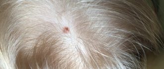
As a rule, atheroma occurs in those places where hair is developed. Therefore, the head, rich in sebaceous ducts, often acts as their incubator.
Atheroma on the head is caused by blockage of the sebaceous glands. The reasons are different - for example, as a consequence of injury or against the background of hormonal imbalance.
However, there is another reason. Remember the words of the heroine Ekaterina Vasilyeva from the movie “An Ordinary Miracle”: “What the hell do I need my head for when it hasn’t been washed for three days?” - she asks rightly. Yes, atheroma of the scalp can also occur due to the banal inability to keep hair clean.
Atheroma of the scalp can be congenital. This is a rare case when it can remain unchanged for many years. Or it may be acquired. Such atheroma occurs at any age and can literally grow from the size of a pea to the size of a chicken egg in just a matter of days.
!
In any case, atheroma is a disease that requires the attention of a doctor. And the sooner the better.
Firstly, the signs of atheroma are very similar to the symptoms of another neoplasm - lipoma; often only a professional diagnostician can distinguish them.
Secondly, a bump on the head attracts a lot of unhealthy attention.
In addition, a rapidly growing large atheroma resembles a cerebral hernia (brain prolapse). So, as is the case with any neoplasms, you cannot wait, nothing itself will ever resolve
!
What to do if warts appear on your head?
The likelihood that warts will disappear on their own in the near future is very low.
They do not go away even under the influence of mysterious spells, medicinal herbs or other methods that the boundless human imagination can come up with.
Perhaps the warts really are regressing.
But this can happen only in a few years, or even decades.
At the same time, they bother most people.
Even if the wart is overgrown with hair and is not visible, in any case it interferes with cutting and combing.
She is easily injured.
It may bleed, and sometimes the resulting wound festeres.
The wart should be removed.
This procedure does not cause pain, takes a few minutes and allows you to forget about the problem once and for all.
If a person does not see a doctor and is not treated, the wart may increase in size.
This does not pose a direct threat to health.
But subsequent removal requires treatment of a larger area of the head.
There is a higher risk that a scar will appear and hair in this area will no longer grow.
Therefore, it is better to remove a wart on the head earlier, while it is still small.
Should my partner get tested for HPV?
- Women should follow routine women's health recommendations, which include regular cervical smears.
- Men do not need to undergo any special examinations or tests as there are no routine or routine HPV tests available for them.
The chance of your partner developing cancer caused by HPV is very low. If your partner has symptoms or concerns, you should discuss this with your doctor.
to come back to the beginning
Warts on the head: is conservative treatment possible?
Conservative treatment of warts on the head is possible, although it is not always effective.
Are used:
- applications of lactic salicylic collodion
- 40% salicylic patch
The use of these techniques is difficult given the location of the wart.
Most people have hair on their heads.
Therefore, applying the patch can be very painful, and also ruins your hairstyle.
Treatment with drugs is also difficult.
They wet the hair.
With a lot of hair, an adequate dose does not always reach the goal.
Therefore, conservative measures to combat warts of the scalp are often ineffective.
It is much easier to remove them using physical methods in one procedure.
Advantages of this approach:
- one-time procedure
- no spread of warts to adjacent areas of skin
- minimal risk of relapse
- 100% effective (after removal, the wart will definitely disappear, which cannot be said about conservative treatment or chemical coagulation)
Curettage is sometimes used under local anesthesia.
Rehabilitation period after removal
The duration of the rehabilitation period after laser removal depends on the area of intervention and averages 7 days. After the procedure, a scab forms at the site of laser treatment, which cannot be removed prematurely. Therefore, the treated area of skin should not be scratched/damaged in any other mechanical way. During the rehabilitation period, sun exposure to the laser treatment area should be avoided and visits to the solarium, swimming pool and sauna should be avoided.
As a rule, the rehabilitation period after surgery to remove moles on the head is quite short, since it is a low-traumatic intervention. The duration of complete recovery depends on the size of the removed lesion and on average takes from 7 to 14 days. During this period, it is necessary to regularly treat the suture in accordance with the doctor’s recommendations, eliminate any mechanical impact on the operated area, sleep in a position in which the pillow does not injure the suture, and avoid visiting the sauna, bathhouse, solarium and swimming pool.
Is it painful to remove warts?
Removing warts using cryodestruction is painful.
Painful sensations are observed both during the period of removal and after it, when an inflammatory reaction is formed.
If electrocoagulation or laser is used, there is no pain.
Because anesthesia is used.
When a wart is located on the scalp, topical anesthesia is usually used.
A cream containing prilocaine and lidocaine is applied to the skin.
After a few minutes, skin sensitivity disappears.
The doctor can remove the wart, and the patient does not feel anything during the procedure.
After its completion, when the anesthesia wears off, there is no pain either.
What about tobacco and alcohol?
People who smoke and drink alcohol are more likely to develop head and neck cancer. However, cancers caused by HPV can develop regardless of whether you drink alcohol or use tobacco products. People with cancer, non-smokers and people who don't drink alcohol have a longer life expectancy and are less likely to develop new types of cancer. For this reason, people with head and neck cancer should stop smoking and limit their alcohol intake. If you need help breaking these habits, MSK can help. Ask your doctor or nurse for more information about our programs, or call the Counseling Center at 646-888-0200.
to come back to the beginning
Kinds
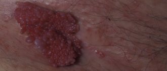
Warts that appear on the head are divided into separate categories. Simple or ordinary types of growths are among the most common types of formations.
In shape they resemble a large mole or genital papilloma.
The color is most often gray-yellow or brown.
Other types of warts on the head include the following:
- Thread-like growths (the name indicates the shape of the growth, and their color is most often pink or flesh-colored).
- Flat types (raise slightly above the skin and have a brownish color).
- Genital condylomas (clearly rise above the surface of the skin; their color can range from flesh-colored to almost black).
- Chicken.
Depending on the type of origin, growths on the head can be keratotic or viral .
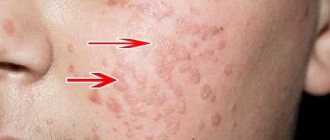
The first group is the category of warts, which are most often called seborrheic or age-related. They usually appear in older people. Visually, they resemble moles of different sizes and shapes.
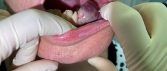
The types described above can appear in women, men and children on:
- legs and feet;
- hands and under the arms;
- face and neck;
- less common on the lips and tongue.
Reasons for development
It is still unclear why soft tissue sarcomas occur, but certain risk factors have been identified.
Genetic predisposition
- nevoid basal cell syndrome (Gorlin syndrome);
- neurofibromatosis (von Recklinghausen disease);
- tuberous sclerosis (Bourneville disease);
- Gardner's syndrome;
- Werner's syndrome.
Carcinogens
The risk of developing angiosarcomas increases significantly with previous exposure to phenoxy herbicides (2,4-dichlorophenoxyacetic acid, 2,4,5-trichlorophenoxyacetic acid) and dioxins.
Immunosuppression
The most striking example is Kaposi's sarcoma in people with AIDS, autoimmune hemolytic anemia, and organ transplant recipients.
Most patients come with a complaint of tumor growth after an injury or bruise. There is no connection between trauma and the development of soft tissue sarcomas.
Diagnosis of skin cancer
An integrated approach to diagnosing skin cancer is the key to timely detection of the tumor process. In order to determine the presence of skin cancer, it is necessary to conduct an examination. The first signs of skin cancer can be detected on your own. For this purpose, the ACORD principle is used.
- A – asymmetry. A lesion that has an uneven and asymmetrical shape requires close attention from a doctor.
- K – edge. A malignant neoplasm of the skin is characterized by jagged edges. The appearance of such a sign requires immediate contact with a dermatologist.
- O – coloring. Skin cancer is characterized by the appearance of a red, dark blue, or black lesion.
- R – size. The tumor process often has a diameter greater than 6 mm.
- D – dynamics. The growth of education in size requires a visit to a doctor.
The appearance of any of the above symptoms requires immediate medical attention. At the appointment, the specialist examines the formation using an epiluminescent microscope, which allows him to differentiate various skin diseases.
- Collecting anamnesis of illness and life. At the doctor's appointment, complaints are collected, the time of occurrence of the pathological formation, and the presence of predisposing factors. Precancerous diseases are determined, and the rate of development of the lesion is determined.
- General analysis of urine and blood. Laboratory diagnostics allows us to determine the presence of an inflammatory process in the body.
- Blood test for tumor markers. Specific markers for the presence of skin cancer are TA 90 and SU 100. Their determination is diagnosed already in the early stages of the tumor process.
- Tissue biopsy of pathological formation. It is one of the most important methods for diagnosing skin cancer. Histological examination makes it possible to differentiate the neoplasm, determine its stage and type. In cases where there is a metastatic lesion, a biopsy of the affected areas is performed.
- MRI and CT diagnostics. Instrumental research methods are carried out to identify metastases.
- PET-CT. This diagnostic method allows you to determine the localization of metastases. The study is carried out using a contrast agent injected into the body before tomography.
- Ultrasound. Ultrasound diagnostics helps to identify damage to the lymph nodes, as well as changes in other organs if metastases are suspected.
Detection of skin cancer at the initial stage of development allows for timely treatment and provides a favorable prognosis. Therefore, if you have the first signs of the disease, you should seek medical help.